Presenting images of a 65-year-old asymptomatic woman with left parotid mass.
1. Benign mixed tumor
2. Non-Hodgkin lymphoma
3. Carcinoma
4. None of the above
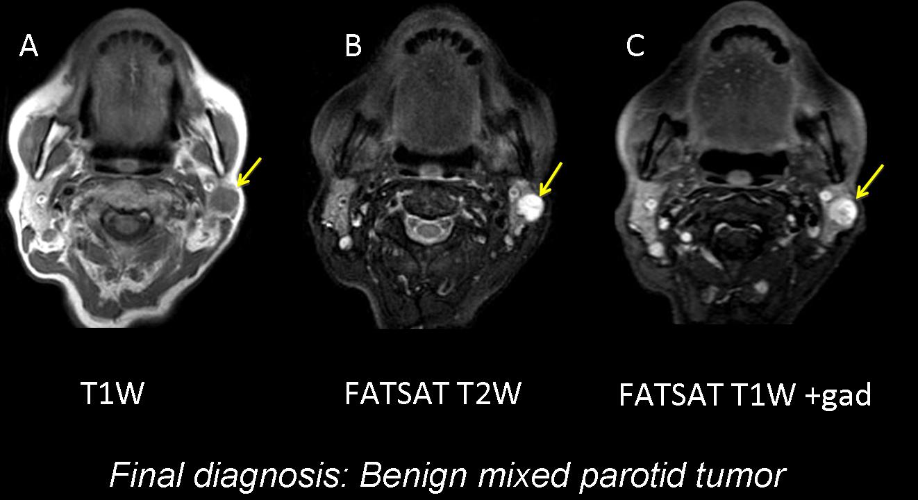
Findings: well-marginated round mass in the superficial lobe of the left parotid (arrows). Hypointense on T1W image (A), uniform high signal on T2W image (B), and homogeneous enhancement with gadolinium (C). Asymptomatic masses arising from the parotid are almost always benign mixed tumours, and the imaging findings support this diagnosis.
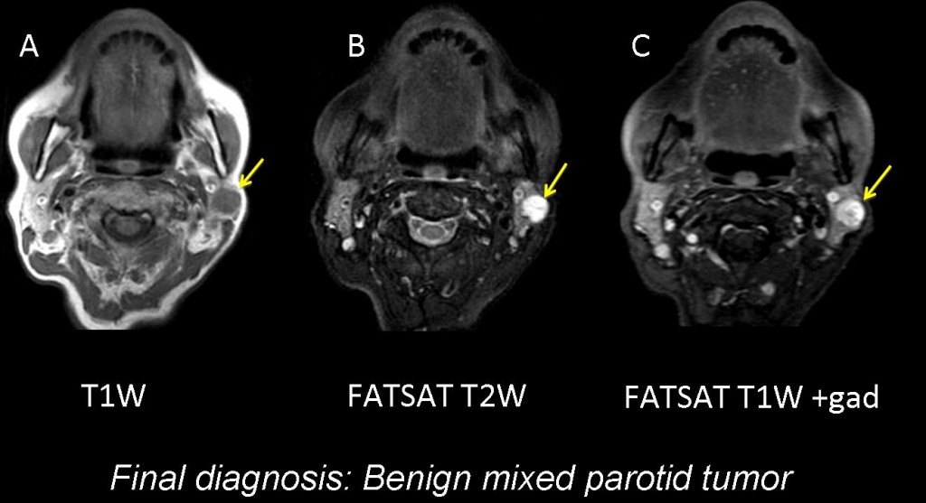
Fig. 1
Benign mixed tumours (pleomorphic adenoma) are the most common parotid tumours. They usually originate in the superficial lobe and occur more often in women.
On MRI, they appear as well-demarcated homogeneous masses with smooth margins that are hypointense on T1W images, hyperintense on T2W images, and show homogeneous enhancement with gadolinium. They can present dystrophic calcification and may have a low signal intensity capsule.
Aside from benign mixed tumours, the differential diagnosis of parotid tumours includes Warthin tumour, non-Hodgkin lymphoma, adenoid cystic carcinoma, mucoepidermoid carcinoma, and nodal metastasis.
Warthin tumour is the second most common benign parotid tumour. 20% are multicentric unilateral or bilateral masses with a cystic component.
Primary lymphomas account for less than 5% of parotid tumours. Patients with Sjögren disease are at an increased risk of developing B-cell non-Hodgkin’s lymphoma. Most of these tumours arise in extranodal mucosal sites, especially the salivary glands.
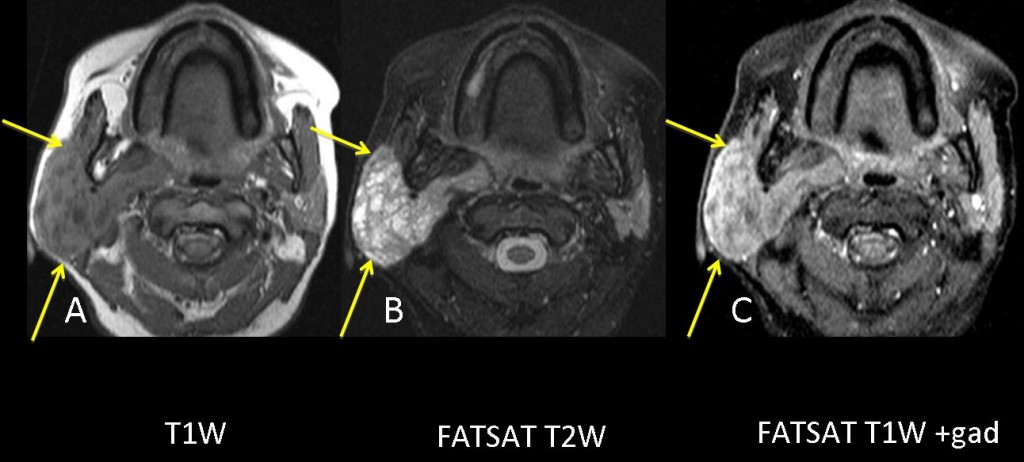
Fig. 2
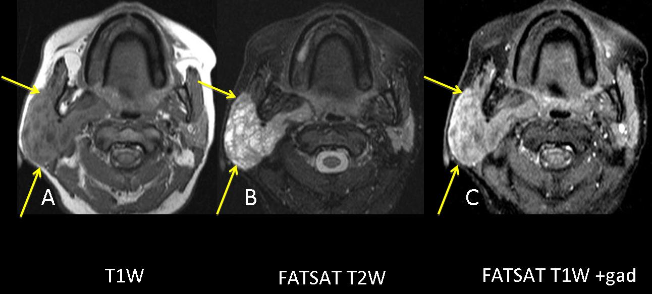
44-year-old woman with Sjögren disease and a right parotid mass. MRI shows enlarged right parotid with multiple intraparotid cystic lesions: hypointense on T1W image (A), hyperintense on T2W image (B), and heterogeneous enhancement with gadolinium (C). Involvement of the superficial and deep lobe of the parotid (arrows). Diagnosis: B-cell non-Hodgkin lymphoma.
Squamous cell carcinomas of the parotid are rare. They presumably arise from the Stensen duct.
Parotid nodes are the first-order nodal site for skin lesions of the upper face. It is not unusual to find metastatic intraparotid lymph node enlargement arising from primary squamous cell tumors of skin.
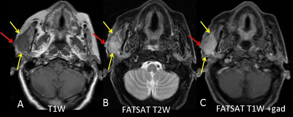
Fig. 3
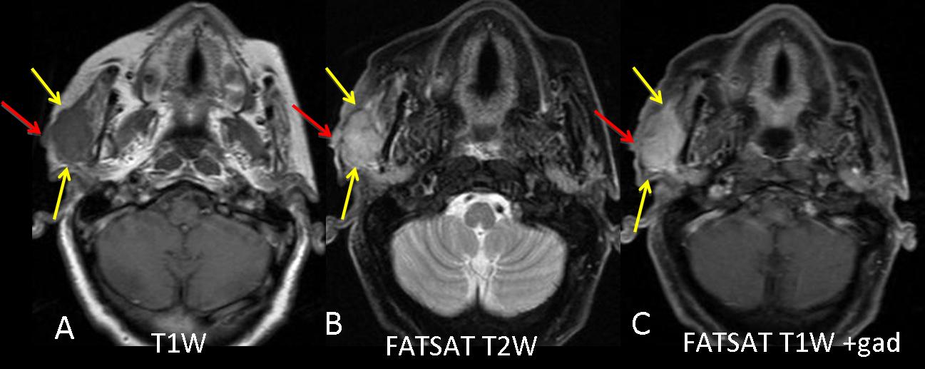
90-year-old woman with a painful parotid mass and skin lesion in the right parotid region. Poorly defined, superficial lobe, intraparotid mass: hypointense on T1W image (A, arrows), intermediate/hyperintense on T2W image (B, arrows) and heterogeneous enhancement with gadolinium (C, arrows). The mass extends to and comes into contact with the skin (red arrows). Diagnosis: squamous cell carcinoma.

Follow Dr. Pepe’s advice:
- The differential diagnosis of parotid masses includes benign mixed tumors, Warthin tumor, carcinoma, lymphoma, and nodal metastasis.
- Benign mixed tumors represent 60% to 70% of parotid tumors. They are more common in women and are usually unilateral with well-defined margins.
- Sjögren syndrome may be associated with non-Hodgkin lymphoma of parotid gland.
Recommended reading:
MR imaging of parotid tumors: typical lesion characteristics in MR imaging improve discrimination between benign and malignant disease. Christe A, Waldherr C, Hallett R, Zbaeren P, Thoeny H. AJNR Am J Neuroradiol. 2011 32:1202-7
CT and MR images of pleomorphic adenoma in major and minor salivary glands. Kakimoto N, Gamoh S, Tamaki J, Kishino M, Murakami S, Furukawa S. Eur J Radiol. 2009; 69:464-72. Epub 2008 Feb 21.
Case presented by Laura Oleaga, MD