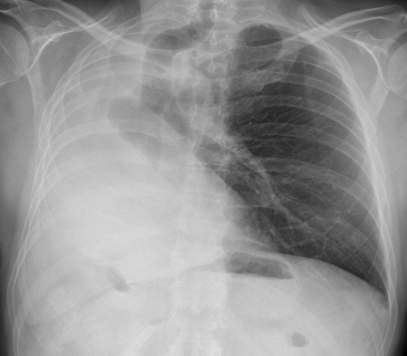
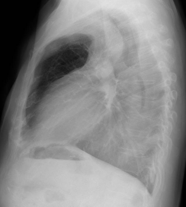
Showing you the last case before Summer vacation (July and August). Presenting radiographs of a 47-year-old man with moderate dyspnea.
1. Collapse of right lung
2. Right pneumonectomy
3. Congenital absence of right lung
4. None of the above
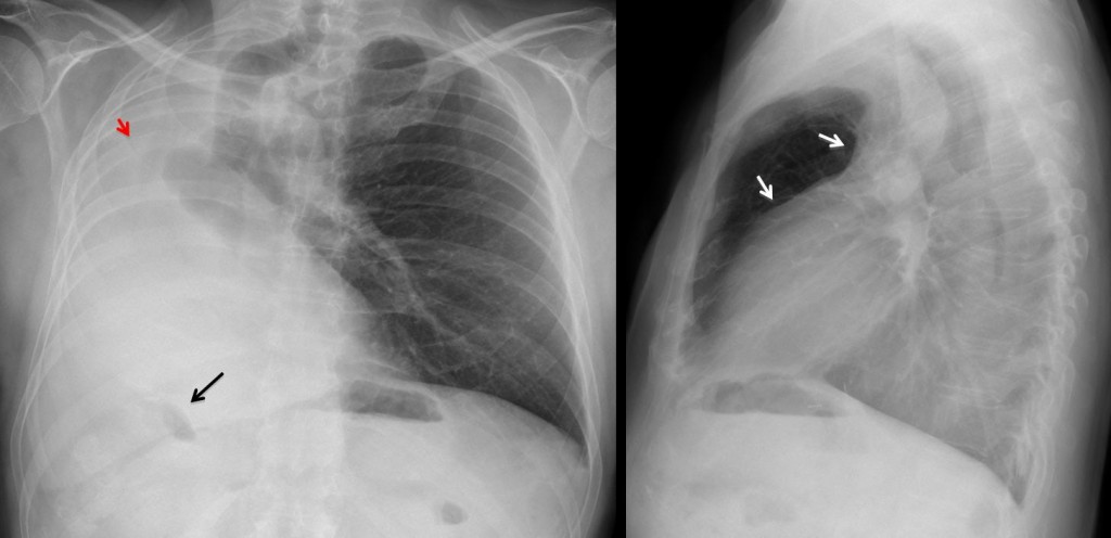
Fig. 1
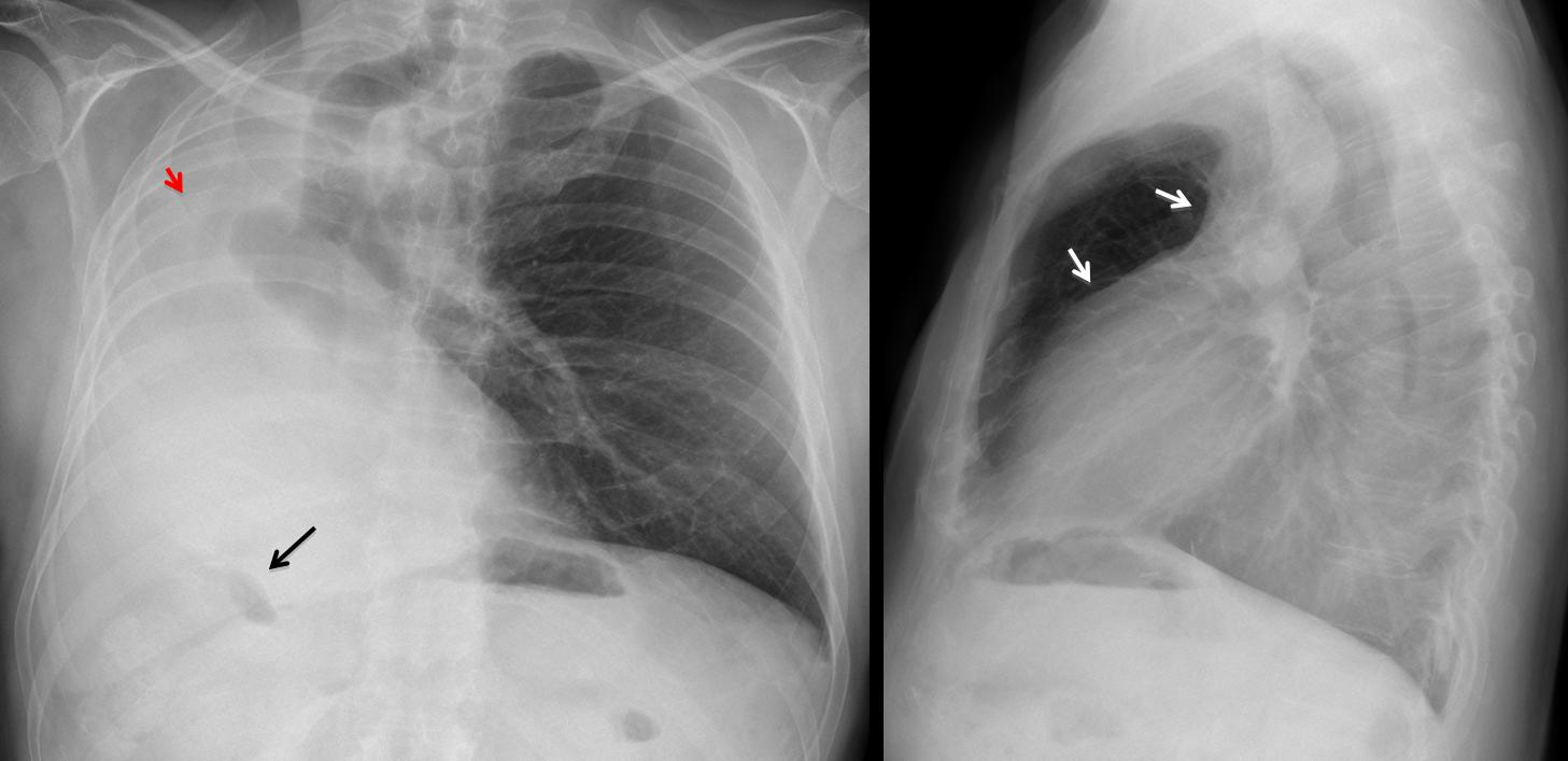
Findings: White hemithorax with marked shift of mediastinal structures. Elevation of right hemidiaphragm, indicated by the high position of the hepatic flexure of colon (arrow). Lateral view shows anterior herniation of opposite lung (arrows). All these findings indicate a considerable volume loss of right lung, which is consistent with all three diagnoses offered. However, in this particular case, the slight positional asymmetry of the sixth rib (red arrow) suggests a previous pneumonectomy.
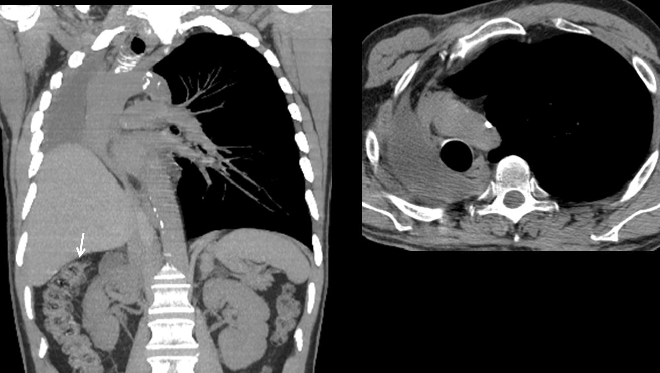
CT shows the post-pneumonectomy space filled with pleural fluid, confirming the diagnosis. The liver partially occupies the right hemithorax. Note the marked elevation of the hepatic flexure (arrow).
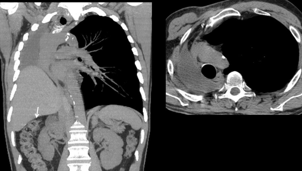
Fig. 2
Final diagnosis: right lung pneumonectomy
The differential diagnosis of a white hemithorax mainly includes lung collapse and massive pleural effusion. The diagnosis is easily established by determining the position of the mediastinal structures, among which the trachea is the easiest to visualize. Massive pleural effusion will displace the trachea towards the opposite side, whereas loss of lung volume will attract the trachea to the affected side.
Other signs of volume loss are diaphragmatic elevation, as signaled by air in the hepatic flexure or in the gastric fornix on the left side and varying degrees of anterior herniation in the lateral view.
Pneumonectomy is one of the most common causes of white hemithorax with volume loss. Aside from the clinical history, look for postoperative changes at the ribs and surgical clips.
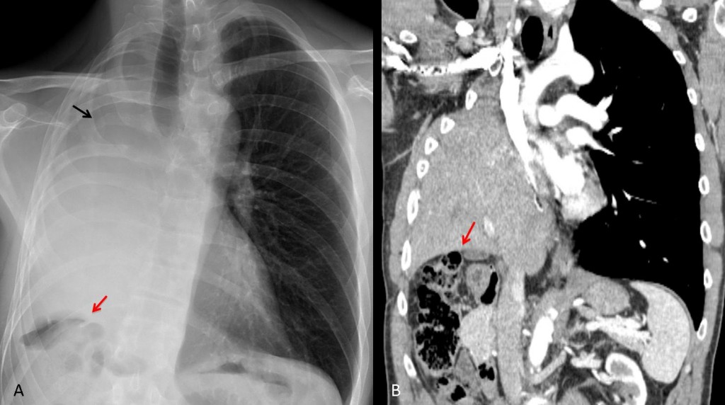
Fig. 3
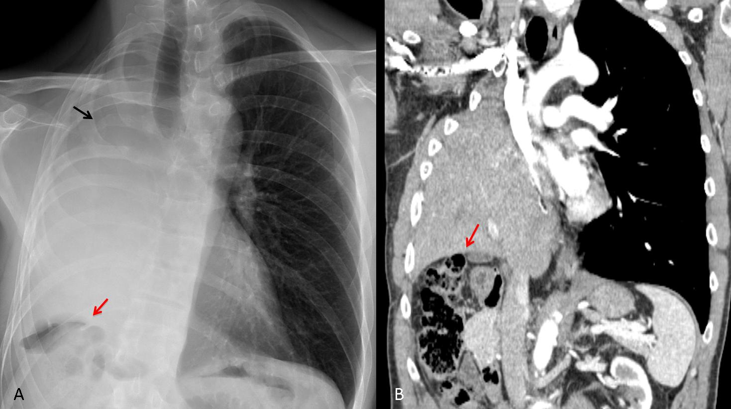
Fig. 3. 54-year-old male after right pneumonectomy with diaphragm resection. The liver is herniated into the right hemithorax. Note the postoperative changes at the right sixth rib (A, black arrow) and the high position of the hepatic flexure (A, B, red arrows)
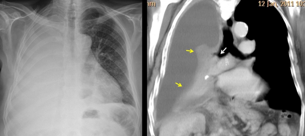
Fig. 4
Fig. 4. In patients who have not undergone surgery, carcinoma of the main bronchus with complete lung collapse is the most common cause of white hemithorax. Mediastinal shift is usually not very marked because the associated pleural effusion compensates for the volume loss. Thus, it is not unusual for the trachea to stay in the midline, as in this case.
CT shows the endobronchial tumour (white arrow), with lung collapse (yellow arrows) and associated pleural effusion.
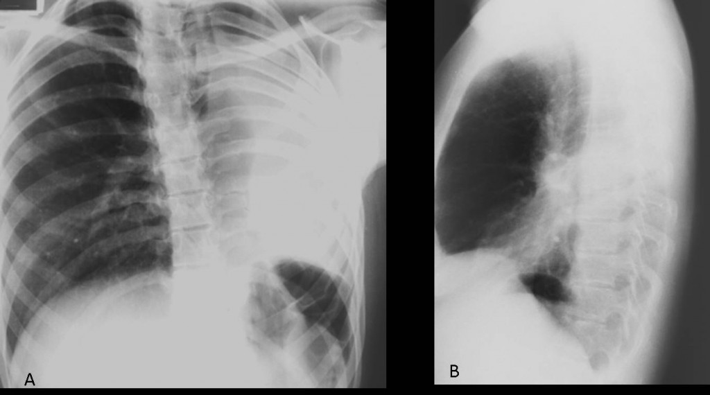
Fig. 5
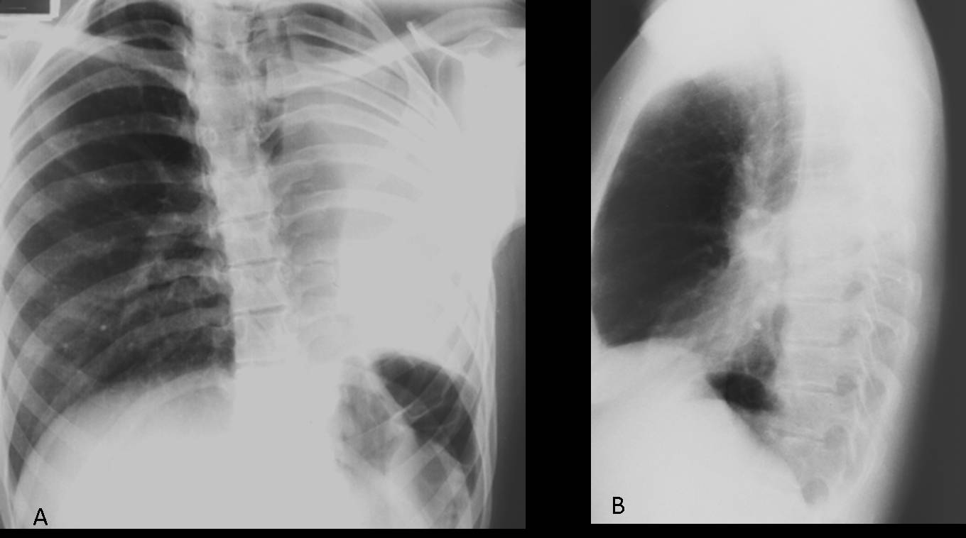
Fig. 5. Congenital lung malformations are not uncommon findings in adults. Agenesis of one lung may affect either side. A small airless hemithorax is seen, with crowding of the ribs and herniation of the contralateral lung across the anterior clear space (A,B arrows).
In an asymptomatic patient with no previous surgery, these features should suggest agenesis of the lung.
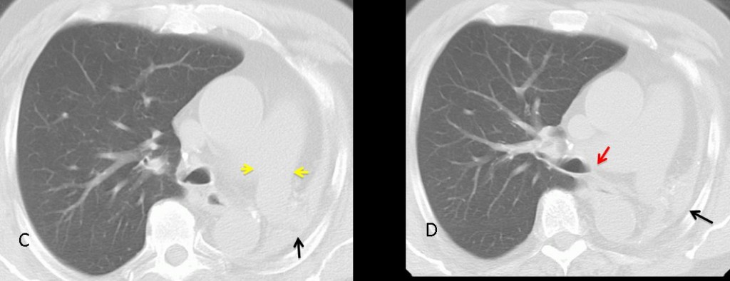
Fig. 6
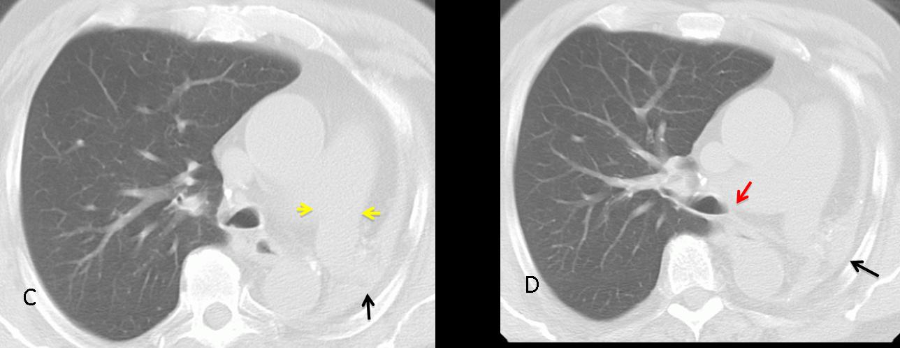
Fig. 6. CT shows a small remnant of hepatized lung tissue (C,D back arrows). The left main bronchus is absent (D, red arrow), although the homolateral pulmonary artery is usually of normal size (C, yellow arrows). Note the herniation of the right lung behind the sternum.
Most patients are asymptomatic and no treatment is necessary. Surgical resection is indicated for the few patients with lung remnants and recurrent infection.

Follow Dr. Pepe’s advice:
- A white hemithorax with mediastinal shift towards the affected side suggests a central carcinoma with secondary collapse or a previous pneumonectomy.
- If no clinical information is available, look for postoperative changes at the ribs and surgical clips.
- In an asymptomatic patient with no previous surgery, consider congenital agenesis of one lung.
Case prepared by Dr. Pepe