Dr. Pepe’s Diploma Casebook: Case 47 – SOLVED!

Dear Friends,
Today I’m showing a vintage case, seen thirty years ago when I was a promising young staffer. The images below belong to a 57-year-old missionary living in Africa and undergoing yearly controls at our institution for unilateral hyperlucent lung.
Leave me your thoughts and diagnosis in the comments section and come back on Friday for the solution.
Diagnosis:
1. Swyer-James/Macleod syndrome
2. Bronchial tumour
3. Pulmonary artery stenosis
4. None of the above

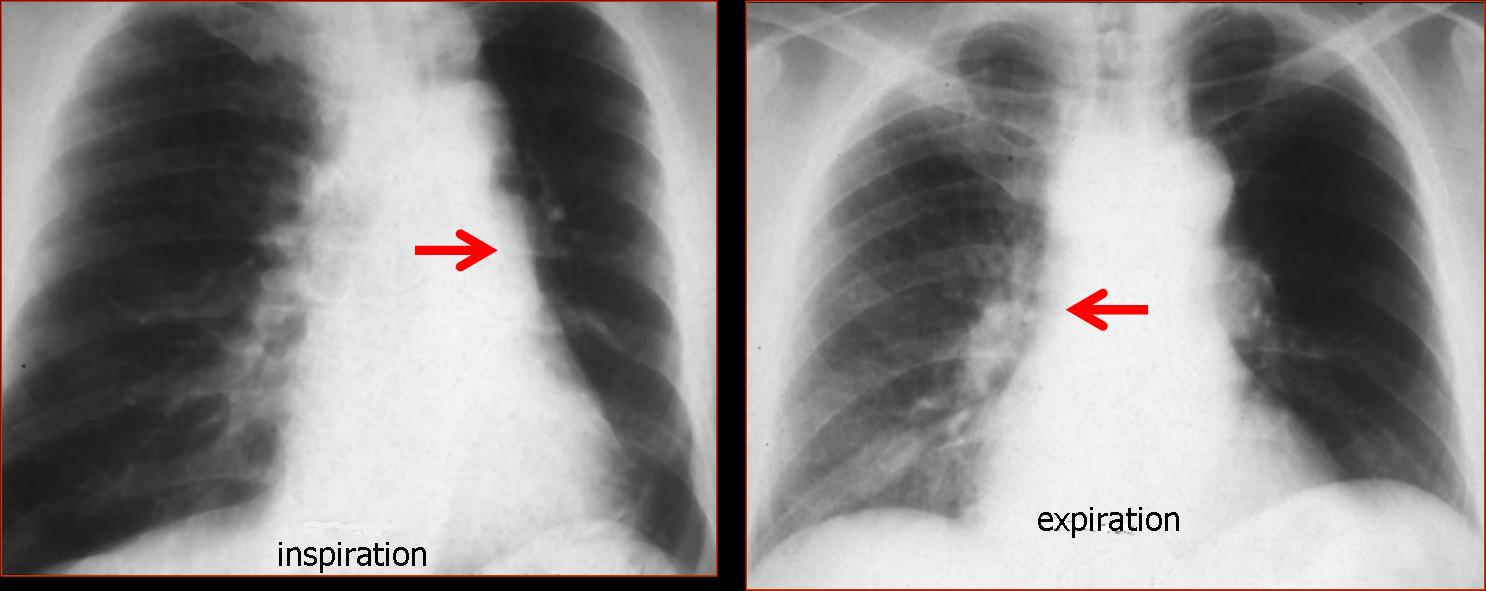
Fig. 1
Lateral view shows a tracheal mass (A, arrows), better seen in the tomogram (B, arrows) and the axial CT (C, arrow). The mass partially obstructed the origin of the left main bronchus; hence, the air-trapping.
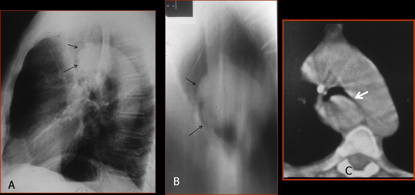
Fig. 2
Final diagnosis: adenoid cystic carcinoma of the trachea and left main bronchus.
This case is a good example of the so-called unilateral hyperlucent lung (UHL), a radiological finding of increased darkness of one lung with or without decreased vascularisation. Unilateral hyperlucent lung is discovered in the PA chest radiograph. The lateral view helps to identify ancillary findings, as in the present case.
The causes of UHL can be classified as follows:
1. Increased lung air
2. Decreased pulmonary vascularity
3. Pneumothorax
4. Chest wall abnormalities
5. Technical factors
In UHL caused by increased lung air, the initial approach is to separate obstructive from non-obstructive causes. This is easily accomplished with inspiratory and expiratory films, which will demonstrate air–trapping in the obstructive cases.
The most common cause of obstructive UHL is an endobronchial lesion causing partial obstruction of the main bronchus with resultant air-trapping. The main offenders are tumours, as in the present case, and foreign bodies in children (Fig. 3).
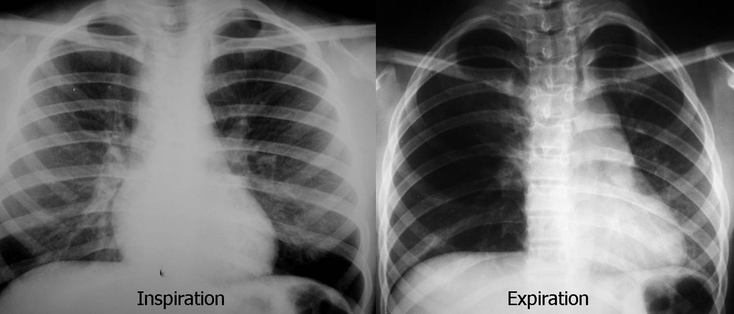
Fig. 3
Fig. 3 (above): foreign body aspiration in a 5-year-old child with dyspnea. Inspiration film looks normal; expiration demonstrates trapping of air in the right lung, with mediastinal shift towards the left. Bronchoscopy demonstrated an aspirated peanut in the right main bronchus.
Inspiration/expiration films are of value for identifying which lung is affected. The inspiration film in the case below (Fig. 4) shows a hyperlucent right lung, making it suspect of disease. On expiration, however, the right lung empties, whereas the left lung remains the same, indicating air-trapping on the left.
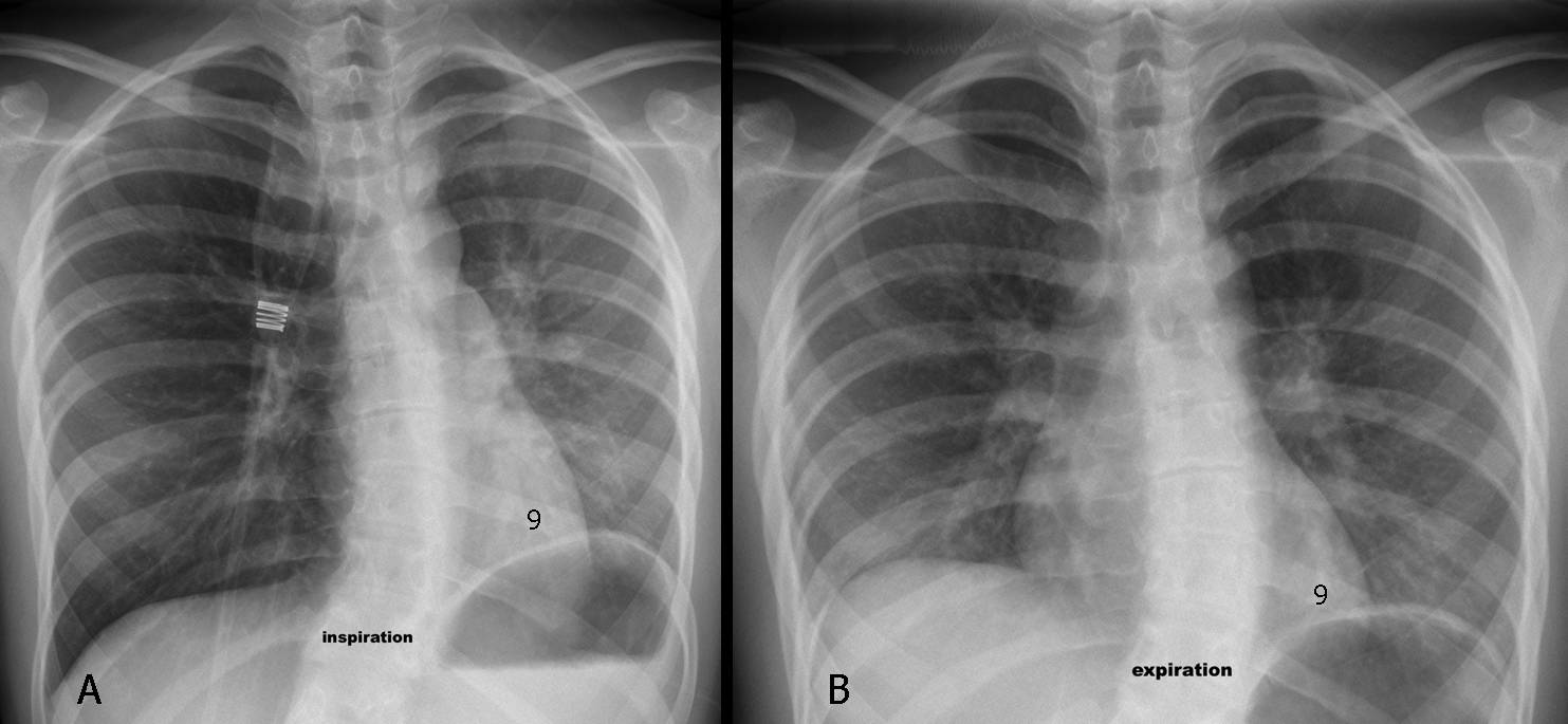
Fig. 4 (above): a 17-year-old girl with asthma and severe dyspnea. Inspiration/expiration films (A,B) show obvious air-trapping of the left lung. Notice that the left hemidiaphragm is always at the level of the 9th rib. Sagittal CT (Fig. 5, below) shows inspissated mucus in the left main bronchus (A, arrows) as the cause of the symptoms. After the mucus cleared, the chest returned to normal (B).
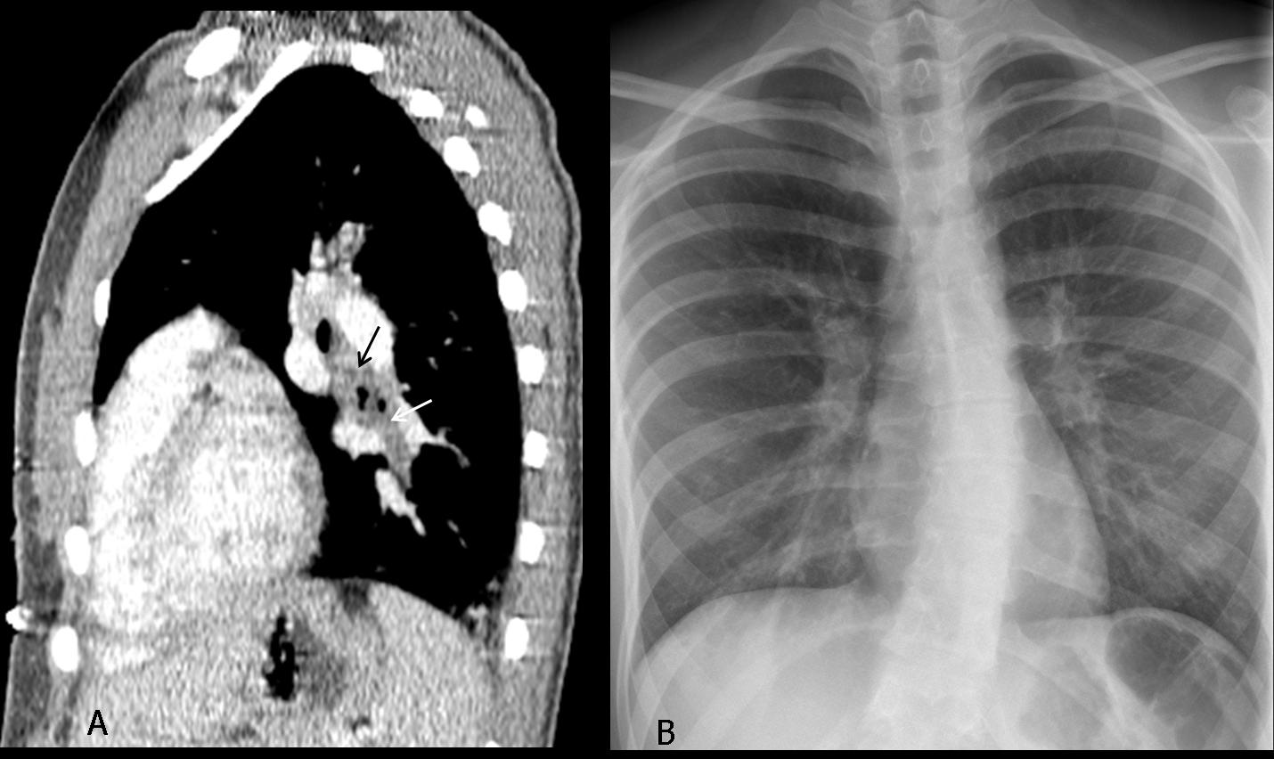
Fig. 5
Swyer-James/McLeod syndrome is a well-recognised, but uncommon, cause of obstructive UHL. In these patients, the main bronchi are permeable and the air-trapping is secondary to peripheral bronchiolitis after viral infection in childhood. Personally, I have seen very few cases of this syndrome in adults (Fig. 6).
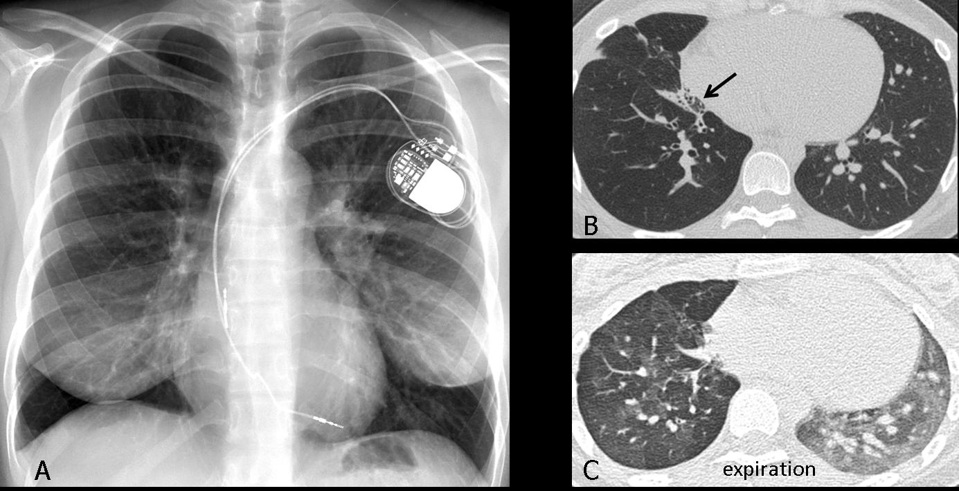
Fig. 6
Fig 6 (above): 47-year-old woman with Macleod syndrome. PA film shows a hyperlucent right lung with decreased vascularity (A). CT images show decreased vascularisation and bronchiectasis (B, arrows). Expiration CT shows air-trapping (C).
Non-obstructive UHL is due to compensatory overinflation secondary to a collapsed lobe or previous lobectomy. Obviously, the expanded lung empties well and does not trap air on expiration. Signs of collapse are usually obvious; if they are not, they should be sought to avoid making an erroneous diagnosis (Fig. 7).
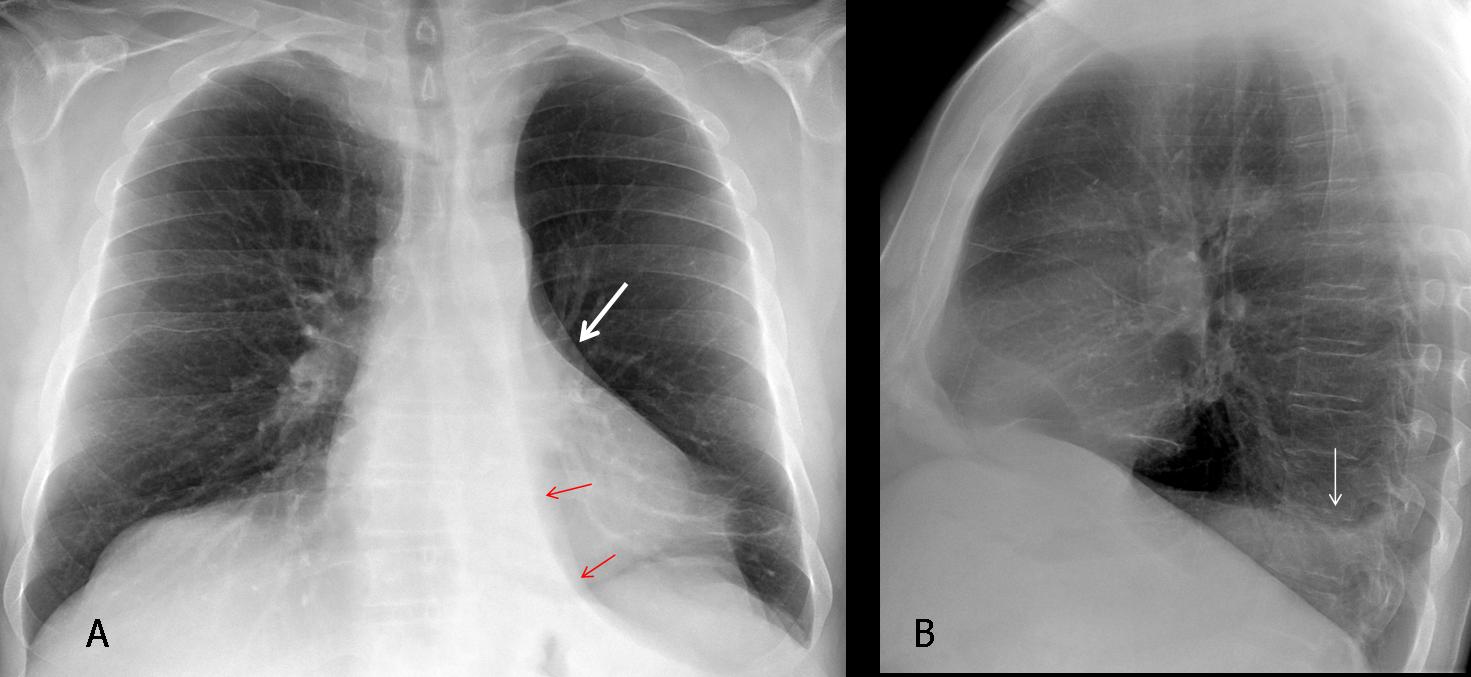
Fig. 7
Fig. 7 (above): 67-year-old man with moderate cough. PA radiograph shows what appears to be a left hyperlucent lung. Close observation shows a markedly descended hilum ( A, white arrow) and a triangular shadow (A, red arrows), which represents the collapsed LLL. The lateral view shows obliteration of the posterior hemidiaphragm by the collapsed lobe (B, arrow).
Axial CT demonstrates the marked LLL collapse (C, arrow), secondary to an endobronchial lesion at the origin of the LLL bronchus (D, arrow). Diagnosis: bronchogenic carcinoma.
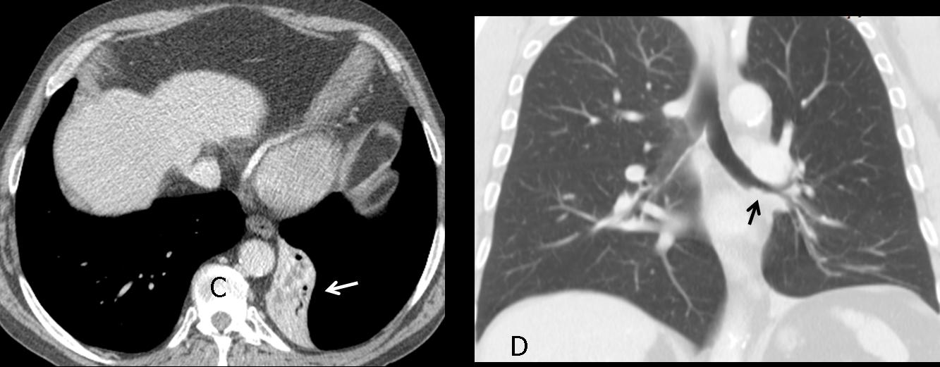
Fig. 8
Time for a test. These radiographs belong to a 23-year-old diabetic male with acute dyspnea. What would your diagnosis be?
1. Pneumothorax
2. Pulmonary embolism
3. Central carcinoid
4. None of the above
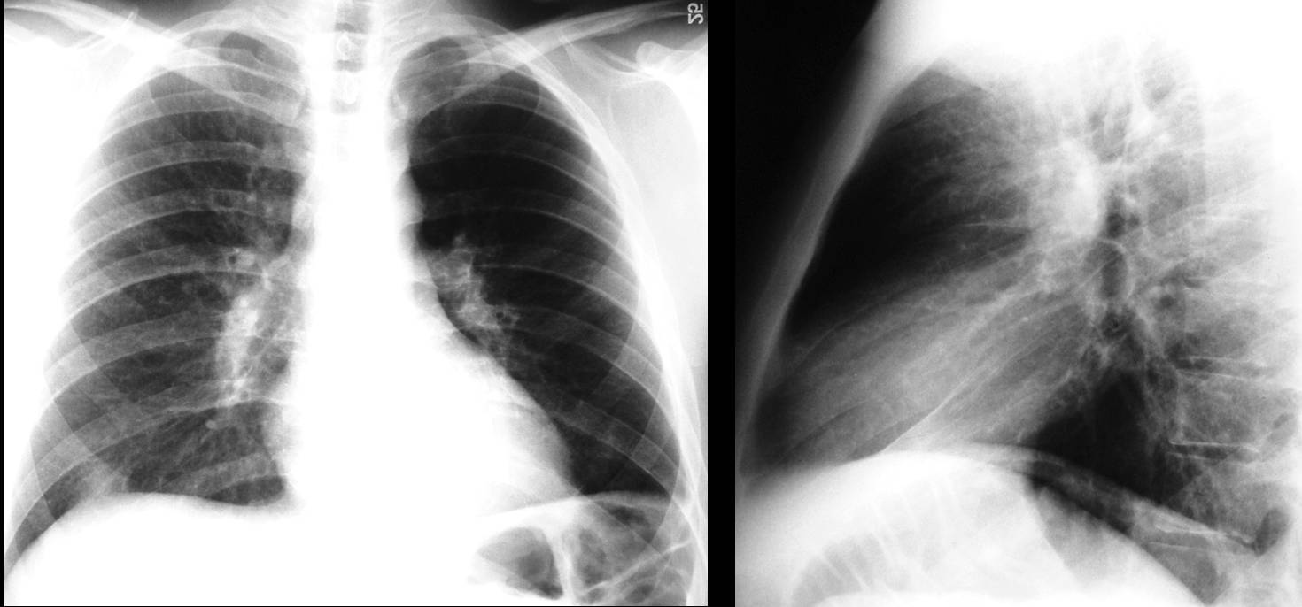
Fig. 9
This is a case of UHL secondary to decreased vascularity. The pulmonary artery and its branches contribute to the opacity of the lungs, and any cause of unilateral decreased pulmonary vascularity (arterial stenosis, pulmonary embolism) will appear to the eye as unilateral hyperlucent lung (Fig. 10). Of course, expiration films will not demonstrate air-trapping.
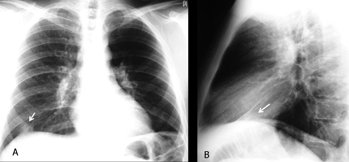
Fig. 10
Fig. 10 (above), findings: hyperlucent left lung with markedly decreased vascularity. There is a pleural-based opacity at the right lung base (A,B, arrows) whose morphology suggests a Hampton hump. CT (Fig. 11, below) confirms bilateral pulmonary emboli (A, arrows) and the typical shape of a pleural-based infarct (B, arrow).
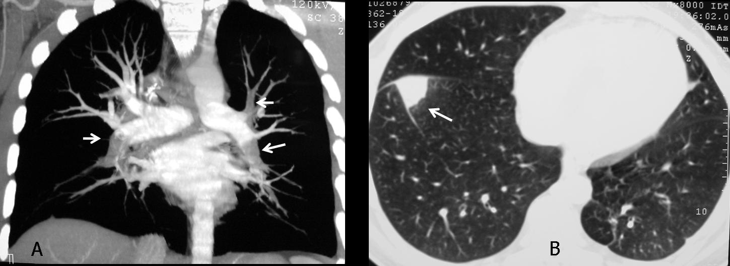
Fig. 11
Pneumothorax is a common cause of hyperlucent lung. The signs of a large pneumothorax are evident and the diagnosis is difficult to miss. The affected hemithorax is hyperlucent, and the collapsed lung appears as a stump in the parahilar area (Fig. 12 A, arrow). In another patient (B), tension pneumothorax is pushing the mediastinal structures towards the opposite side, hiding the pulmonary stump. Note in this case the telltale V sign, typical of pneumothorax (B, arrows).
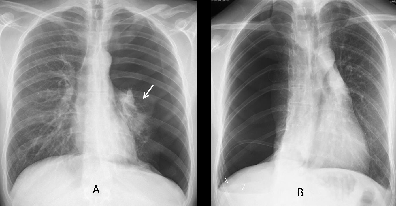
Fig. 12
Chest wall abnormalities, such as mastectomy or the rare congenital absence of the pectoralis major muscle (Poland syndrome), are responsible for increased unilateral lung lucency by decreasing the thickness of the superimposed soft tissues.
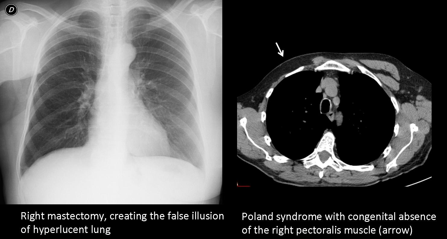
Fig. 13
Last, but not least, technical factors such as slight oblique positioning of the patient and/or a mis-centered x-ray beam can cause spurious increased unilateral lucency of one lung. In my experience, this is probably the most common cause of UHL and should be excluded before engaging in a more detailed investigation.
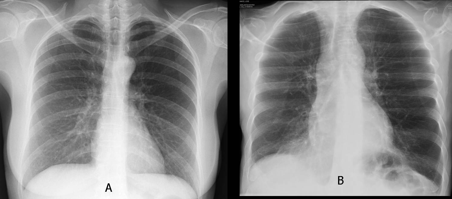
Fig. 14
Fig. 14 (above): two patients with hyperlucent left lung due to a miscentered x-ray beam. In A, note how the soft tissues of the shoulder are also darker, suggesting unequal penetration of the x-ray beam.

Follow Dr. Pepe’s advice
Follow Dr. Pepe’s advice:
- Obstructive UHL is caused by endobronchial lesions or, rarely, by peripheral bronchiolitis.
- To investigate obstructive UHL, inspiratory/expiratory films are mandatory.
- Other pulmonary causes include compensatory overinflation and decreased pulmonary vascularisation.
- The most common causes of UHL are technical factors, mastectomy, and pneumothorax.


