Dr. Pepe’s Diploma Casebook: Case 49 – SOLVED!

Dear Friends,
The first case of 2014 belongs to a 40-year-old male with a mild cough. What would you suspect?
1. Tuberculosis
2. Carcinoma
3. Enlarged left pulmonary artery
4. None of the above
Check out the images below and leave your thoughts and diagnosis in the comments section.
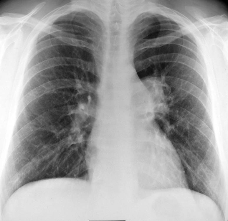
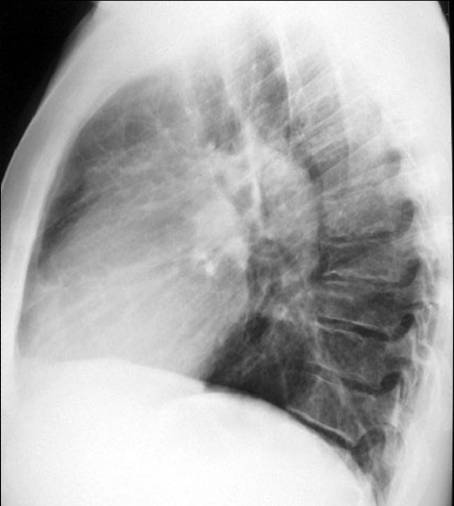
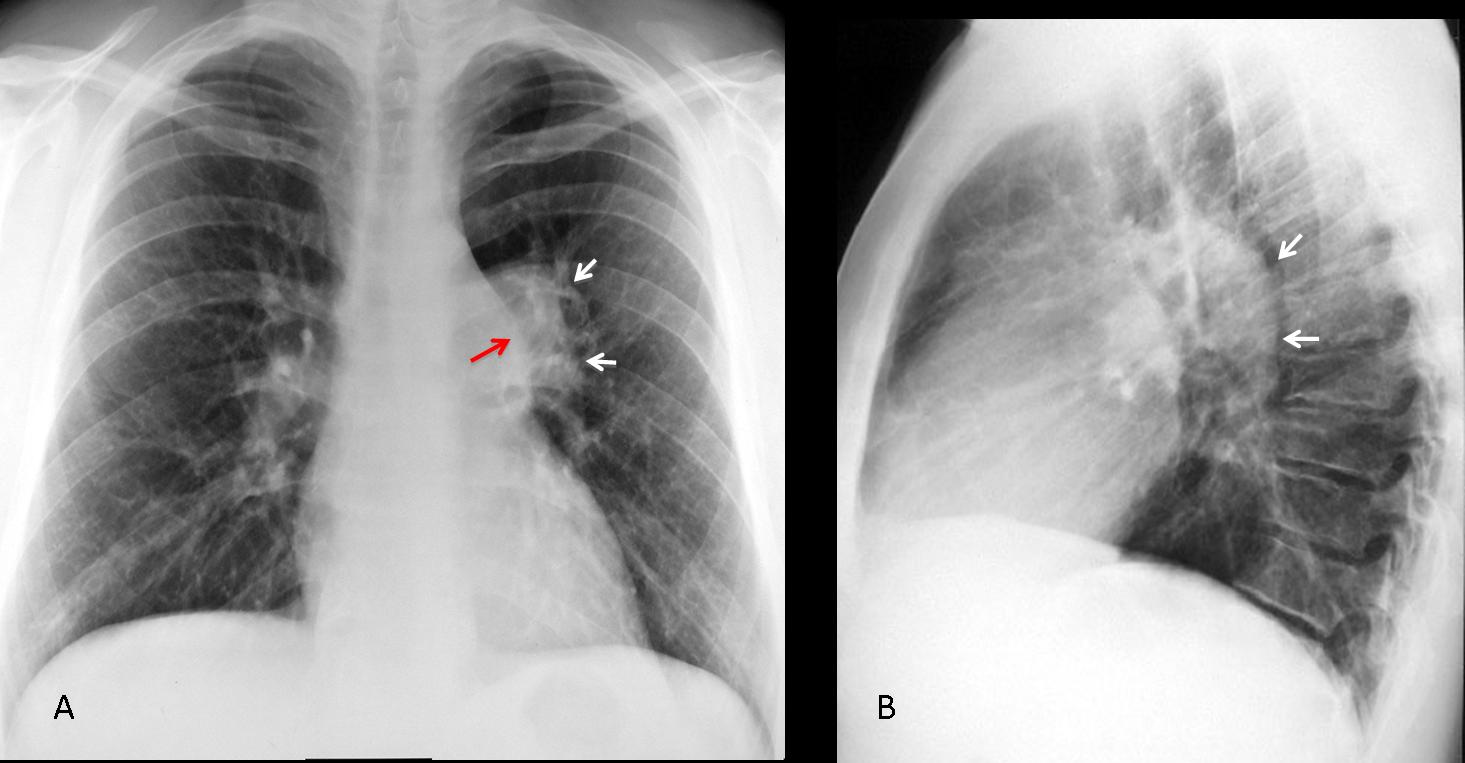
Fig. 1
Findings: the PA view shows that the left hilum is larger and more opaque than the right (A, white arrows). The pulmonary arch is prominent (A, red arrow). The lateral view show an increase in size of the left pulmonary artery (B, arrows). This combination of signs is highly suggestive of pulmonary valve stenosis in which the jet strikes the main pulmonary trunk and left pulmonary artery, enlarging both.
Enhanced CT confirms marked enlargement of the left pulmonary artery (C, arrows).
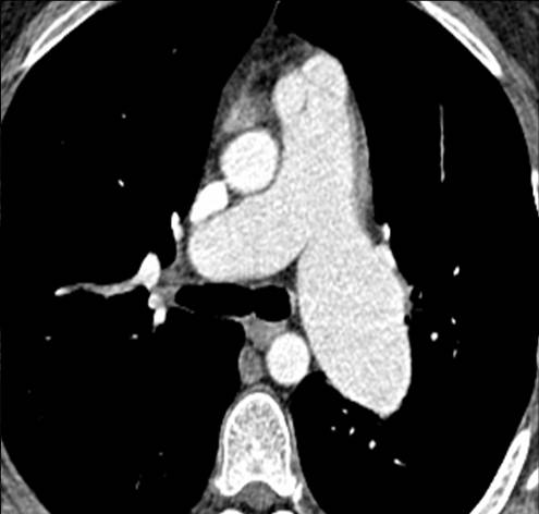
Fig. 2
Final diagnosis: congenital pulmonary valve stenosis, with secondary enlargement of the left pulmonary artery.
This case is presented to discuss unilateral hilar enlargement. This feature is easily recognisable by comparing the size and opacity of the two hila. In my experience, increased hilar opacity is more reliable than increased size.
Considering that the hila has 3 components ─ the pulmonary artery, bronchi, and minute lymph nodes ─ the possible causes of hilar enlargement are:
1. Enlarged pulmonary artery (A)
2. Bronchogenic carcinoma (B)
3. Lymphadenopathy (C)
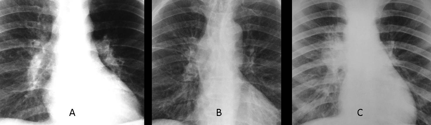
Fig. 3
Before discussing hilar pathology, we should review the normal hilar anatomy. In the PA view, the right and left pulmonary hila include the right and left main pulmonary arteries, respectively.

Fig. 4
In the lateral view, the right main pulmonary artery appears as a nodular shadow (Fig. 4 A,B, white arrow) in front of the trachea (T), whereas the left main pulmonary artery has the shape of a comma behind the posterior tracheal wall (Fig. 4 A,B, red arrow). Thus, an enlarged artery is easily recognisable in the lateral film, as in the initial case presented. When a solid mass is present, the hilar components are ill-defined (Fig. 4 C, arrows).
A word of caution: if the lateral view is not straight, normal hilar anatomy may not be recognisable. In case of doubt, always perform an enhanced CT.
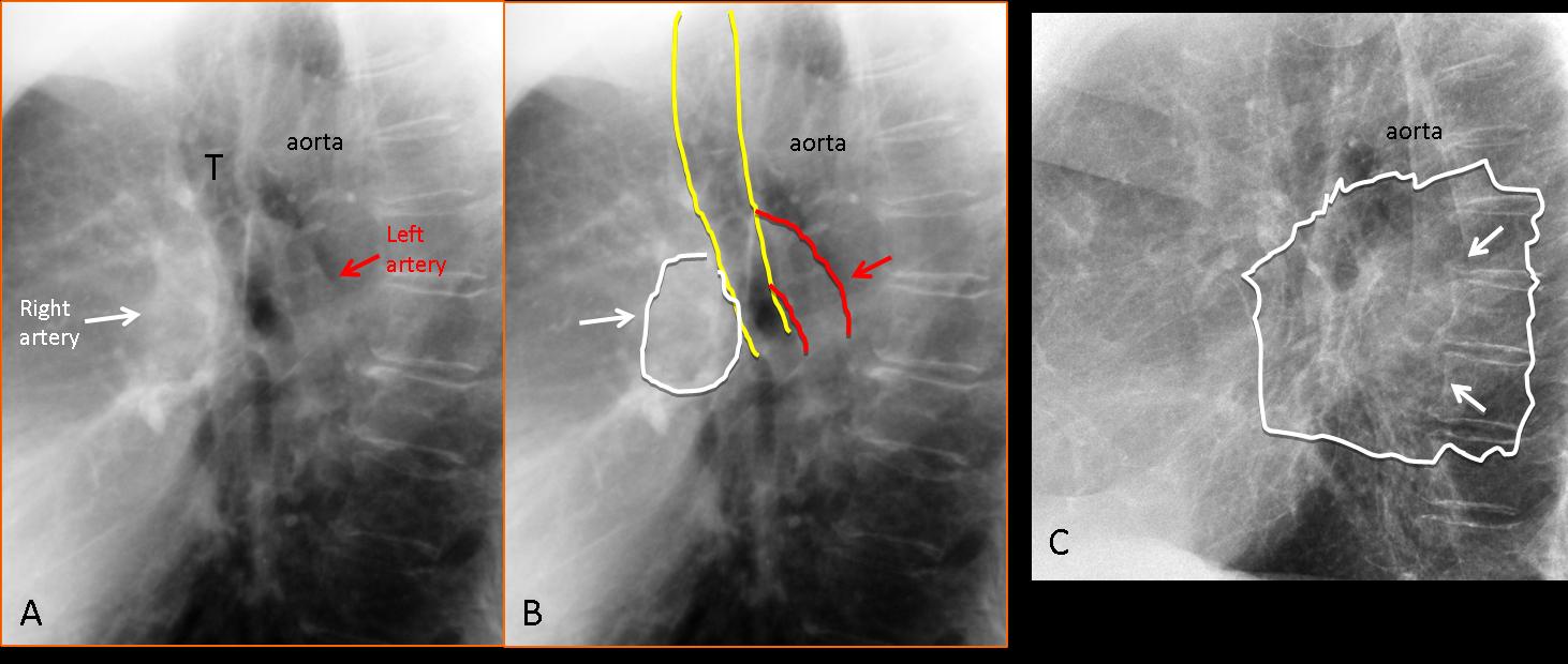
Fig. 5
TIP: the enlarged/dense hilum is identified in the PA view. The lateral view is useful to determine if the cause is an enlarged artery or a mass.
Congenital pulmonary valve stenosis is an unusual cause of hilar enlargement and affects only the left hilum. Another cause of vascular hilar enlargement is central pulmonary embolism. The embolus lodged in the central artery causes an increase in size and opacity of the hilum and may affect either one (Fig. 6).
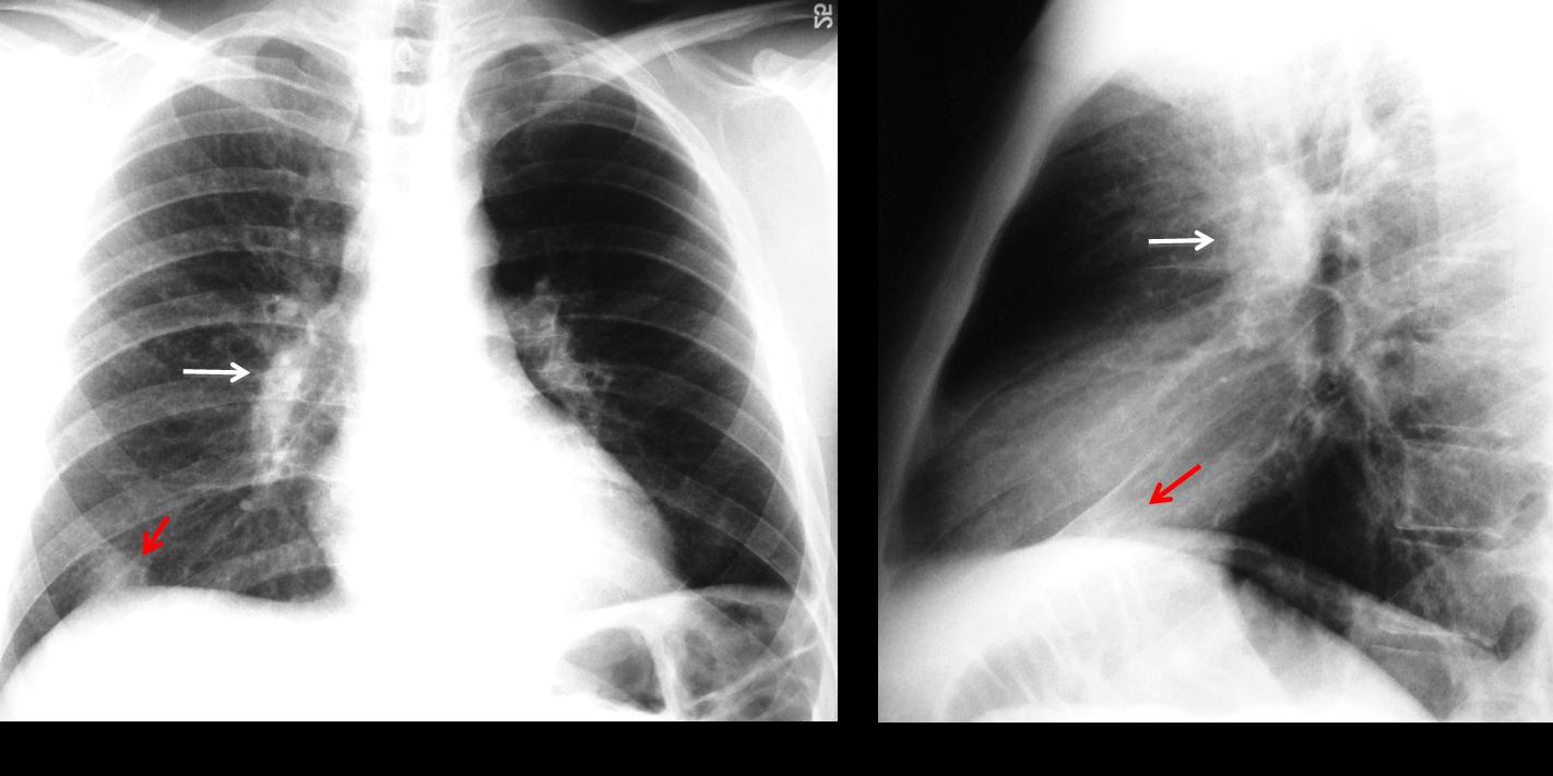
Fig. 6
Fig. 6 (above): 23-year-old diabetic man with pulmonary embolism. Note the enlarged right hilum in the PA and lateral view (white arrows). Pleural-based opacity at the right lung base (red arrows) typical of a Hampton hump.
In pulmonary embolism, when the anatomic contour of the hilum is not well defined, enhanced CT is needed to differentiate embolism from a hilar mass (Fig. 7).

Fig. 7
Fig. 7 (above): 47-year-old male with hypernephroma and abnormal right hilum (A, arrow). Metastases were suspected. Standard enhanced CT shows a central embolus in the right pulmonary artery (B, arrow).
Solid lesions, such as bronchogenic carcinoma or lymphadenopathy, are by far the most common cause of unilateral hilar enlargement. It is difficult to differentiate between these conditions, and enhanced CT should always be performed (Figs. 8 and 9).

Fig. 8
Fig. 8 (above): increased opacity of a normal-sized left hilum (A, arrow). Lateral view shows a normal left pulmonary artery (B, white arrow) and an anterior, rounded mass (B, red arrow). CT confirms a lung mass in the left hilum (C, arrow). Diagnosis: bronchogenic carcinoma.
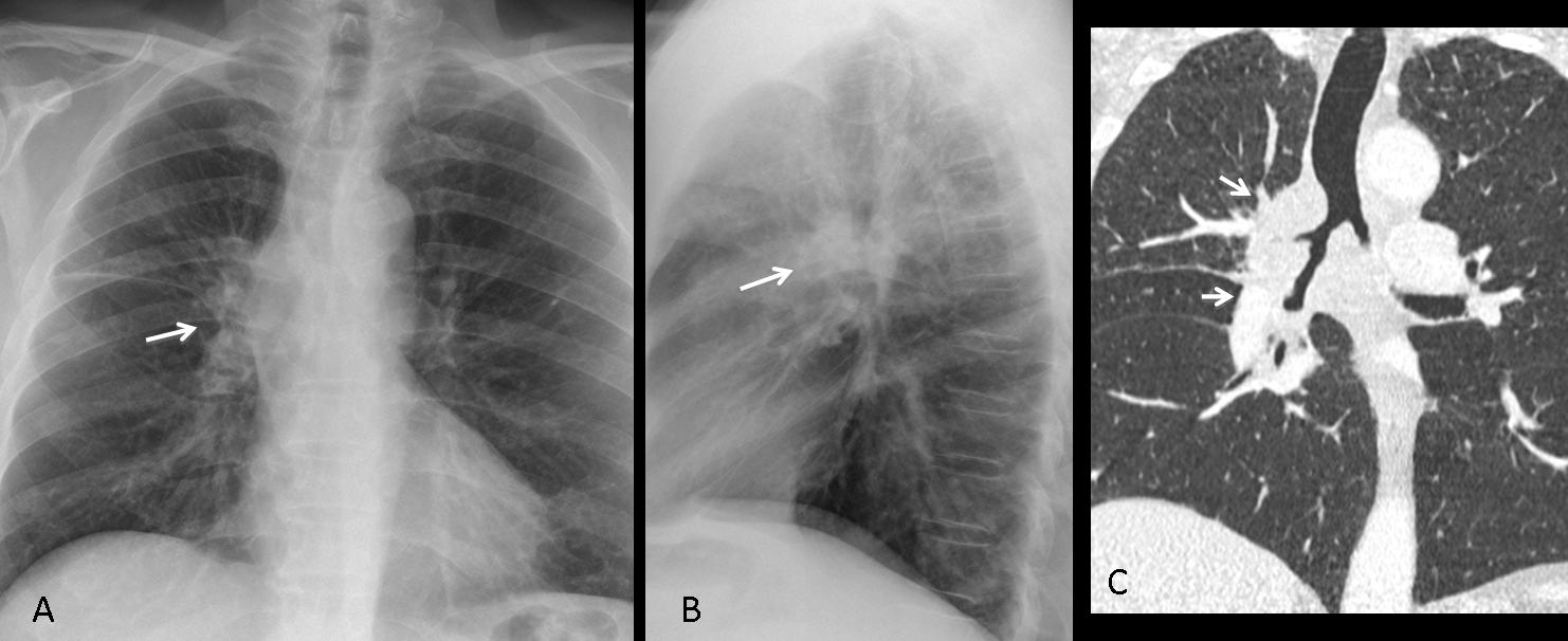
Fig. 9
Fig. 9 (above): abnormal right hilum, which is enlarged and increased in opacity compared to the left one (A, B, arrows). Coronal CT shows a large hilar mass narrowing the bronchi (C, arrows). Diagnosis: central bronchogenic carcinoma.
Lymphadenopathy is another cause of unilateral enlarged hilum. The most common origin is metastasis from bronchogenic carcinoma (Fig. 10), with tuberculosis as a distant second (Figs. 11 and 12).
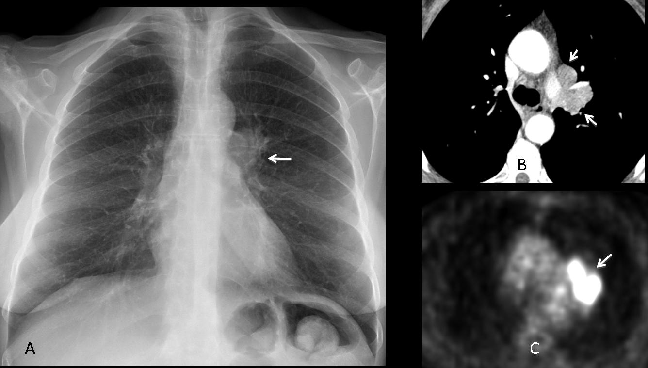
Fig. 10
Fig. 10 (above): 61-year-old woman with prominent left hilum (A, arrow). Axial CT shows enlarged lymph nodes (B, arrows) with high metabolic rate on PET (C, arrow): Small-cell carcinoma.
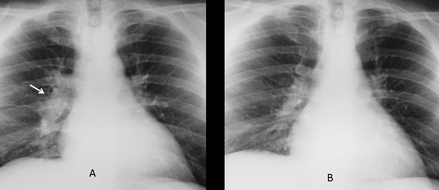
Fig. 11
Fig. 11 (above): 35-year-old man with unilateral enlargement of right hilum secondary to tuberculous lymphadenopathy (A, arrow). Radiograph after treatment (B) shows decreased size and density of the right hilum.
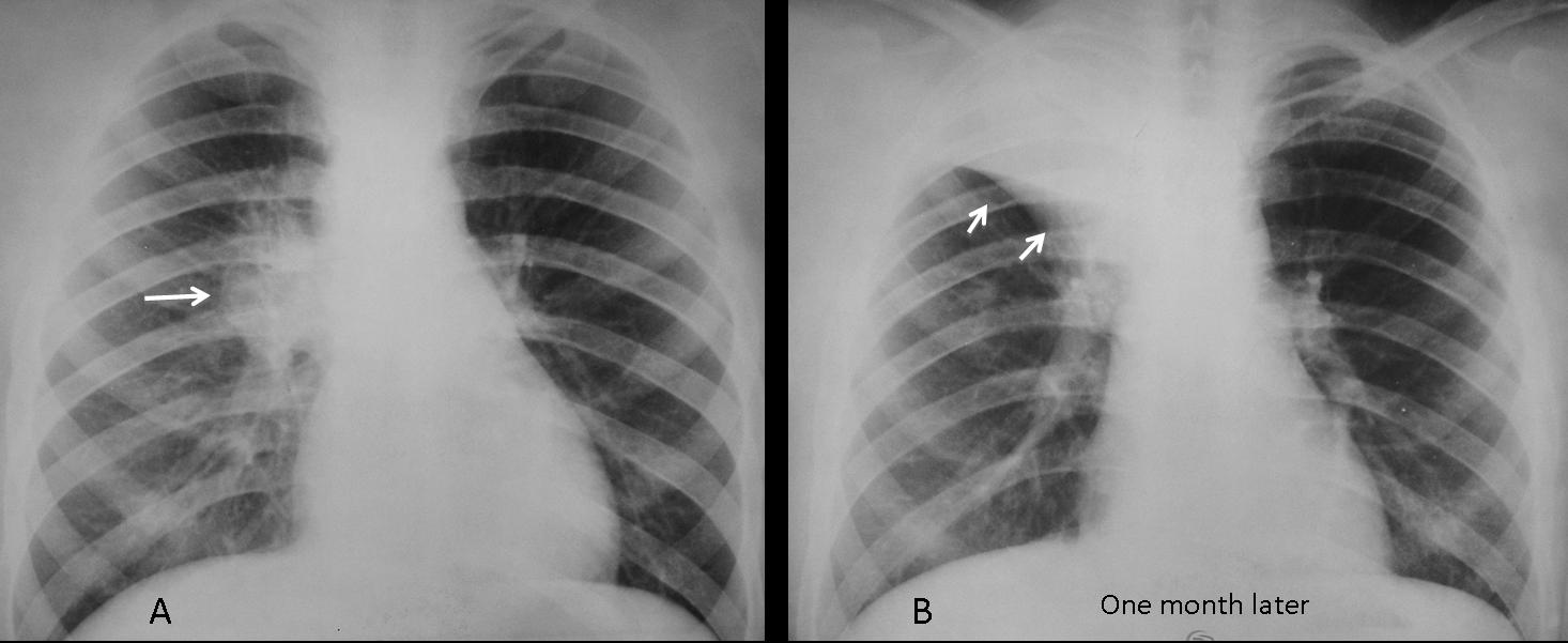
Fig. 12
Fig. 12 (above): 16-year-old male with tuberculous lymphadenopathy of the right hilum (A, arrow). One month later, the enlarged lymph nodes have caused RUL collapse (B, arrows).
Time for a test. The radiographs below are of a 49-year-old male, smoker, with fever. Diagnosis:
1. Carcinoma
2. TB
3. Lymphadenopathy
4. None of the above
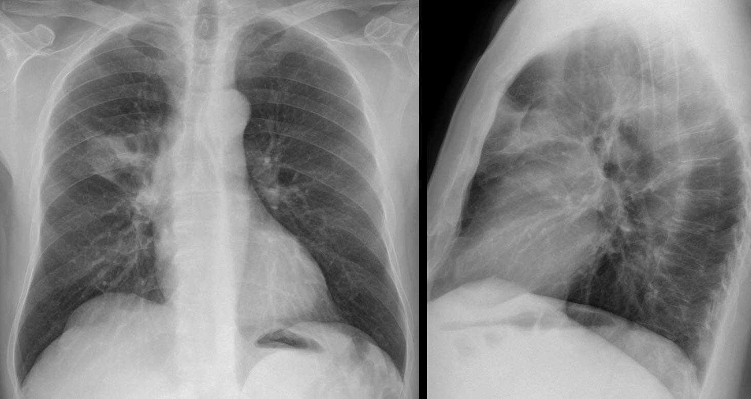
Fig. 13
Aside from the already mentioned causes of enlarged hilum, there is a spurious one caused by superimposition of a pulmonary lesion. Any process in front of or behind the hilum (tumor, infection, etc.) may simulate a dense hilum. In this particular case there is a pneumonic infiltrate in the anterior RUL, simulating a dense right hilum (Fig. 14 A, arrows). The lateral view shows the anterior pneumonia (B, white arrow) and the intact hilum (B, red arrow).
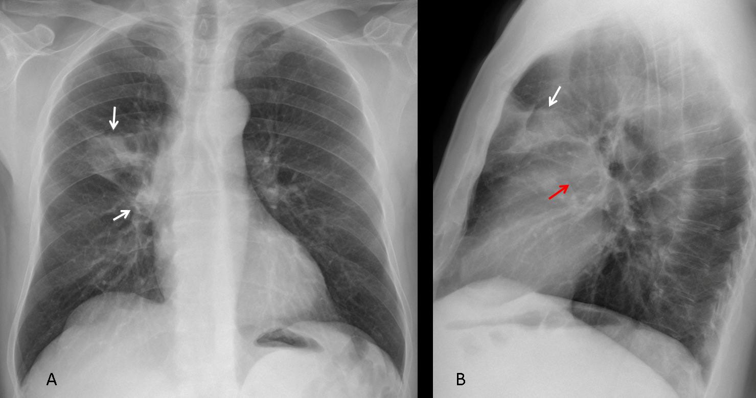
Fig. 14
Another example, in which a peripheral nodule in the apical segment of the LLL simulates a dense left hilum (Fig. 15). This case emphasises the importance of obtaining a lateral radiograph to better evaluate possible hilar disease.
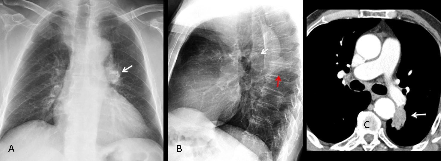
Fig. 15
Fig. 15. PA chest film shows an apparently dense left hilum (A, arrow). Lateral view shows a normal left hilum (B, white arrow) and a pulmonary nodule projected over the spine (B, red arrow). CT confirms the absence of hilar disease and the pulmonary nodule (C, arrow): bronchogenic carcinoma.
For completeness’ sake, I should mention increased density of the left hilum by a superimposed elongated aorta (Fig. 16, arrow). This is a common mistake of first-year residents.
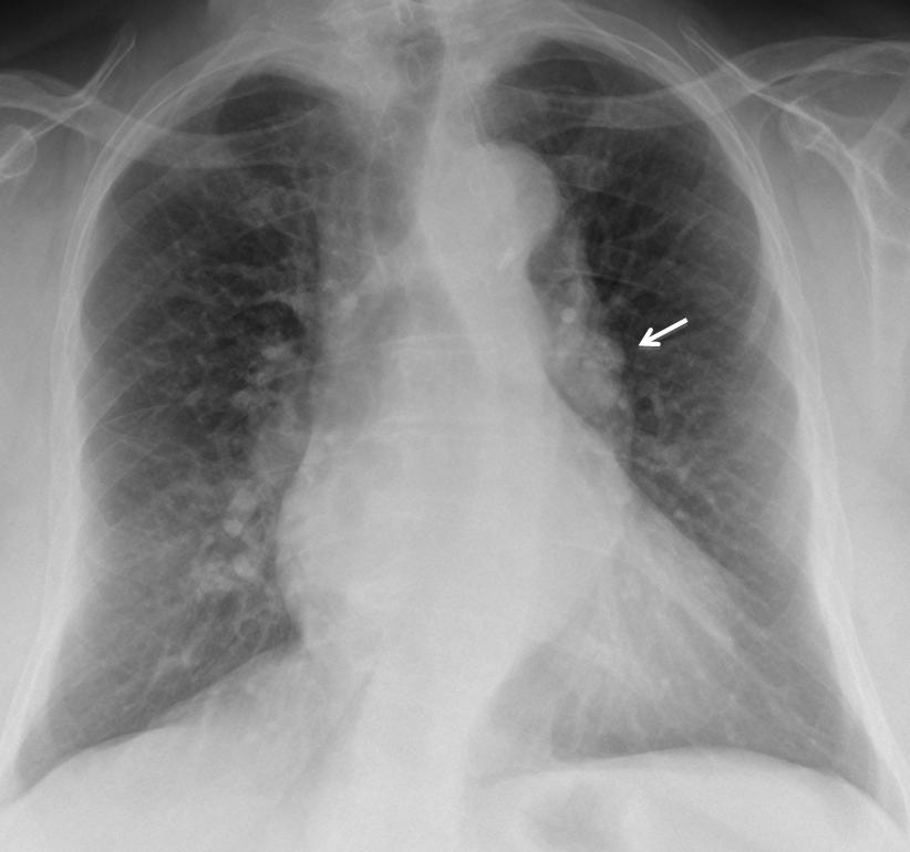
Fig. 16

Follow Dr. Pepe’s advice
Follow Dr. Pepe’s advice:
- Bonchogenic carcinoma is the most common cause of unilateral enlarged hilum
- Occasionally it may be due to TB lymph nodes or an enlarged pulmonary artery
- The lateral view helps to identify enlarged pulmonary arteries
- The lateral view identifies superimposed lung processes that simulate an enlarged hilum in the PA view


