Dr. Pepe’s Diploma Casebook: Case 52 – SOLVED!

Dear Friends,
Today I am presenting radiographs of a 43-year-old woman with moderate dyspnoea. Disregard the widening of the right superior mediastinum, secondary to long-standing goiter. Leave me your thoughts and diagnosis in the comments sectiona and come back on Friday for the answer.
Diagnosis:
1. Thymoma
2. Heart disease
3. Lymphoma
4. None of the above
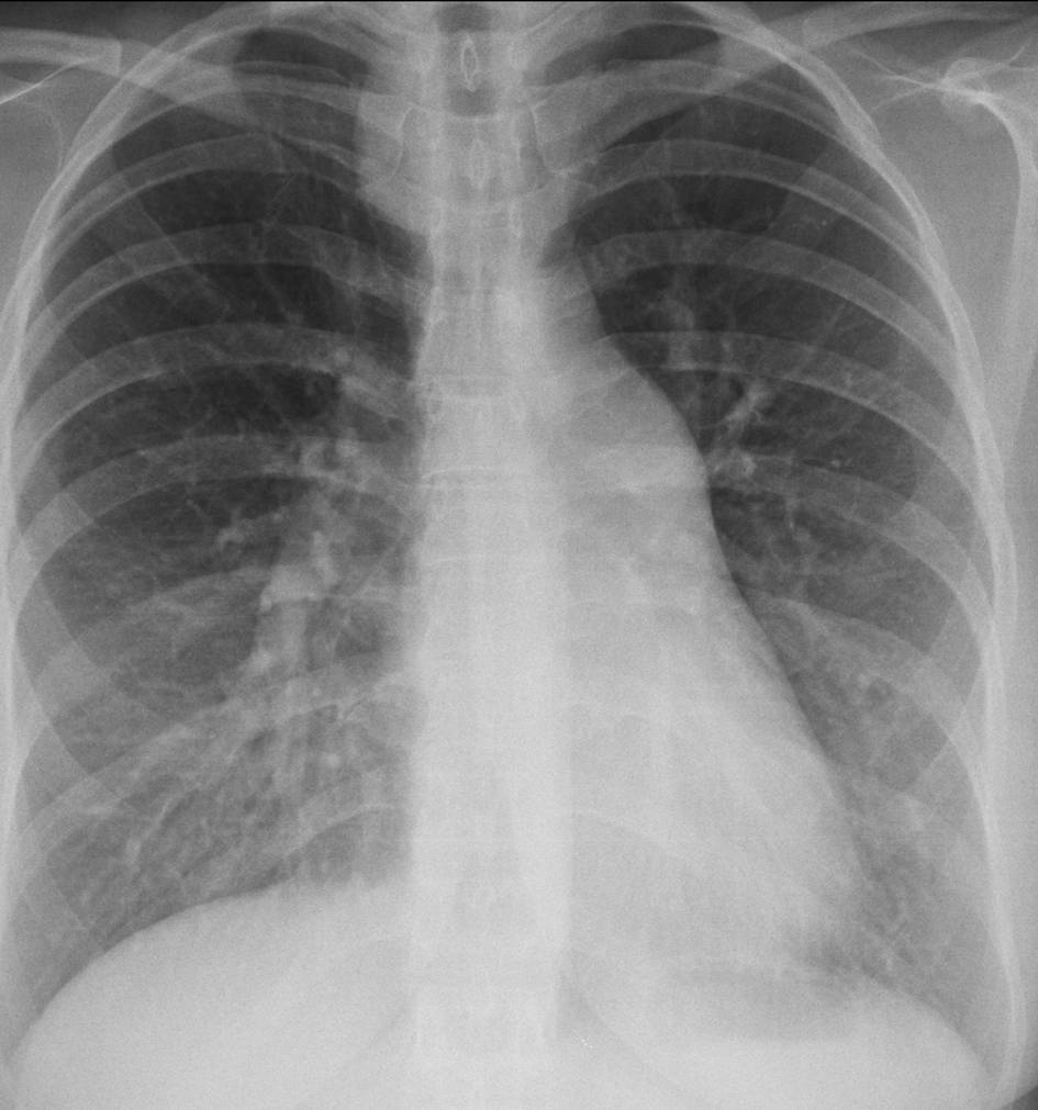
43-year-old woman with moderate dyspnoea
Findings: PA chest film shows bulging of the main pulmonary artery contour (A, arrow), accompanied by moderately increased arterial circulation of the lungs (note increased diameter of vessels in both lower lobes and left upper lobe). This combination of findings suggests a left-to-right shunt. MRI shows communication (B, arrow) between the right atrium (RA) and the left (LA).
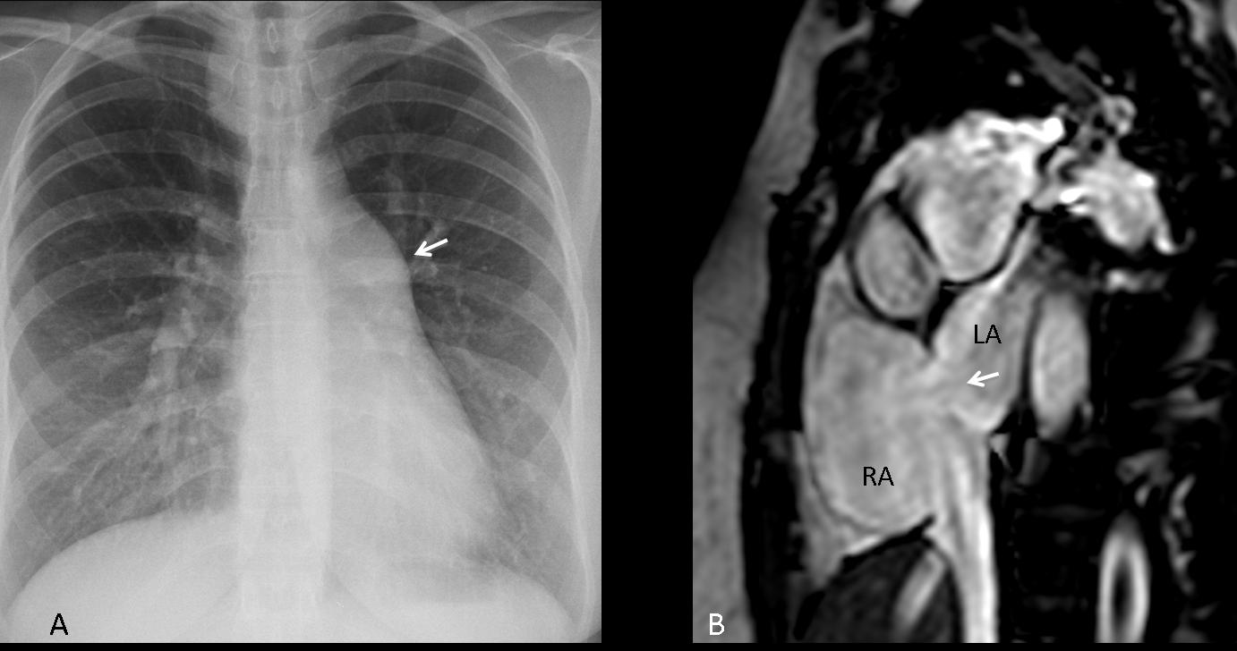
Fig. 1
Final diagnosis: septum secundum atrial septal defect with left-to-right shunt
This case is presented to discuss prominence of the pulmonary arch. The mediastinum has three bumps on the left contour: the uppermost is the aortic knob (A, arrow). The second corresponds to the main pulmonary artery (A, red arrow) and the third represents the left ventricle (A, yellow arrow). There is a virtual bulge between the second and the third arch which, when present, represents the atrial appendage in mitral disease (B, arrow).
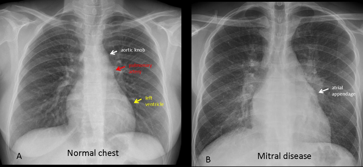
Fig. 2
Bulging of the pulmonary arch usually indicates dilatation of the main pulmonary artery. Three conditions cause dilatation of the artery. The most common is pulmonary arterial hypertension, followed by left-to-right shunt and pulmonary valve stenosis. These conditions can be suspected on chest radiographs if one knows their imaging manifestations.
Pulmonary arterial hypertension (PAH) is not uncommon in daily practice. In the chest plain film, PAH is recognized by a prominent pulmonary arch (A, white arrow) with enlargement of the central pulmonary arteries, visible in the PA and lateral views (A,B, red arrows), accompanied by abrupt tapering of the peripheral pulmonary vessels.
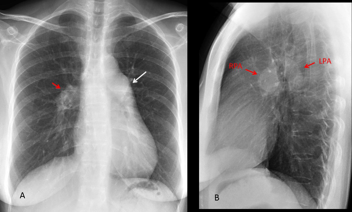
Fig. 3
Axial and coronal CT images show marked enlargement of the central pulmonary arteries (A, arrows), with tapering of the peripheral vessels. The CT criterion for diagnosing PAH is main pulmonary artery diameter greater than or equal to 29mm (B, arrow).

Fig. 4
The etiology of PAH is varied. The most frequent cause is chronic pulmonary disease (mainly COPD). Other causes include obstruction of vessels (chronic pulmonary embolism), inverted left-to-right shunt (Eisenmenger syndrome), and idiopathic PAH, which occurs in young adults and more commonly affects women.
All cases of PAH show similar imaging findings on plain films, regardless of the cause. The following signs may suggest the etiology: evidence of pulmonary disease (COPD or others) (Fig. 5) or scars and blunting of the costophrenic angles, suggestive of previous pulmonary emboli. In advanced cases, CT may show calcification of pulmonary artery walls, a sign of long-standing PAH (Fig. 6).
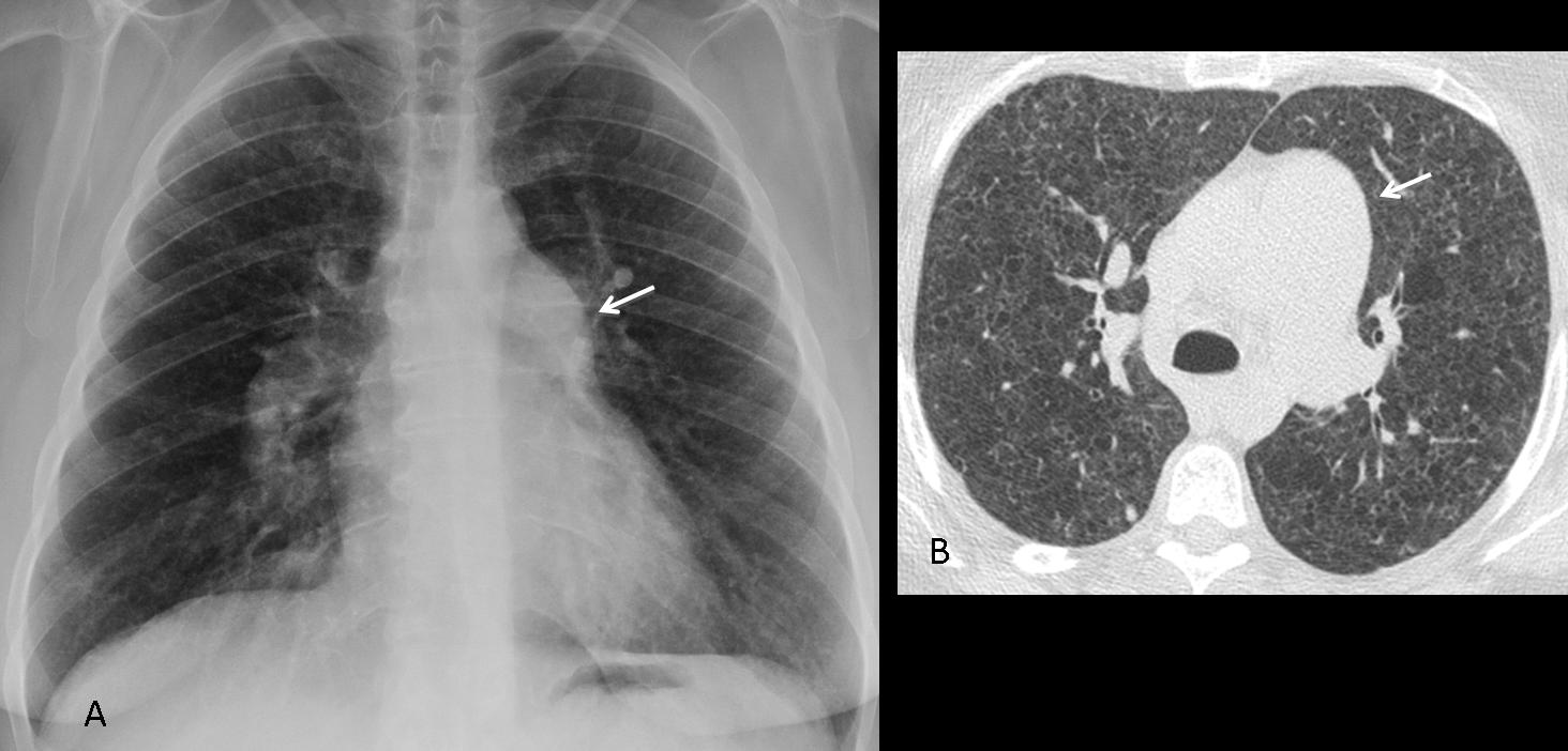
Fig. 5
Fig. 5 (above): 44-year-old woman with Langerhans cell histiocytosis. PA chest film shows the typical signs of PAH: a prominent pulmonary arch (A, arrow) and enlarged hila with tapering vessels. Axial CT shows diffuse lung disease and a markedly enlarged pulmonary artery (B, arrow).
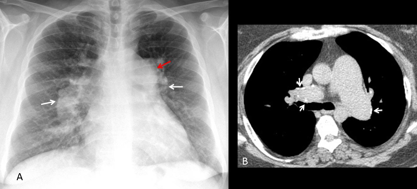
Fig. 6
Fig. 6 (above): 45-year-old man with long-standing idiopathic PAH. PA chest film depicts prominent hila (A, white arrows) with abrupt tapering of distal arteries and bulging of the pulmonary arch (A, red arrow). CT shows the marked size disparity between the aorta and pulmonary arteries, which have calcified walls (B, arrows).
Isolated left-to-right shunts are another cause of a prominent pulmonary arch. They may occur at the level of the atria, ventricles, or through a persistent ductus arteriosus. The last two are usually discovered in infancy, whereas atrial septal defect may go into adulthood without significant symptoms and be discovered in a routine chest examination. Suggestive signs of shunts are a prominent pulmonary artery and increased arterial circulation in both lungs (Fig. 7). The heart is usually enlarged.
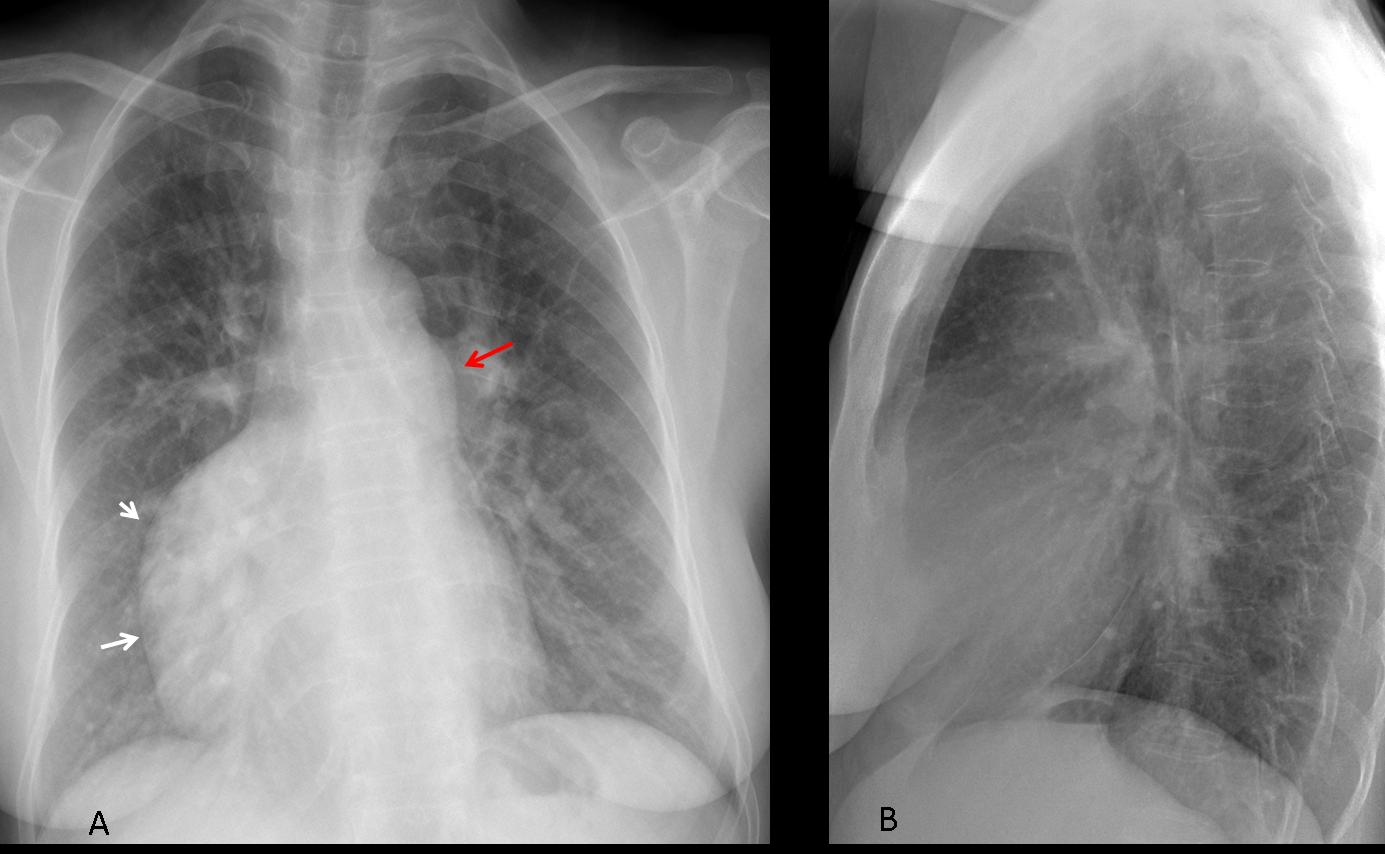
Fig. 7
Fig 7 (above): 47-year-old female with ASD. Note the markedly enlarged right atrium (A, arrows) secondary to circulation overload. The pulmonary arch is prominent (A, red arrow) and the pulmonary arterial circulation is increased. Diagnosis: septum secundum atrial septal defect.
The third cause of enlarged central pulmonary artery is congenital pulmonary valve stenosis. In this condition, the jet of blood caused by the stenosis leads to dilatation of the main and left pulmonary arteries. The pulmonary vasculature is either normal or decreased (Fig. 8). Patients are usually asymptomatic and the abnormality is discovered in the 3rd or 4th decade.
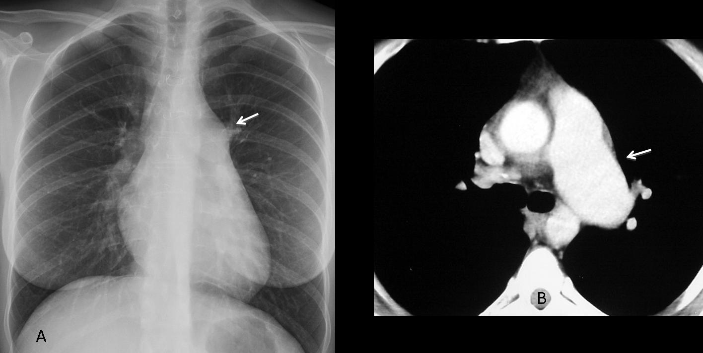
Fig. 8
Fig. 8 (above): 34-year-old female with pulmonary valve stenosis. PA chest film shows a prominent pulmonary arch (A, arrow) and decreased circulation in both lungs. Axial CT confirms the dilatation of the main and left pulmonary arteries (B, arrow).
To summarize: the imaging findings accompanying dilatation of the pulmonary arch are:
- Enlarged hila and abrupt tapering of pulmonary arterial circulation: Pulmonary arterial hypertension (A)
- Enlarged hila and increased circulation of lungs: Isolated left-to-right shunt (B)
- Normal or decreased pulmonary circulation: Pulmonary valve stenosis (C)
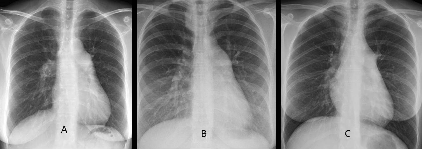
Fig. 9
A less common cause of prominent pulmonary arch is an anterior mediastinal mass superimposed on the pulmonary segment. In these cases, the hila and pulmonary vessels are unremarkable and the lateral view may show the mass in the anterior clear space (Fig. 10)
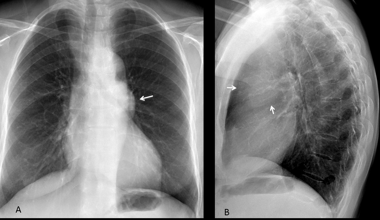
Fig. 10
Fig. 10 (above): Preoperative chest radiograph in a 45-year-old woman showing a prominent pulmonary arch (A, arrow). Hila and pulmonary vessels are normal. Lateral chest film depicts a large mass in the anterior clear space (B, arrows).
Unenhanced CT (Fig. 11, below) confirms the presence of a large soft-tissue mass (A,B, arrows) projected over the pulmonary artery arch. Final diagnosis: thymoma.

Fig. 11
Moderate prominence of the pulmonary arch is common in adolescents. It is believed to correspond to thymic tissue and should not be considered an abnormal finding (Fig. 12).
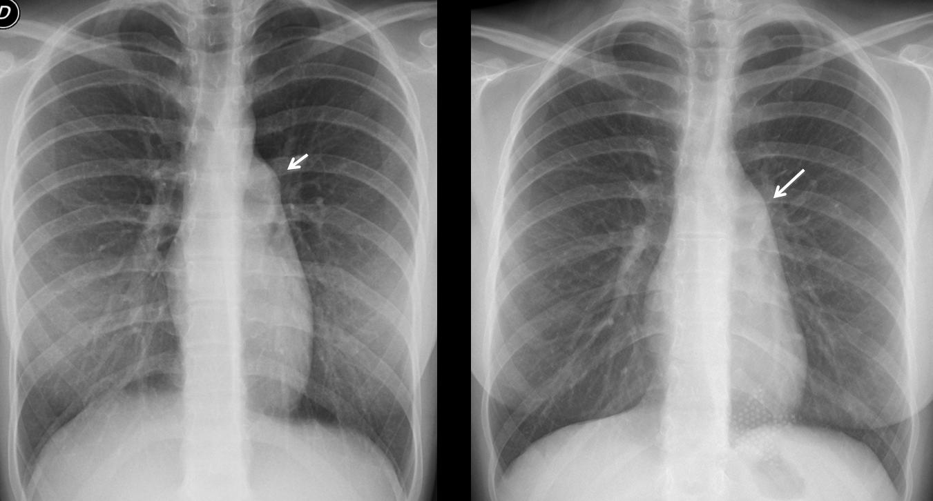
Fig. 12
Fig. 12. PA radiographs of two asymptomatic female teenagers. Both have prominent pulmonary arch (A,B, arrows).

Follow Dr. Pepe’s advice:
- Prominence of the pulmonary arch is abnormal, except in teenagers.
- The most frequent cause is increased size of the main pulmonary artery due to PAH, left-to right-shunt, or pulmonary valve stenosis
- Anterior mediastinal masses may simulate increased size of the pulmonary arch. Look at the anterior clear space in the lateral view.


