Dr. Pepe’s Diploma Casebook: Case 53 – SOLVED!

Dear Friends,
Today I am presenting radiographs of a 36-year-old woman with shortness of breath increasing over the last three months. Examine the image below and leave me your thoughts and diagnosis in the comments. Come back on Friday for the answer.
Diagnosis:
1. Metastatic disease
2. Sarcoid
3. Interstitial pneumonia
4. None of the above
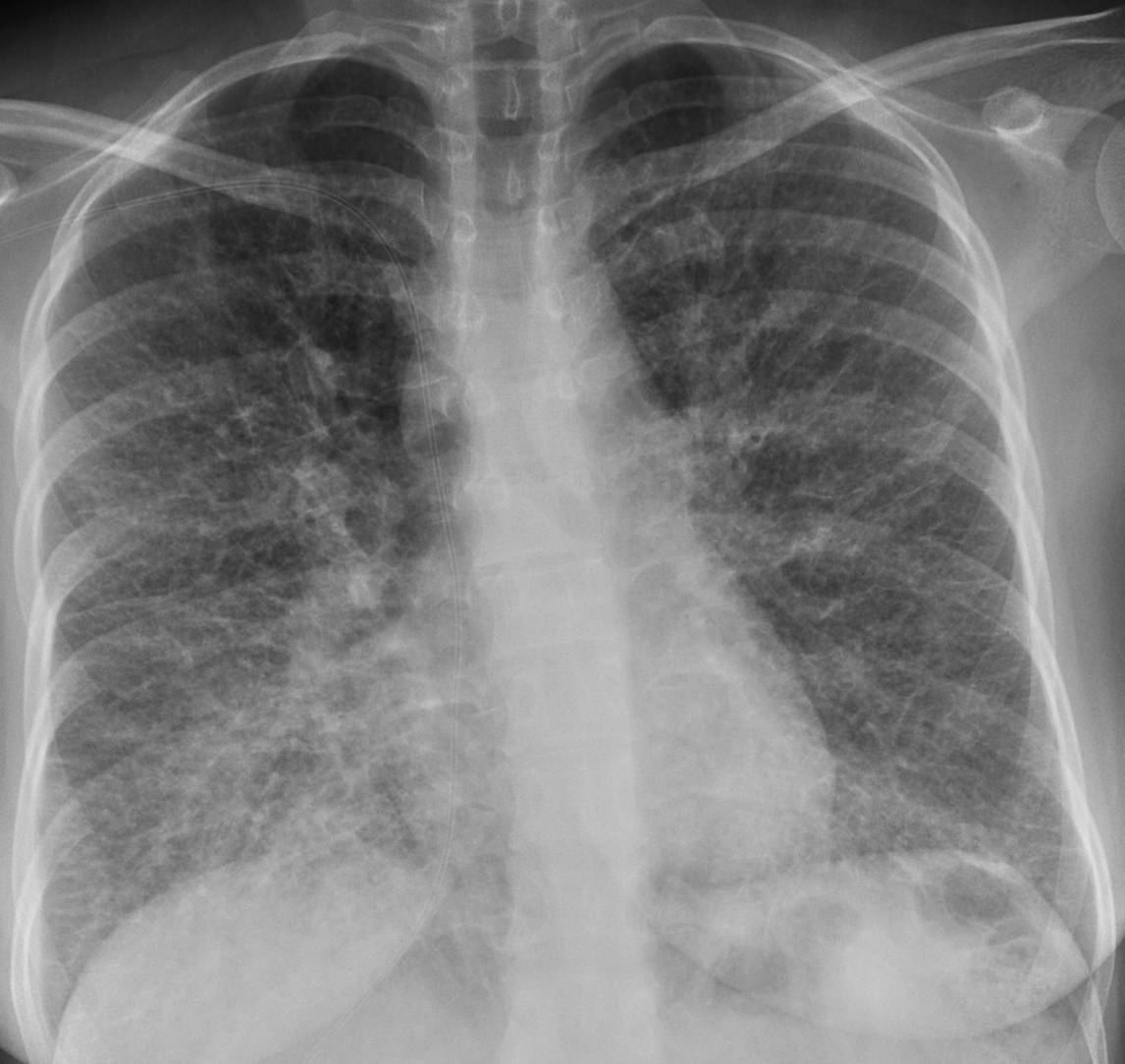
36-year-old woman with shortness of breath
Findings: chest radiographs show widespread involvement of the lung with numerous Kerley B lines in both lower lobes (Fig. 1 A, B arrows), confirmed with CT (Fig. 1 C, arrows). In the presence of widespread Kerley lines in a chronic setting, the first consideration should be lymphangitic metastasis to the lung. In this patient, endoscopy discovered an advanced carcinoma of the stomach.
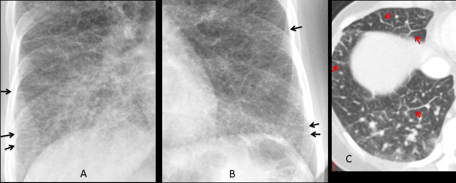
Fig. 1
Final diagnosis: carcinoma of the stomach with lymphangitic lung metastasis.
This case is presented to discuss Kerley lines. This finding is important because their discovery considerably narrows the differential diagnosis of pulmonary processes affecting the interstitium.
From a practical approach, Kerley lines can be classified as A and B. Kerley A lines are thin and non-branching, radiate from the hila, and are better seen in the middle lung areas in the PA view and in the anterior clear space in the lateral view (Fig. 2 A, arrows). Kerley B lines are short and horizontal, and are better seen in the periphery of the lung bases at the costophrenic angles (Fig. 2 B, arrows). A and B lines are equally significant, but B lines are easier to identify. A lines are nearly always accompanied by B lines.

Fig. 2
Fig. 2 (above): a 37-year-old woman with vasculitis. Prominent Kerley A lines are seen in the anterior clear space in the lateral view (A, arrows). PA view shows multiple Kerley B lines, particularly in the costophrenic angle (B, arrows).
Prominent lymphatic vessels and thickened interlobular septa are the anatomic substrata of Kerley lines. CT identifies them much better than plain radiographs (Fig. 3).
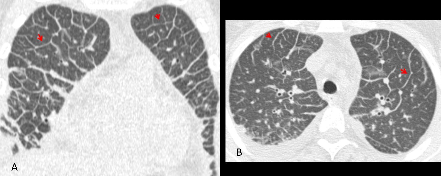
Fig. 3
Fig. 3 (above): CT images of the same patient show the prominent septa to better advantage (A,B, arrows).
From the diagnostic viewpoint, Kerley lines can be classified as transient or persistent. It is important to know that this distinction cannot be made with a single study. Follow-up films or clinical information (acute vs. chronic) are needed. The main characteristic of transient Kerley lines is that they disappear on follow-up studies (Fig. 4).

Fig, 4
Fig. 4 (above): a 47-year-old patient with heart failure. Initial film shows a prominent hilum and Kerley B lines (A, arrows). After treatment, the B lines are less conspicuous (B).
Pulmonary oedema of any etiology (Fig. 5) is the most common cause of transient Kerley lines. Less common ones are vasculitis, allergic reactions, and initial stages of viral pneumonia (Fig. 6).
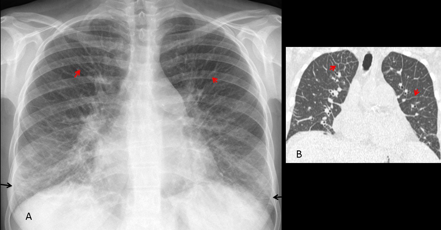
Fig. 5
Fig. 5 (above): 58-year-old man with cardiac pulmonary oedema. PA chest film (A) shows numerous A lines (red arrows) and B lines (black arrows), confirmed with CT (B, arrows). Compare with the initial case of lymphagitic carcinomatosis and notice that the appearance is identical. To differentiate between them we need clinical information or serial films.
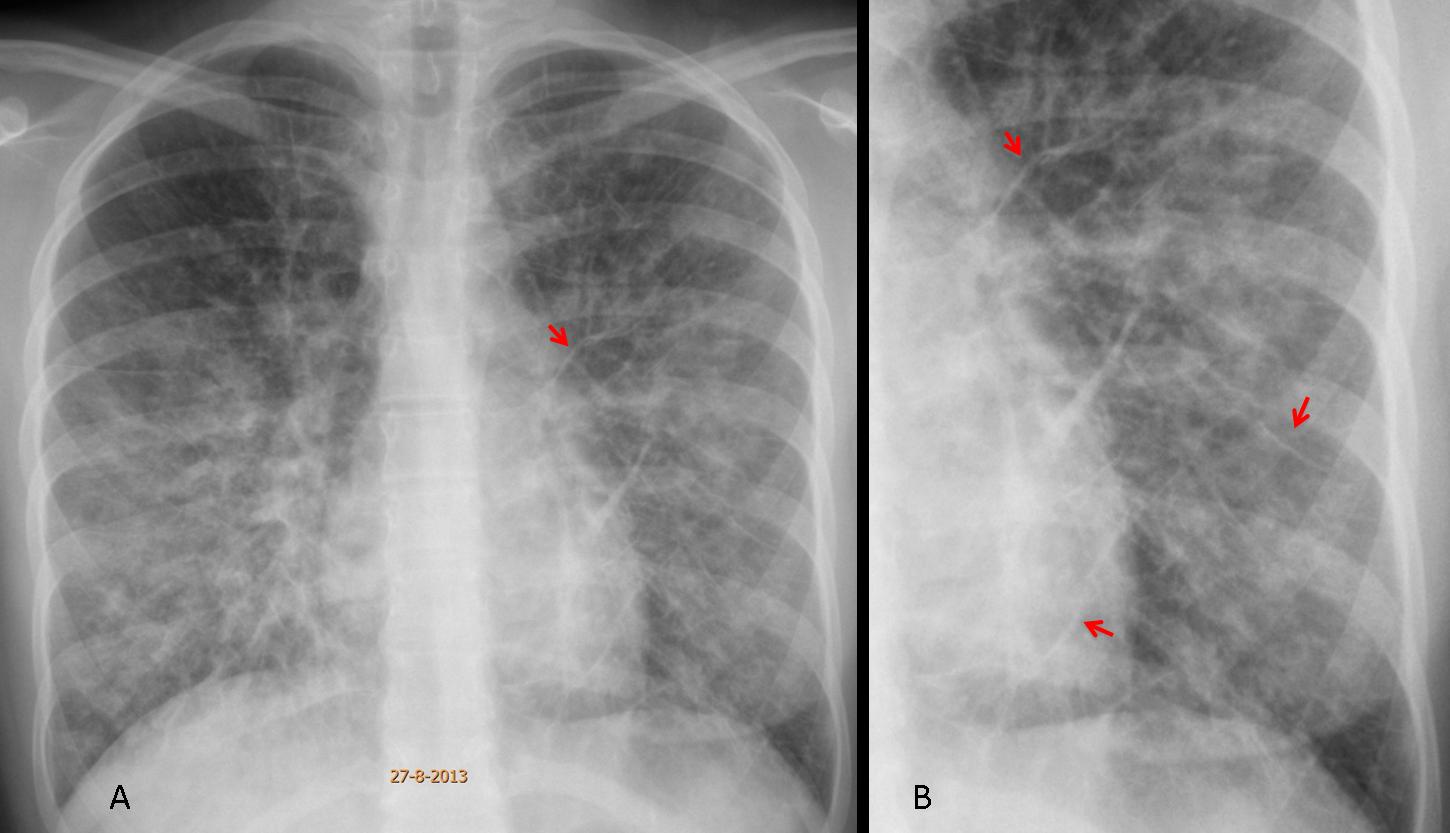
Fig. 6
Fig. 6 (above): 16-year-old girl with viral pneumonia. Notice the widespread air-space disease with prominent A lines (A, arrows), better seen in the close-up view (B, arrows).
Persistent Kerley lines are almost always due to lymphangitic metastasis secondary to the arrival of hematogenous tumour emboli to distal arterial branches and subsequent spread along the pulmonary lymphatics. Any tumour can cause lymphangitic metastasis, but the most common are lung and breast (usually unilateral) (Fig. 7) and GI malignancies: stomach, pancreas, and colon (usually bilateral) (Fig. 8).

Fig. 7
Fig. 7 (above): 41-year-old man with carcinoma of the lung. PA chest film shows an infiltrate along the right mediastinal border, with A lines better seen in the insert (B, arrows). Coronal and axial CT confirm paramediastinal spread of the tumour and septal thickening (C,D, arrows).

Fig. 8
Fig. 8 (above): 57-year-old woman with progressive dyspnea. PA chest film shows widespread Kerley lines (A, arrows), confirmed with CT ( B, arrows). Further studies demonstrated a carcinoma of the colon.
Aside from lymphangitic carcinomatosis, chronic interstitial lung disease may show Kerley lines, although they are not the relevant imaging finding (Fig. 9). Formerly, mitral disease was a classic exponent of Kerley B lines, but nowadays mitral disease is rarely seen in developed countries.

Fig. 9
Fig. 9 (above): 32-year-old man with lupus and interstitial lung disease (A). There are a few Kerley B lines at the left costophrenic angle (B, arrows).

Follow Dr. Pepe’s advice:
1. Kerley lines can be classified as transient or persistent.
2. To differentiate transient Kerley lines from persistent ones, we need serial studies or correlation with clinical findings (acute vs. chronic).
3. The most common cause of transient Kerley lines is pulmonary edema of any cause.
4. The most common cause of persistent Kerley lines is lymphangitic carcinomatosis.


