MRI of the heart is now a standard tool in diagnostic imaging. This week, I want to show you an MRI examination of a 62-year-old man with dyspnoea.
1. Hypertrophic cardiomyopathy
2. Cardiac amyloidosis
3. Cardiac sarcoidosis
4. None of the above
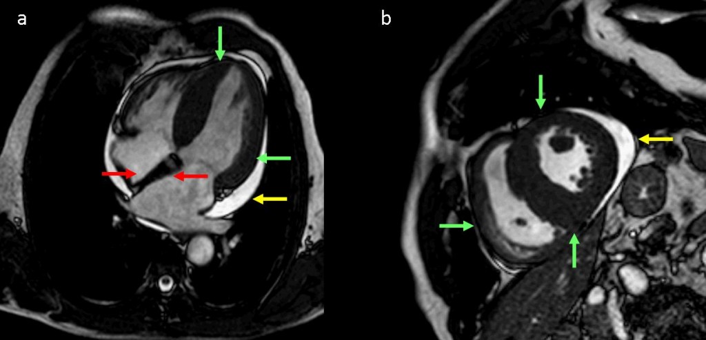
Fig. 3
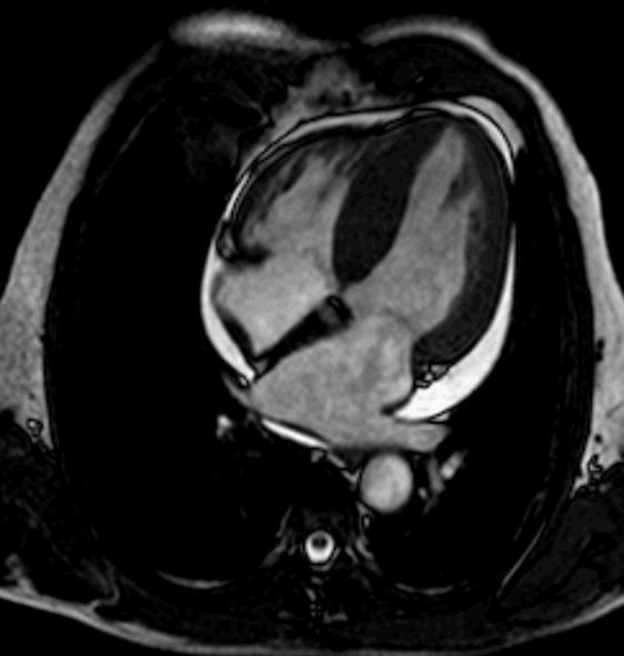
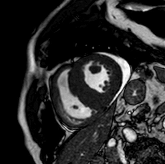
Findings: Four-chamber (a) and short-axis (b) steady-state free precession cine-MRI images show diffuse thickening of myocardial ventricles (green arrows) and atrial septum (>8 mm) (red arrows), together with pericardial effusion (yellow arrows).
- Thickening of the myocardium can be due to hypertrophic cardiomyopathy (HCM), hypertension, valvular stenosis, or deposition diseases. In acute cardiac sarcoidosis, focal myocardial thickening due to oedema is often seen. In very unusual cases of cardiac sarcoidosis, massive infiltration may lead to diffuse myocardial thickening
- Of these causes, the associated findings of thickened ventricular walls with non-dilated left and right ventricles and mild pleural or pericardial effusion suggest cardiac amyloidosis. Atrial septal enlargement >6mm appears to be specific for amyloid infiltration.
- Final diagnosis: cardiac amyloidosis
The heart is the chest organ most commonly affected by amyloidosis. The myocardium, the atrial septum and atrial walls, the valve leaflets and pericardium can be involved. Increased myocardial wall thickness leads to systolic and diastolic dysfunction, and cardiac arrhythmia.
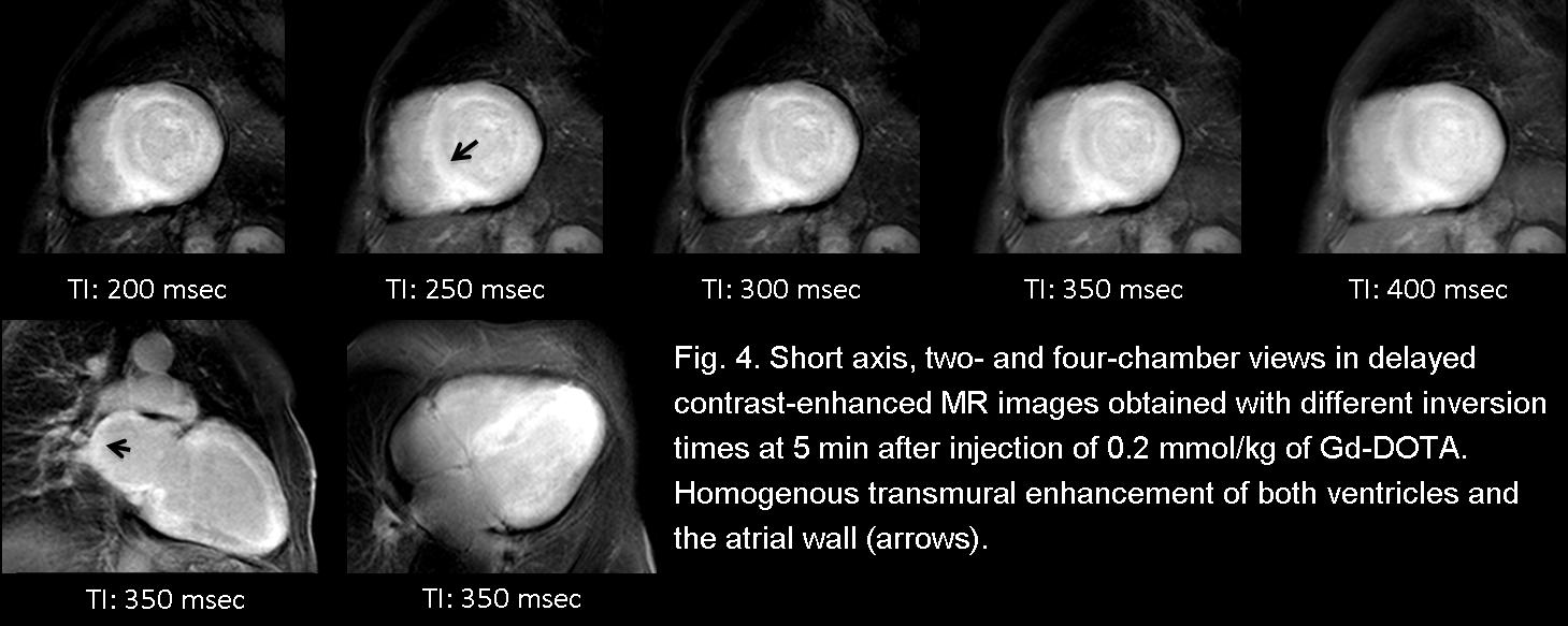
Delayed contrast enhancement is a frequent finding in cardiac amyloidosis (Fig. 4). Global transmural or subendocardial delayed enhancement is the most common pattern, although suboptimal myocardial nulling and focal patchy areas may also be seen. These findings can occur as an early cardiac abnormality in patients with amyloidosis, even when left ventricular thickness is normal.
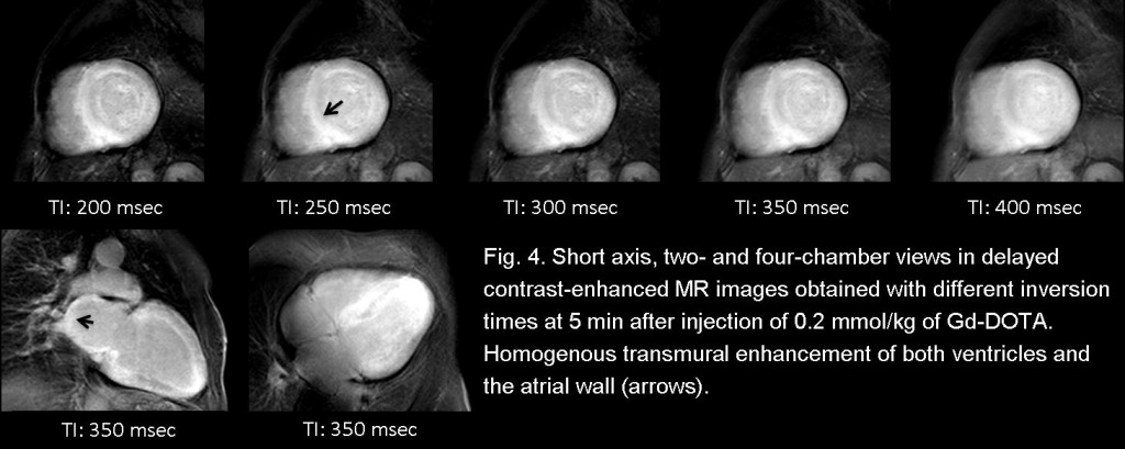
Fig. 4
In hypertrophic cardiomyopathy, delayed enhancement is usually seen as patchy, bright, intramyocardial areas, related to myocardial disarray and fibrosis (Fig. 5).
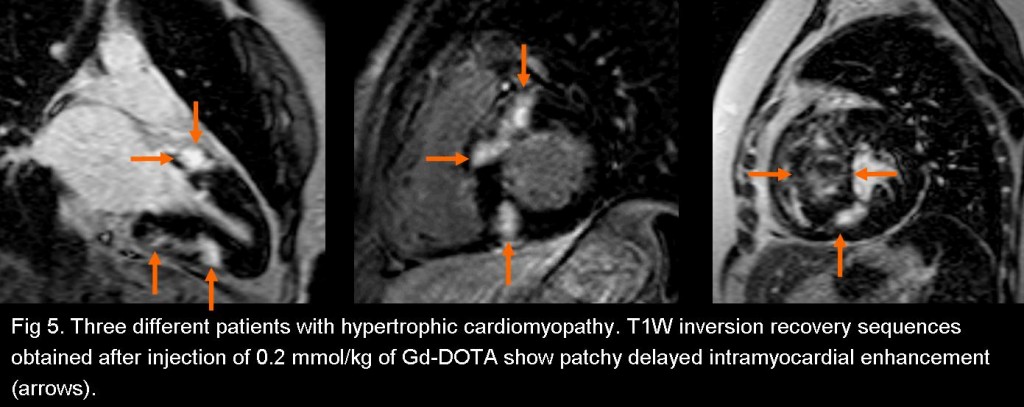
Fig. 5
Cardiac sarcoidosis in the acute phase is characterised by focal or diffuse enlargement, contraction abnormalities, and increased signal intensity on both T2-weighted images and early and delayed images after gadolinium contrast administration, due to oedema associated with granulomatous inflammation.
In the post-inflammatory, replacement scarring phase of cardiac sarcoidosis, there is delayed enhancement resulting from fibrosis.
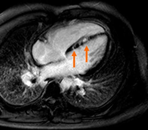
Fig. 6
Fig. 6 shows a 29-year-old man with a history of pulmonary sarcoidosis who presented with arrhythmia. The septal wall shows delayed intramyocardial contrast enhancement without myocardial thickening (arrows) on T1W inversion recovery sequence obtained 10 minutes after contrast injection. Note enhancement of fibrous tissue in lower lobes of the lung.

Follow Dr. Pepe’s advice
Follow Dr. Pepe’s advice:
- Thickened ventricular walls with non-dilated left and right ventricles and mild pleural or pericardial effusion strongly suggest cardiac amyloidosis.
- Atrial septal enlargement >6mm appears to be specific for amyloid infiltration
- Delayed gadolinium enhancement is common and detects interstitial expansion from amyloid deposition
Suggested reading:
- Syed IS, Glockner JF, Feng D, et al. Role of cardiac magnetic resonance imaging in the detection of cardiac amyloidosis. JACC Cardiovasc Imaging. 2010;3:155-64. PMID: 20159642.
- Van Geluwe F, Dymarkowski S, Crevits I, De Wever W, Bogaert J. Amyloidosis of the heart and respiratory system. Eur Radiol 2006;16:2358-65. PMID: 16703313
Case prepared by Rafaela Soler MD