Comprehensive personalised imaging transforms cardiothoracic disease management

ECR2015_ECRToday_Blog

Watch this session on ECR Live: Sunday, March 8, 8:30–10:00, Room E1
Tweet #ECR2015E1 #NH17

Besides personalised imaging, a new paradigm is emerging in radiology that should re-shape clinical practice and benefit the patient immensely. Supported by new technologies that enable radiologists to image the body faster and better, radiologists are now trying to broaden their focus during examinations.

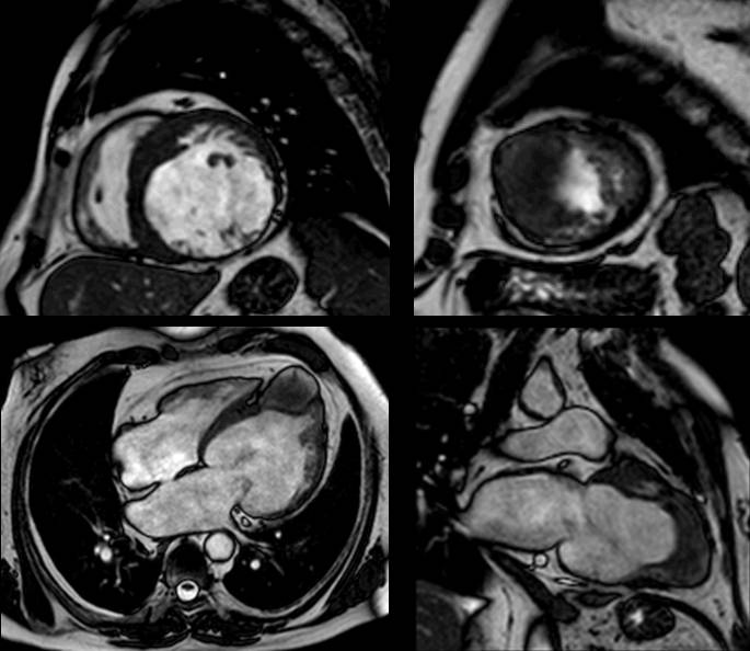
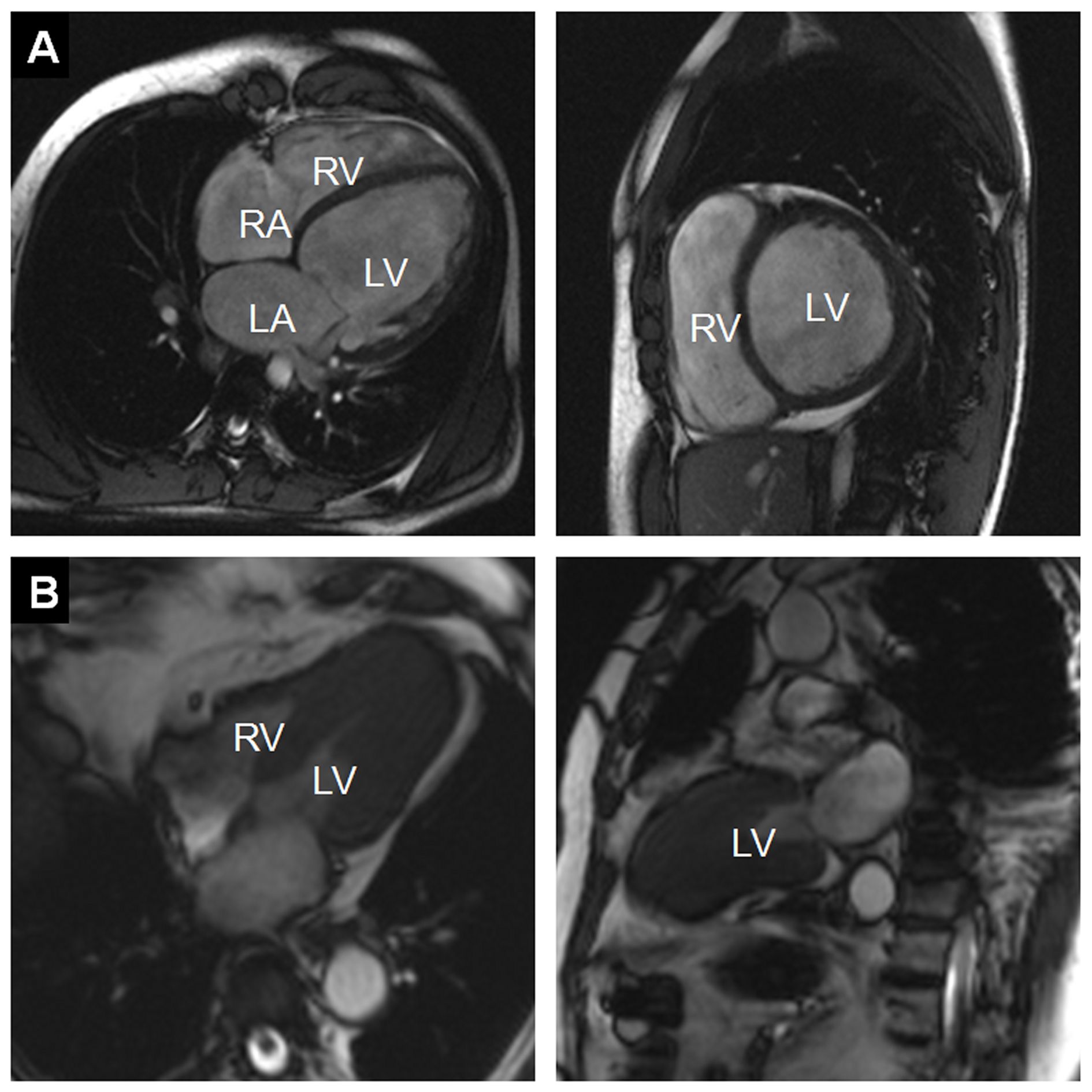
If there is a field where these advances make a tremendous difference, it is cardiothoracic imaging, an area where diseases are more often than not intertwined. Cardiovascular and chest radiologists will explain how the comprehensive personalised approach impacts their work and try to convince radiologists on both sides to take an interest in the other, in a New Horizons session on Sunday at the ECR.

For years, the trend was for radiologists to subspecialise as much as they could. Cardiovascular radiologists and chest radiologists would focus on their own area with little or no interest beyond that. But among these subspecialists, an increasing number are now changing their approach, as mounting evidence shows that diseases of the heart and chest are very often related, according to Dr. Christian Loewe, deputy head of the section of cardiovascular and interventional radiology at the Medical University of Vienna, Austria.

Dr. Christian Loewe is deputy head of the cardiovascular and interventional radiology section at the Medical University of Vienna, Austria.

Dr. Christian Loewe is deputy head of the cardiovascular and interventional radiology section at the Medical University of Vienna, Austria.

“In the past patients were investigated by either focusing on chest or cardiac diseases. This choice was mainly driven by their first clinical examination. However, there are a lot of situations and diseases where chest problems are caused by cardiac diseases and vice versa. There’s a huge interaction between heart and chest, and that’s why it’s interesting and important to look at this relationship in more detail today,” he said.

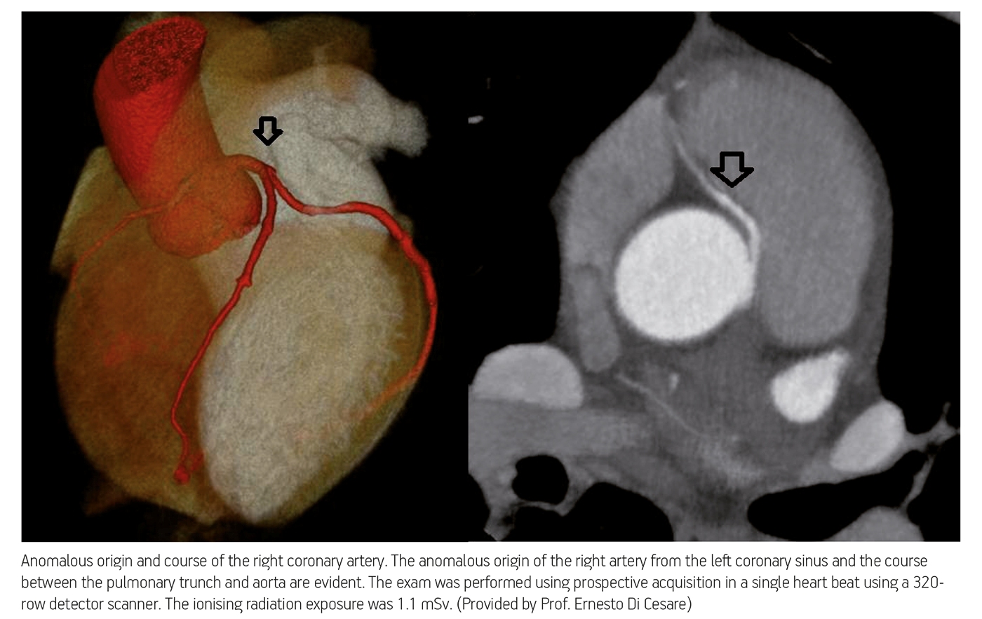
To prove his point, Loewe, a cardiovascular radiologist, will talk about acute and chronic chest pain during the session. Some of the most severe causes of chest pain are due to cardiovascular diseases, including myocardial infarction or acute aortic diseases. However, acute chest pain can also be caused by a number of pulmonary diseases, including pulmonary embolism, pneumonia and others. Therefore, radiologists must learn the different life-threatening disorders that cause chest pain, whether they are respiratory or cardiovascular.

Read more…

ECR 2013 Rec: Coronary artery imaging from a chest CT examination: when and how #SF14b #A451

ECR2013Rec_Blog_Cover_SF14b_A451l

A-451 Coronary artery imaging from a chest CT examination: when and how

R. Marano | Sunday, March 10, 14:00 – 15:30 / Room G/H

The continuous technological evolution of multi-detector CT scanner characterized by larger detector array with increased anatomical coverage per rotation, faster rotation and table speed, and shorter acquisition time have made reliable to perform chest imaging with reduced cardiac motion artifacts, improving the assessment of heart and contiguous structures in the course of routine thorax CT. Further, the larger anatomic coverage of detectors and the availability of scan protocols with lower radiation dose have also made reliable to apply ECG-synchronization to chest CT study, and therefore to couple cardiac/coronary imaging with chest imaging. Different clinical queries requiring a chest CT imaging may underlie cardiac or coronary source that is not clinically evident; similarly, patients scheduled for thoracic surgery, staging or studied in emergency setting may present unexpected heart or coronary artery findings that can be detected in the course of pre-operative CT or may change the treatment and prognosis. Therefore, the capability to perform the assessment of both the heart and chest by a single diagnostic tool is becoming progressively significant because the evaluation of the heart often can provide clinically relevant information in the course of routine or emergency chest CT that is not otherwise easily available.

Dr. Pepe’s Diploma Casebook: Case 45 – SOLVED!

Diploma_casebook_case45

Dear Friends,

Today I am presenting the case of a 73-year-old woman who had preoperative radiographs for haemorrhoid treatment.

Check the two images below and leave me your comments and diagnosis in the comments. Come back on Friday for the answer!

Diagnosis:
1. Cyanotic heart disease
2. Acyanotic heart disease
3. No heart disease
4. None of the above

Read more…

ECR 2013 Rec: Cardiomyopathies #A093 #MC422

ECR2013Rec_Blog_Cover_MC422_A093

A-093 Cardiomyopathies

P. Sipola | Friday, March 8, 08:30 – 10:00 / Room L/M

Cardiac magnetic resonance imaging (CMRI) is highly valuable in the differential diagnosis of cardiomyopathies. MRI diagnosis is based on cine imaging of cardiac function, T2-weighted imaging of oedema and late gadolinium-enhanced (LGE) patterns of scar tissue. Hypertrophic cardiomyopathy (HCM). Left ventricular hypertrophy (LVH) is typically located in basal septum and anterior wall but has variable expression (diffuse, localized). Associated abnormalities include left ventricular (LV) high-ejection fraction (EF), mitral valve abnormalities, apical aneurysm, and right ventricular (RV) hypertrophy. Scattered intramyocardial LGE may occur in various patterns. The differential diagnoses in patient with hypertrophic phenotype include pressure overload hypertrophy, amyloidosis, sarcoidosis, and Fabry’s disease. LGE patterns is useful in differentiation. Dilated cardiomyopathy (DCM): Dilated LV end-systolic volume and impaired EF% are characteristics. Non-ischaemic DCM typically shows no LGE (in contrast to ischaemic cardiomyopthy). Sometimes faint midwall enhancement can be observed, which has prognostic value. Presence of extensive non-compacted myocardium indicates non-compaction cardiomyopathy. Arrhythmogenic right ventricular cardiomyopathy (ARVC): The RV volume is enlargened and akinetic RV segments can be seen. Local bulging or dyskinesia in conjunction with fatty infiltration and LGE is typical. Restricted cardiomyopathy (RCM): Enlargened atrias and normal sized ventricles with preserved EF% and no LGE are characteristics. Myocarditis: LV systolic function is typically lowered but may be normal. T2 images may show increased signal. LGE limited to the subepicardial myocardium is highly suggestive of myocarditis. Iron overload cardiomyopathy: Cine imaging is used to assess LV global function and T2*-weighted imaging to quantitate ventricular iron deposition.

05
Sep 2013
POSTED BY
DISCUSSION 0 Comments

Dr. Pepe’s Diploma Casebook: Case 26 – SOLVED

Diploma_casebook_case26

Dear Friends,

Today I’m showing a case of a 63-year-old man with left heart dysfunction and angor.

one

Read more…

ECR Today – Cardiac imaging in 2020: reaching new heights

ECR2013_ECRToday_RC303

Watch this session on ECR Live: Thursday, March 7, 16:00–17:30, Room D2

Over the past decade, technological improvements have led to the widespread use of imaging modalities in the prediction, diagnosis and follow-up of coronary disease. Radiologists now have the ability to obtain information on the structure of cardiac muscle with MRI and evaluate cardiac arteries with CT, while hybrid imaging will soon allow them to do both. Cardiac CT will also provide more functional information in the future, and its use will continue to grow. Experts will present the newest and upcoming possibilities of cardiac imaging today at the ECR.

Advances in cardiac CT have brought its use in clinical routine to unprecedented levels. The main reason is that image acquisition optimisation strategies allow radiologists to assess blood vessels with the same efficiency as coronary angiography, non-invasively and almost instantaneously.

ECR_2013_ECRToday_CardiacImaging_1

Read more…

06
Mar 2013
POSTED BY
POSTED IN ,
DISCUSSION 2 Comments

ECR 2013 Focus: Organs From A to Z: Heart

ECR2013_Focus_8_AZHeart

Heart disease affects a very large number of people worldwide, and the consequences can be serious and even lethal. Here, and perhaps more than in many other areas of medicine, imaging has helped to improve treatment and prevention. It does so by detecting the disease at an early stage, sometimes even before its emergence, especially in patients at risk of ischaemic heart disease.

Today, diagnosing cardiac patients has become routine for many radiologists. However, some of them may not know of recent developments in this field and they may need to refresh their knowledge. A panel of experts will update both general and specialised radiologists with the latest information available on cardiac imaging, during the dedicated Mini Course ‘Organs from A to Z: Heart’ at ECR 2013. After an introduction to heart anatomy and the main imaging protocols, the course will focus on valvular diseases and cardiomyopathies; two pathologies commonly encountered in radiology practices.

Figure 1

Figure 1:
A) Example of a dilated cardiomyopathy (DCM). Cine-MR images in four-chamber view (left) and short-axis view (right) at end-diastole show significant dilatation of the LV cavity. Ejection fraction was <35% in this patient. (RA = right atrium; LA = left atrium; RV = right ventricle; LV = left ventricle)
B) Example of an asymmetrical, apical hypertrophic cardiomyopathy (HCM). Cine-MR images in a four-chamber (left) and two-chamber view (right) in systole show a markedly thickened left ventricular myocardium predominantly of the apex, as compared with the basal segments (RV = right ventricle; LV = left ventricle).

Read more…

Dr. Pepe’s Diploma Casebook: Case 7 – SOLVED!

Dear Friends,

MRI of the heart is now a standard tool in diagnostic imaging. This week, I want to show you an MRI examination of a 62-year-old man with dyspnoea.

Read more…

Dr. Pepe’s Diploma Casebook: Case 3 – SOLVED!

Dear Friends,

Moving forward on our journey through the systems, our next stop is the heart. I want to put your diagnostic skills to the test with the following cardiac case.

Our patient is a 60-year-old male with dyspnea and fatigability.

Read more…