Dr. Pepe’s Diploma Casebook: Case 70 – SOLVED!

Dear Friends,
Today I am showing chest radiographs of a 34-year-old male with dyspnea. Check the images below, leave your diagnosis in the comements section and come back on Friday for the answer.
Diagnosis:
1. Mitral disease
2. Pulmonary arterial hypertension
3. Interstitial pneumonia
4. None of the above
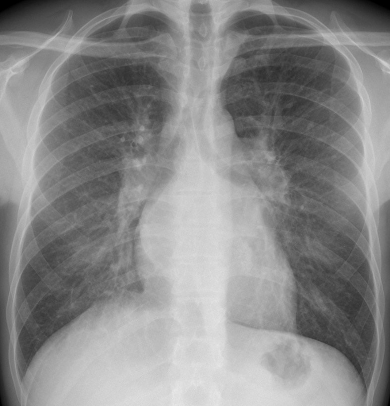
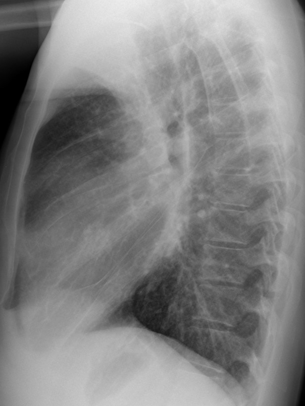
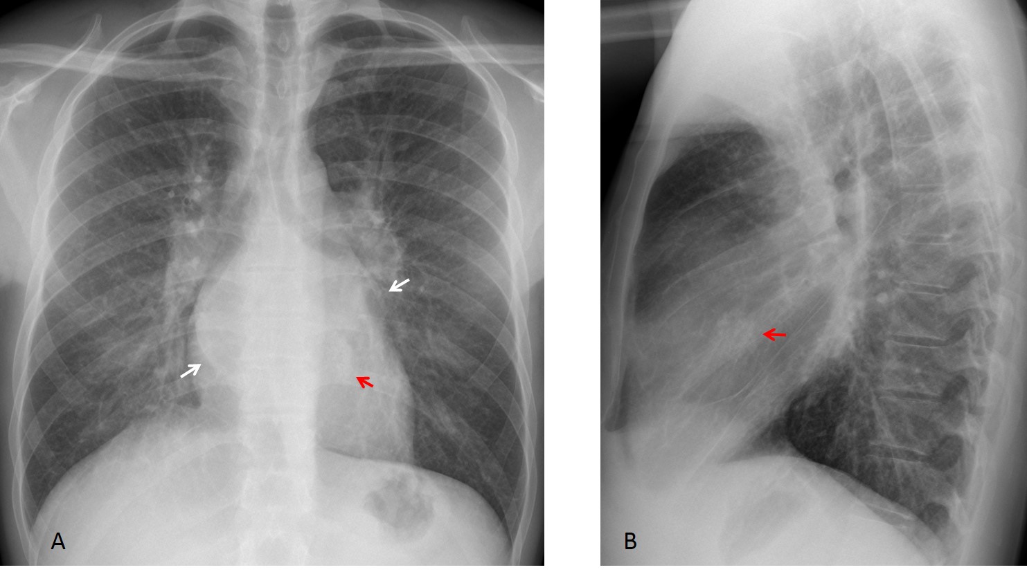
Findings: PA radiograph shows redistribution of the pulmonary vessels. The hila are enlarged and blurry. The left atrium and atrial appendage have a double contour (A, white arrows). Calcium is visible in the area of the mitral valve (A,B, red arrows).
CT confirms the left atrial enlargement (C,D white arrows), as well as calcium in the mitral valve (C,D, red arrows).
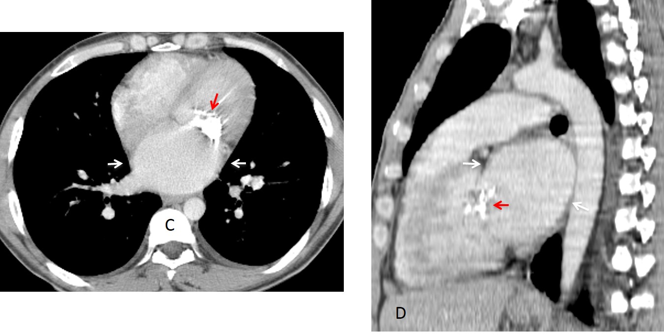
Final diagnosis: calcified mitral stenosis with vascular redistribution.
This case is presented to discuss redistribution pattern of the pulmonary vessels.
In the normal erect PA view, the vessels of the upper lobes are smaller than those of the lower ones (Fig. 1 A). In redistribution, the upper vessels are larger than the lower ones (Fig. 1 B).
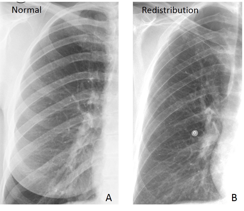
Fig. 1
Fig. 1: Normal vascular pattern (A) and redistribution (B). Note the small upper vessels in A compared with the lower ones. In B, the appearance is reversed. Redistribution is better seen in the right lung because the left lower vessels are obscured by the heart.
I am using the word redistribution as a purely anatomical term, without specifying the etiology. The great majority of cases have a cardiovascular etiology, secondary to increased left atrial pressure, causing the blood to back up into the pulmonary veins. As a consequence, the arteries of the lower lobes constrict, redistributing the flow to the upper vessels, which then appear larger than the lower ones.
Recognising redistribution needs a certain degree of expertise, but it is rewarding because it may reveal a cardiovascular problem before clinical findings are evident.
Although most cases of redistribution have a cardiovascular etiology, obstruction of lower lobe arteries and basal emphysema may give similar findings.
Cardiovascular redistribution can be classified as obstructive or non-obstructive.
The main cause of non-obstructive redistribution is left-sided heart failure. When the left heart fails, blood collects in the pulmonary veins, causing redistribution as an early sign. If failure progresses, pulmonary oedema develops (Fig. 2).
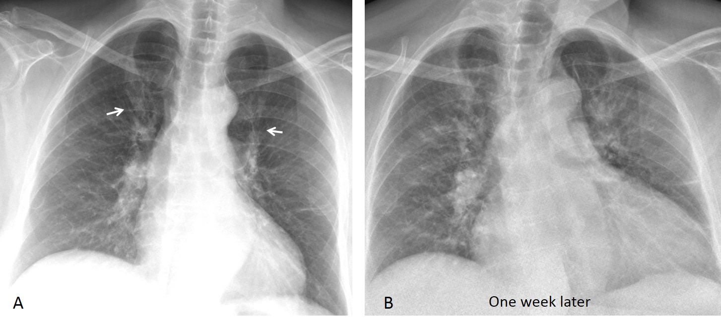
Fig. 2
Fig. 2: 65-year-old man with cardiomyopathy. Initial film shows borderline cardiac enlargement and redistribution (A, arrows). One week later, the heart has enlarged and there is pulmonary oedema (B).
A relatively common cause of non-obstructive redistribution is atrial fibrillation, which occurs in 5% of individuals older than 65. Consider atrial fibrillation when you see left atrial enlargement and vessel redistribution in an elderly patient (Figs. 3 and 4). The diagnosis is important because embolic stroke is a common complication.
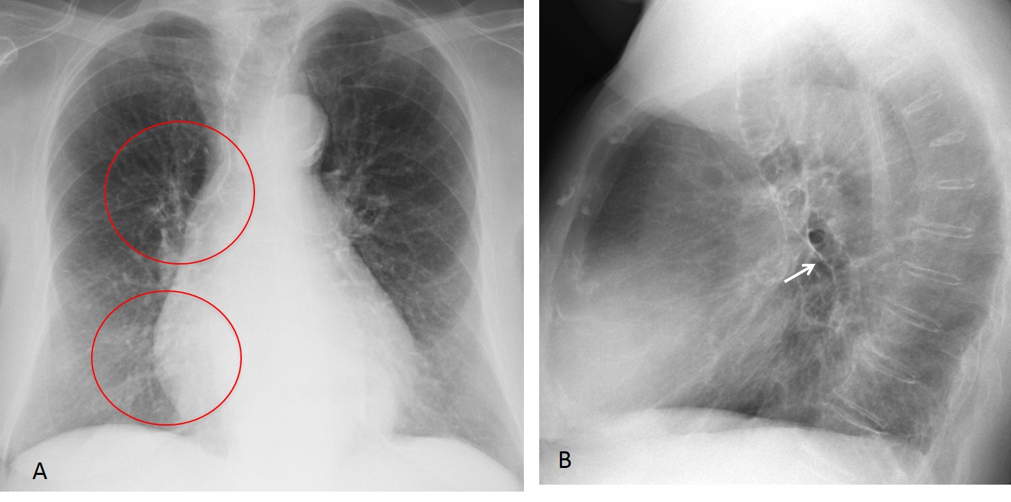
Fig. 3
Fig. 3: 72-year-old woman with dizzy spells. PA chest film shows moderate cardiomegaly and redistribution (A, circles). Lateral view shows posterior displacement of the left bronchus (B, arrow), a sign of atrial enlargement. Diagnosis: atrial fibrillation.
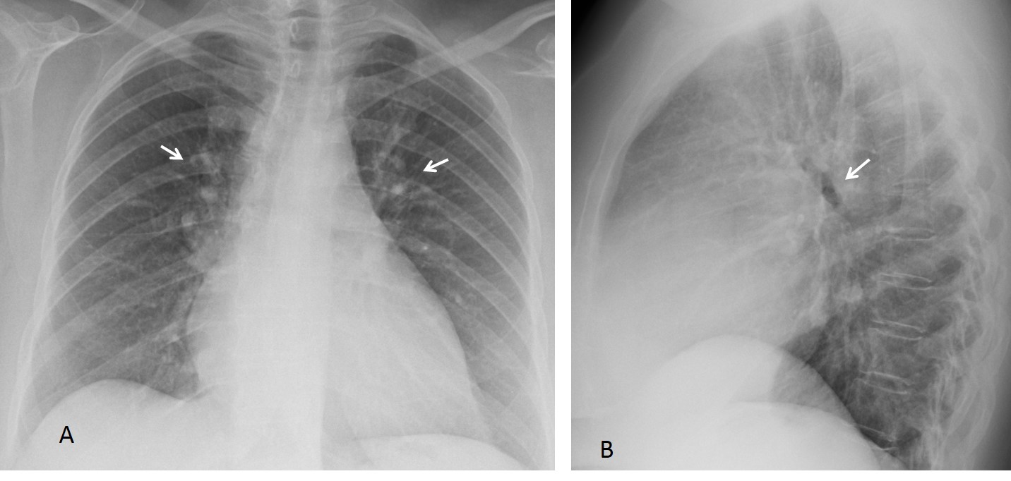
Fig. 4
Fig. 4: 68-year-old woman with moderate cardiomegaly, redistribution (A, arrows) and posterior displacement of left bronchus (B, arrow). Again, atrial fibrillation.
The main causes of obstructive cardiovascular lesions are mitral valve disease, aortic valve stenosis, and aortic coarctation. Rheumatic mitral disease used to be the most frequent obstructive cause of redistribution. It is now uncommon in developed countries, but is still seen in citizens of less favored ones. The findings are well known and are described in Fig. 5.
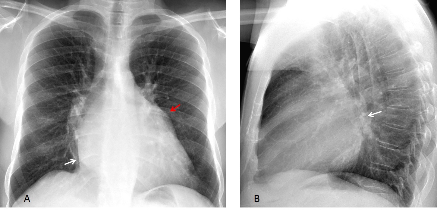
Fig. 5
Fig. 5: 28-year-old immigrant with typical findings of mitral disease. PA view shows redistribution, double contour of left atrium (A, white arrow), and prominent appendage (A, red arrow). There is posterior prominence of left atrium (B, arrow).
Stenotic lesions of the aorta also increase pressure in the left side of the heart, resulting in redistribution. The most common is coarctation, although any aortic obstruction can produce similar findings (Fig. 6).
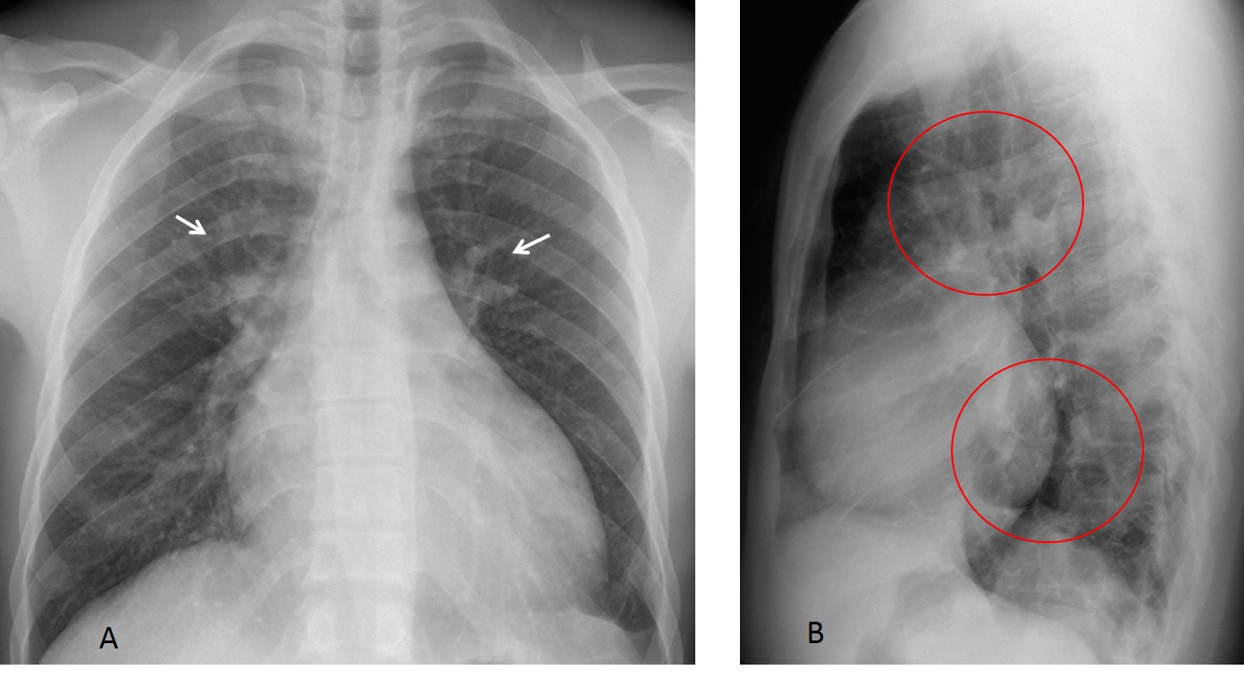
Fig. 6
Fig. 6: 27-year-old man with elevated blood pressure. PA chest film shows cardiomegaly and redistribution (A, arrows). In this particular case, redistribution is also seen in the lateral view (B, circles).
3D reconstruction shows thinning and interruption of the thoracic aorta (C, arrows) with marked collateral circulation. A thoracic-to-abdominal aortic bypass was placed (D, arrow). Post-op radiograph shows decrease in heart size and in redistribution. Note the outline of the graft (E, arrows). Diagnosis: mid-aortic syndrome, secondary to Takayasu disease.
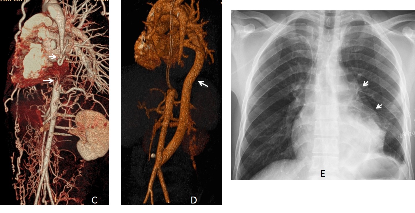
Fig. 6
Obstruction of lower lobe pulmonary arteries is an uncommon cause of redistribution. It can occur in pulmonary embolism (Fig. 7) or in vasculitis (Fig. 8).
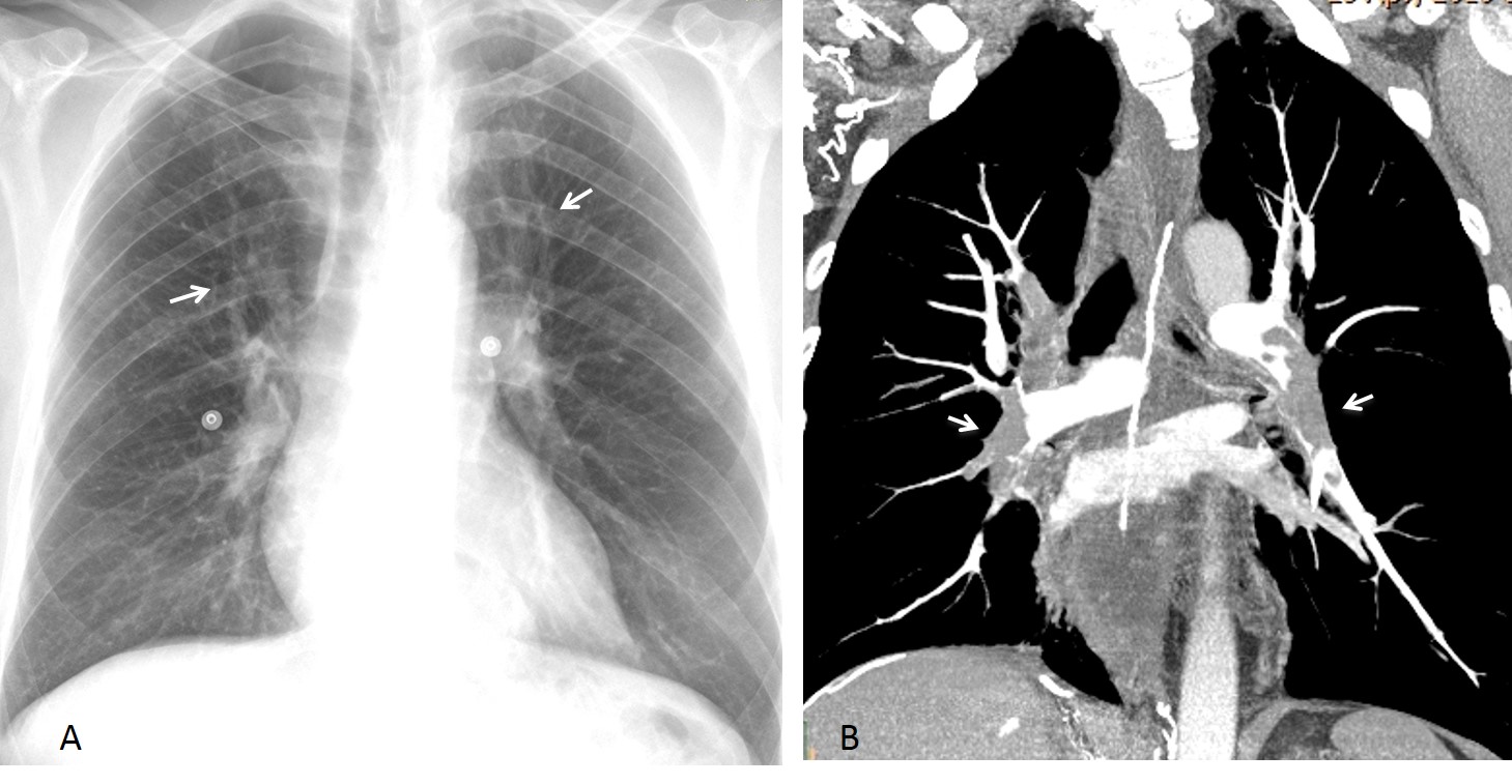
Fig. 7
Fig. 7: 45-year-old man with lower limb phlebitis and severe dyspnea. PA chest film shows redistribution (A, arrows) with a normal-sized heart. CT angiogram confirms the redistribution, secondary to multiple emboli, mainly to lower lobe arteries (B, arrows).
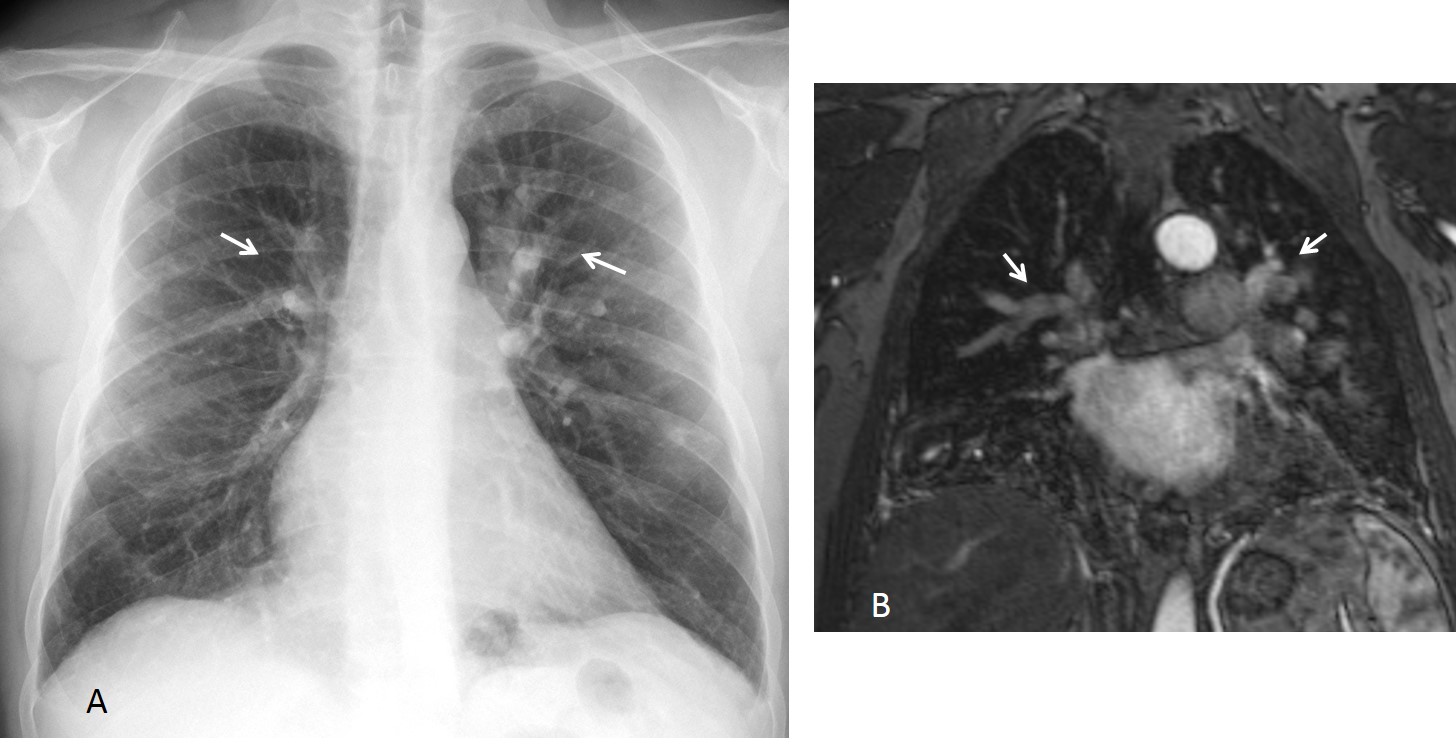
Fig. 8
Fig. 8: 48-year-old man with cognitive problems. PA chest film depicts obvious redistribution (A, arrows). MR angiogram shows that blood flow is decreased in the lower pulmonary arteries and increased in the upper ones (B, arrows). Diagnosis: Takayasu
Emphysema of lower lobes diminishes the blood flow and increases the flow to the upper vessels. In these cases, the etiologic diagnosis is obvious (Fig. 9).
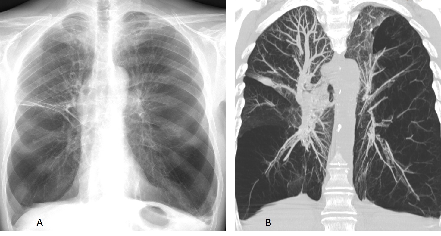
Fig. 9
Fig. 9. 68 y.o. man with alpha 1 antitrypsin emphysema. Marked emphysema of lower lobes causes redistribution (A), confirmed on coronal CT (B).

Follow Dr.Pepe’s Advice:
1. Redistribution may reveal underlying heart problems before clinical symptoms are obvious.
2. Non-obstructive causes of redistribution are left heart failure and atrial fibrillation.
3. Nowadays, the most common obstructive cause of redistribution is aortic valve stenosis.
4. Uncommon causes of redistribution are obstruction of lower lobe arteries and lower lobe emphysema.


