Dr. Pepe’s Diploma Casebook: Case 79 – SOLVED

Dear Friends,
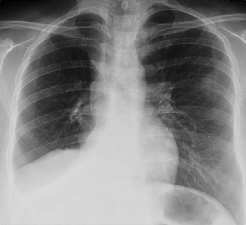
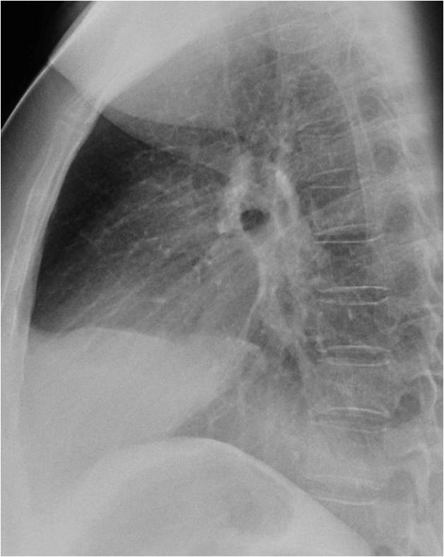
I would like to start the fourth season with an easy case. Showing pre-op radiographs for facial surgery of a 31-year-old woman. Check the images below, leave me your thoughts in the comments section, and come back on Friday for the answer.
Diagnosis:
1. Carcinoma of the lung
2. Subpulmonary fluid
3. Aspiration pneumonia
4. None of the above
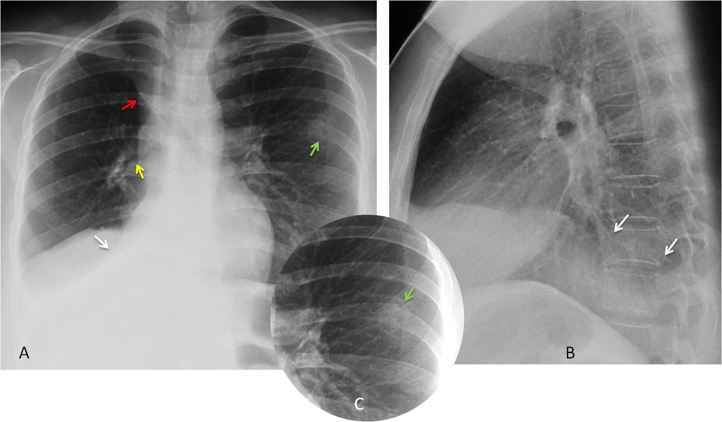
Findings: PA radiograph shows a triangular opacity in the right lower lung (A, white arrow) and an upper triangle sign (A, red arrow). These findings are highly suspicious of RLL collapse. There is also a mass in the right hilum, with narrowing and irregularity of the main bronchus (A, yellow arrow). A pathological fracture is visible in the left sixth rib (A,C, green arrow). The lateral view shows blurring of the posterior right hemidiaphragm (B, arrows).
Unenhanced coronal CT confirms RLL collapse (C, D, white arrows) and the right hilar mass, with irregularity and narrowing of the main bronchus (C, D, red arrows).
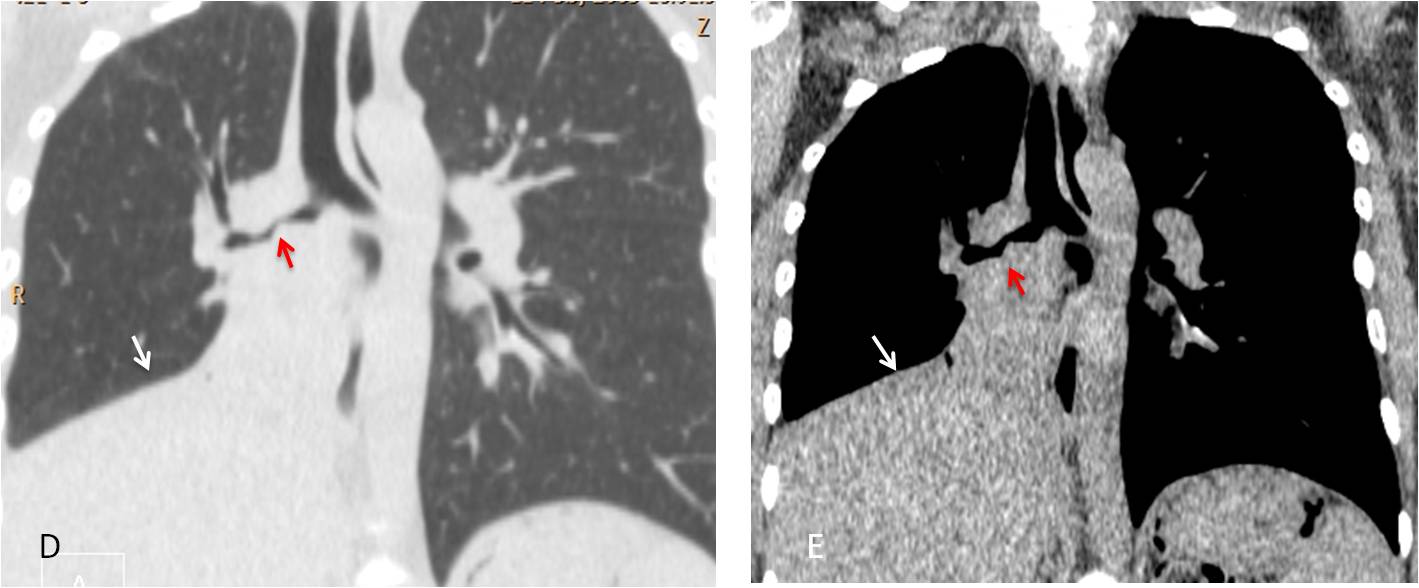
Final diagnosis: bronchogenic carcinoma with RLL collapse
In this presentation I’d like to discuss common and uncommon manifestations of RLL collapse. RLL collapse is easy to recognise when typical signs are present. However, it can have an unusual appearance and be confused with other conditions, leading to a delay in the diagnosis. A prompt diagnosis is important because the main cause of RLL collapse is carcinoma of the lung, followed by inflammatory bronchiectasis.
The typical signs of RLL collapse in the PA view are a well-defined triangular opacity in the lower lung, accompanied by downward displacement of the hilum. The lateral view shows a posterior opacity that blurs the posterior diaphragmatic outline. The diagnosis is easily confirmed by CT (Fig. 1).
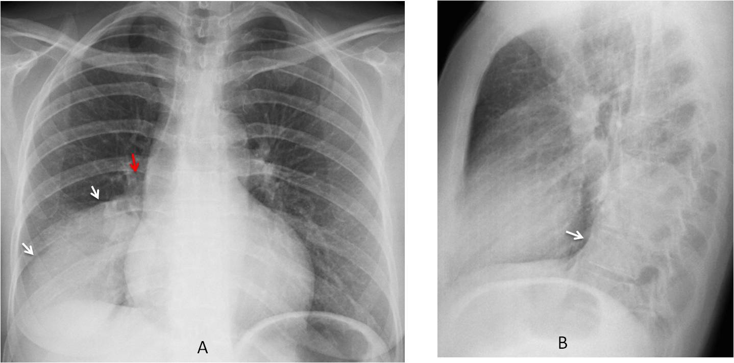
Fig. 1
Fig. 1: 55-year-old woman with carcinoma and partial RLL collapse. PA radiograph shows the triangular shape of the collapsed lobe (A, white arrows). The hilum is descended (A, red arrow). The lateral view shows a posterior opacity and concave major fissure (B, arrow).
Enhanced coronal and sagittal CT images confirm the collapsed lobe. The major fissure is displaced downward in the coronal view (C, white arrow) and is convex in the sagittal reconstruction (D, arrow). The obstructed bronchus is clearly visible (C, red arrow).
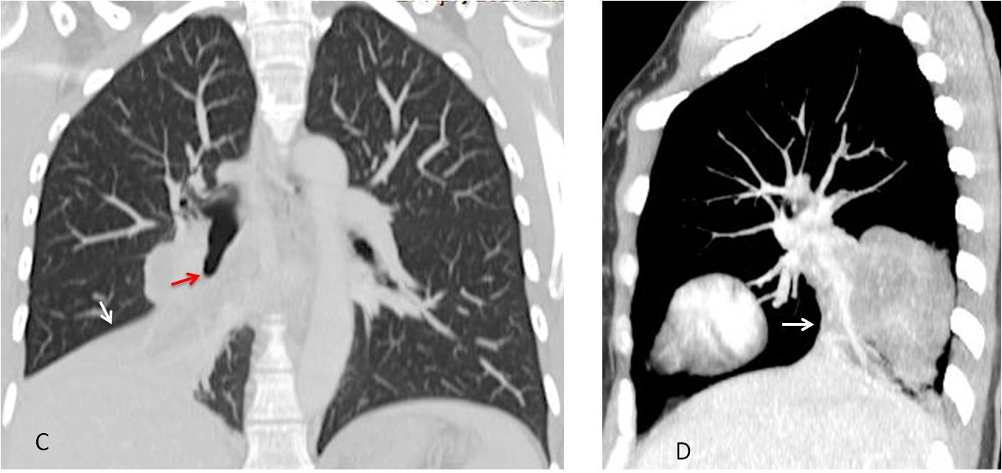
Fig. 1
A central mass causing RLL collapse may be visualized as an S, a variant of the Golden sign (Fig. 2).
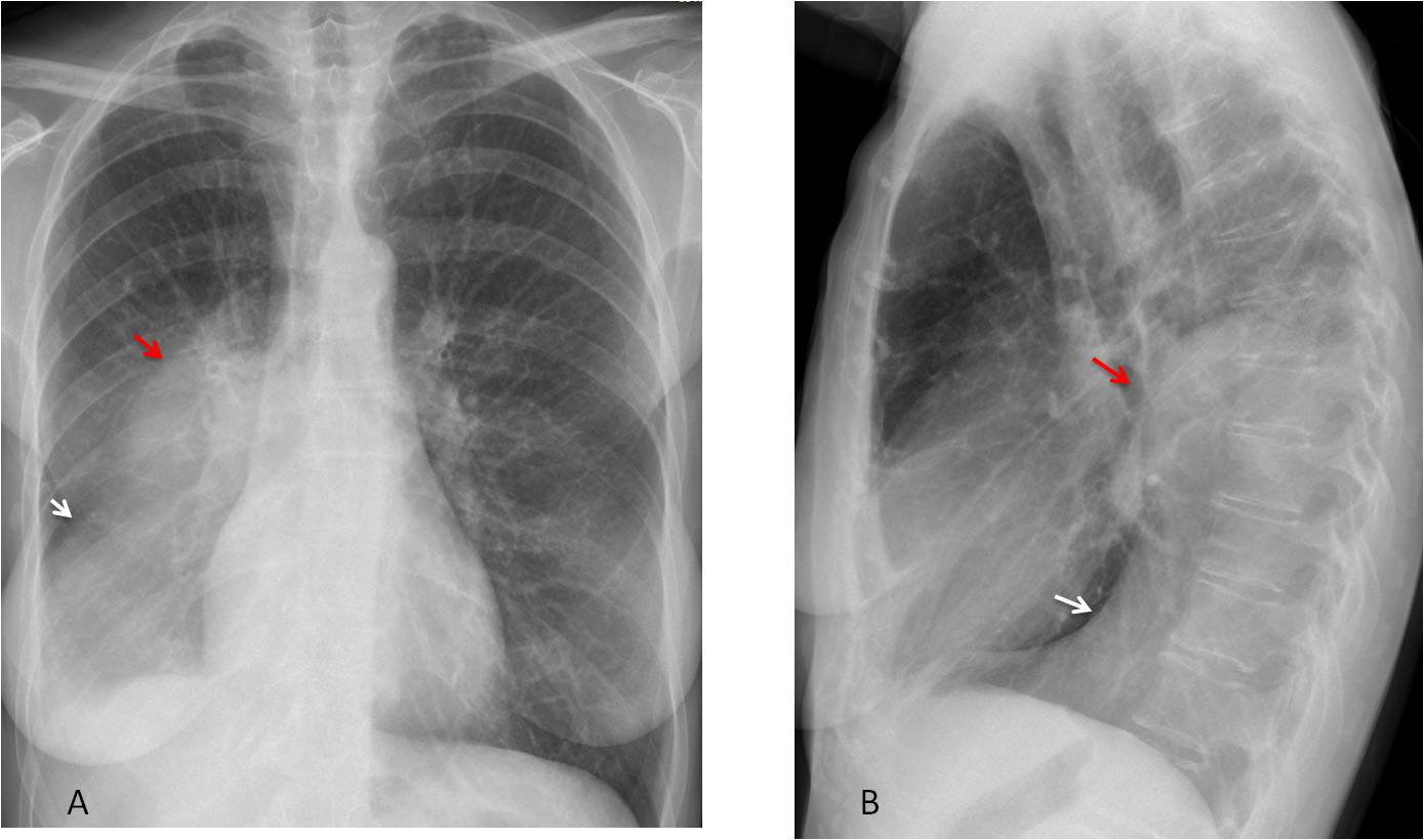
Fig. 2
Fig. 2: Carcinoma of RLL with Golden sign (A, B, red arrows). Note the oblique course of the displaced major fissure (A, white arrow), which is convex in the lateral view (B, white arrow)
Enhanced coronal and sagittal CT images confirm the central mass (C, D, red arrows), as well as the distal collapsed lobe (C, D, white arrows)
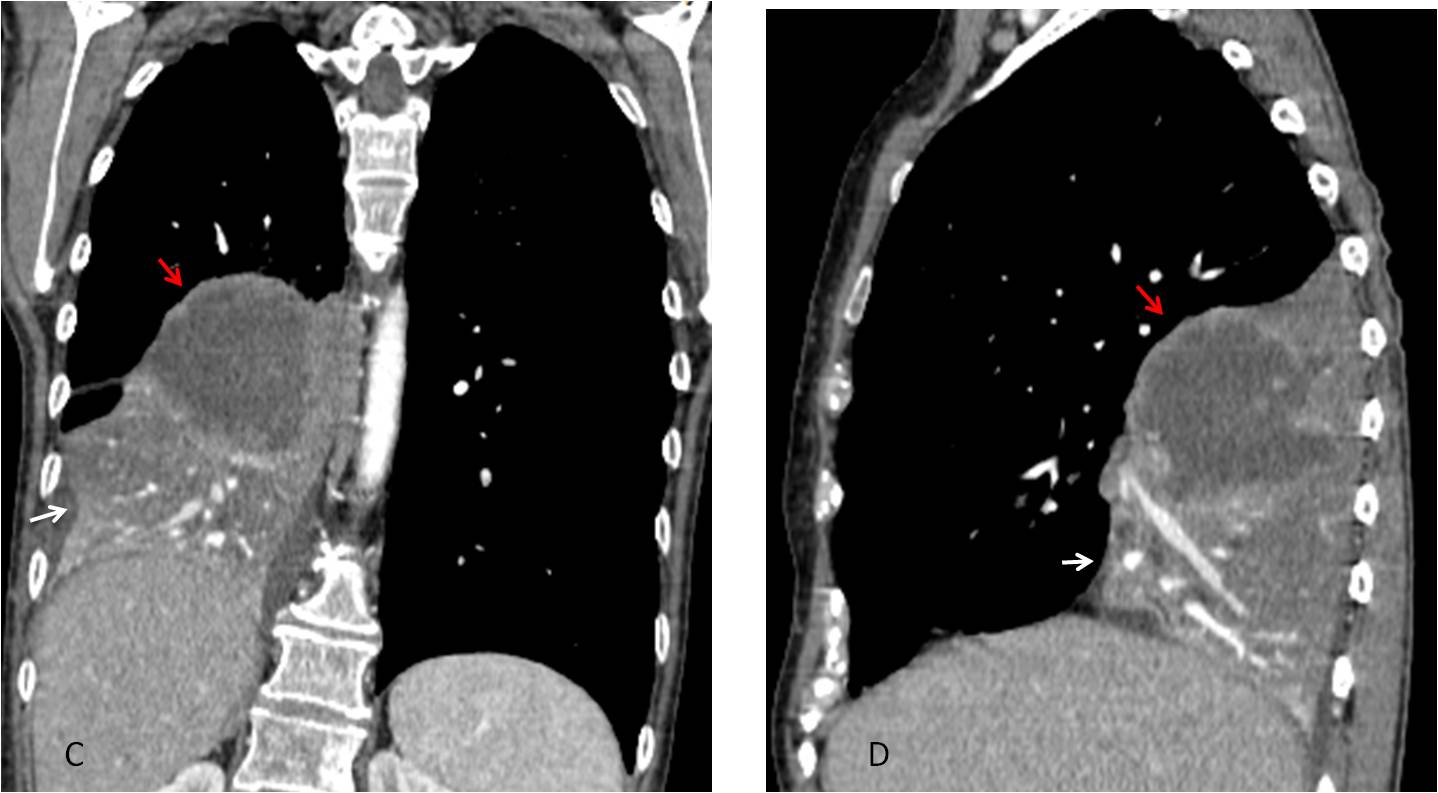
Fig. 2
In my experience, RLL collapse simulating pleural fluid is not uncommon. In this presentation form, the peripheral lung obliterates the costophrenic sinus in both views. The sharp interface of the major fissure helps to create the impression of pleural effusion. The clue to the correct diagnosis is the downward displacement of the hilum (Figs. 3 and 4).
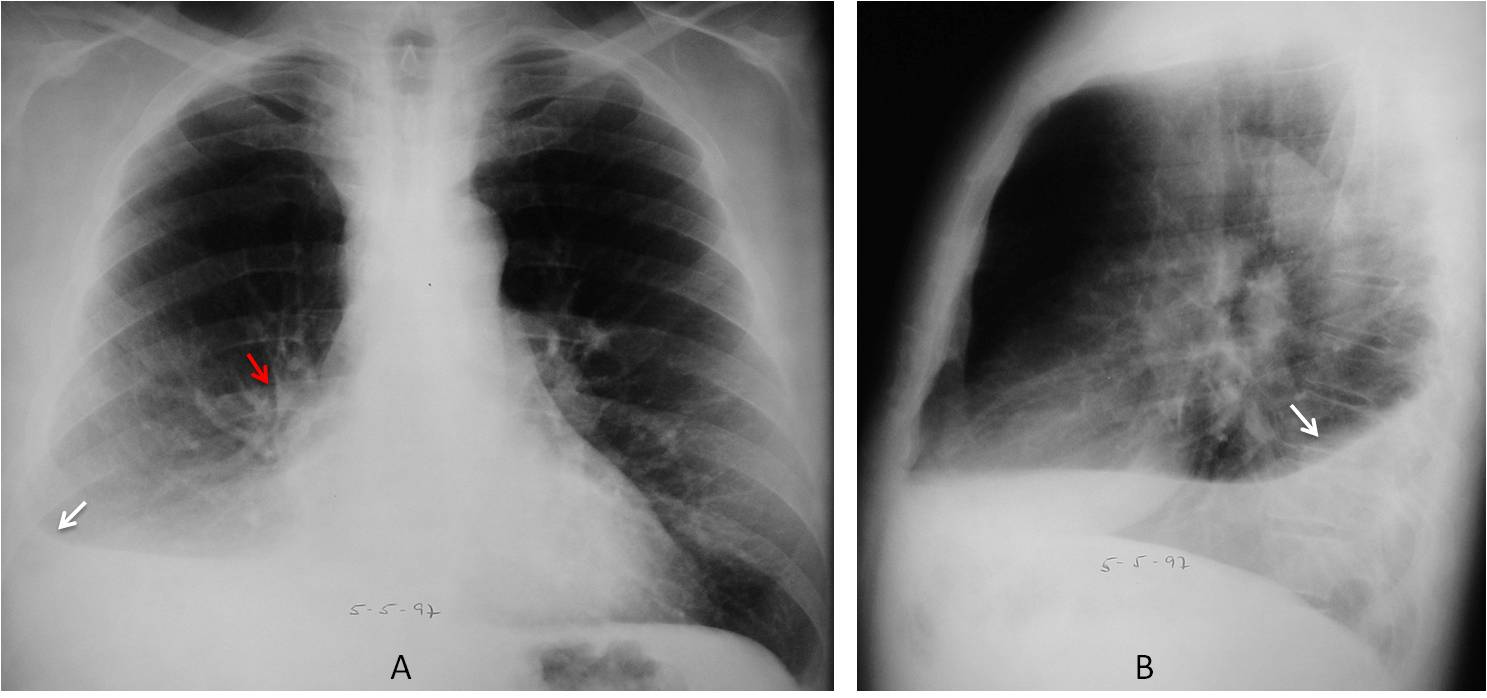
Fig. 3
Fig. 3: RLL collapse simulating pleural effusion. There is blunting of the costophrenic sinus in the PA radiograph with a horizontal major fissure (A, white arrow). The collapsed lobe in the lateral view simulates free fluid (B, arrow). Note the descended right hilum (A, red arrow).
Unenhanced axial CT shows marked narrowing of the RLL bronchus (C, arrow). The distal lobe is visible in the posterior costophrenic sulcus and shows an air bronchogram (D, arrow). There is no evidence of pleural fluid.
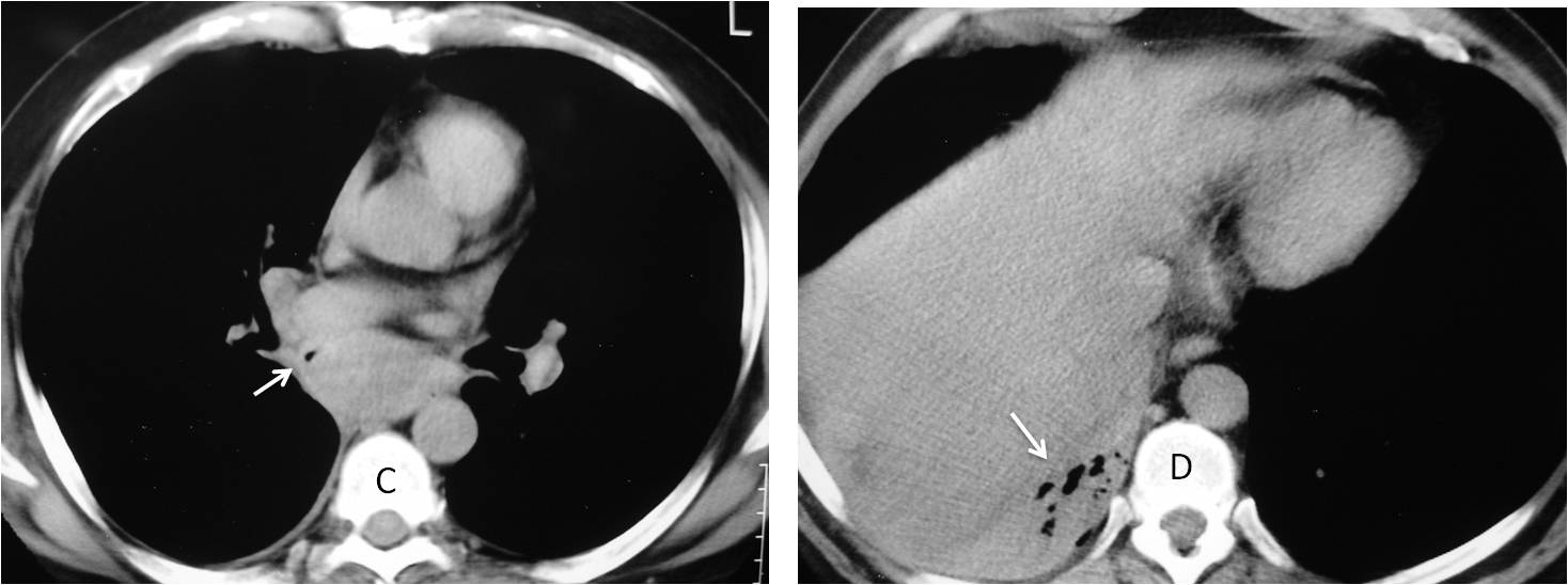
Fig. 3
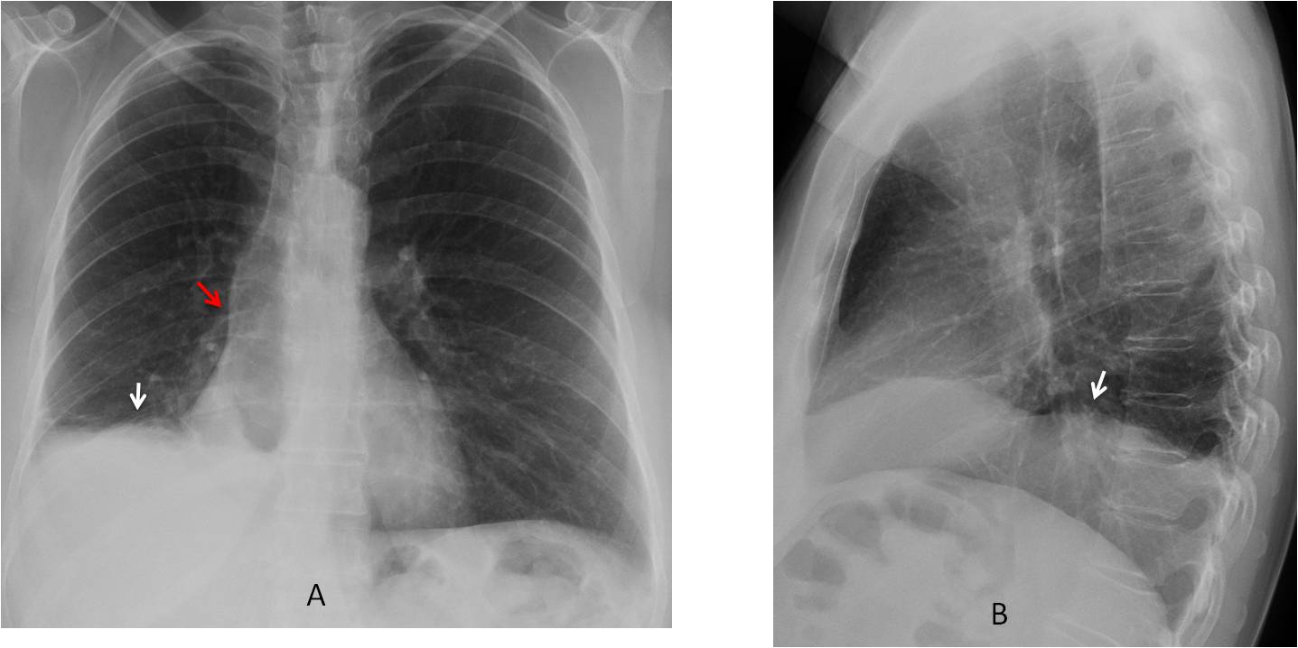
Fig. 4
Fig. 4: 44-year-old woman with haemoptysis. PA and lateral radiographs depict an irregular right hemidiaphragm with the appearance of subpulmonary fluid (A, B, white arrows). However, the right hilum is descended (A, red arrow), which raises the possibility of RLL collapse.
Post-contrast CT demonstrates an enhancing sliver of RLL that simulates pulmonary fluid (C, D, arrows). A more cephalad slice shows occupation of he RLL bronchus (E, arrow). Bronchoscopy demonstrated a blood clot occluding the bronchus, with no evidence of tumour.
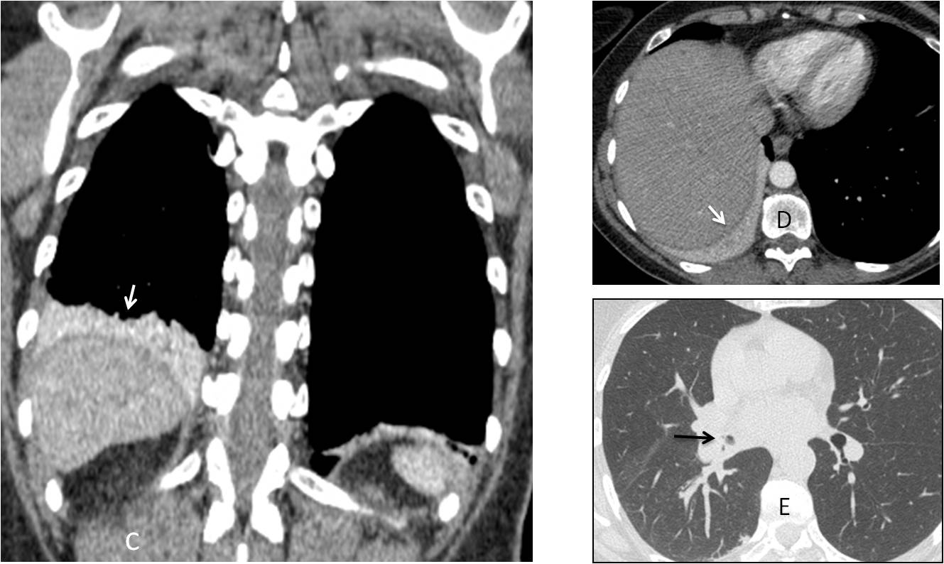
Fig. 4
On chest radiography a few days after bronchoscopy, the chest had a normal appearance. Note the normal aspect of the diaphragm and correct location of the right hilum (F, G, arrows).
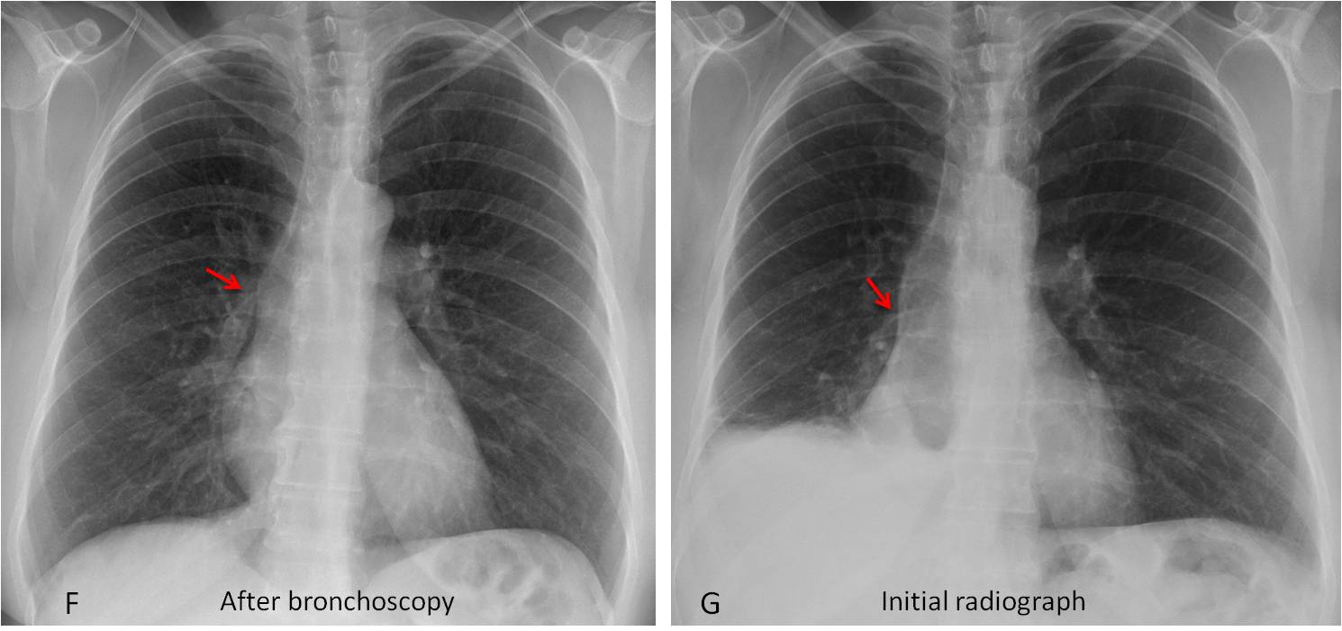
Fig. 4
In aerated RLL collapse the lobe loses volume but there is no increase in opacity, making the collapse less obvious. The descended hilum and major fissure are clues to the diagnosis (Figs. 5 and 6). The lack of opacity may be secondary to incomplete obstruction of the lobar bronchus, collateral ventilation through an incomplete major fissure, or both.
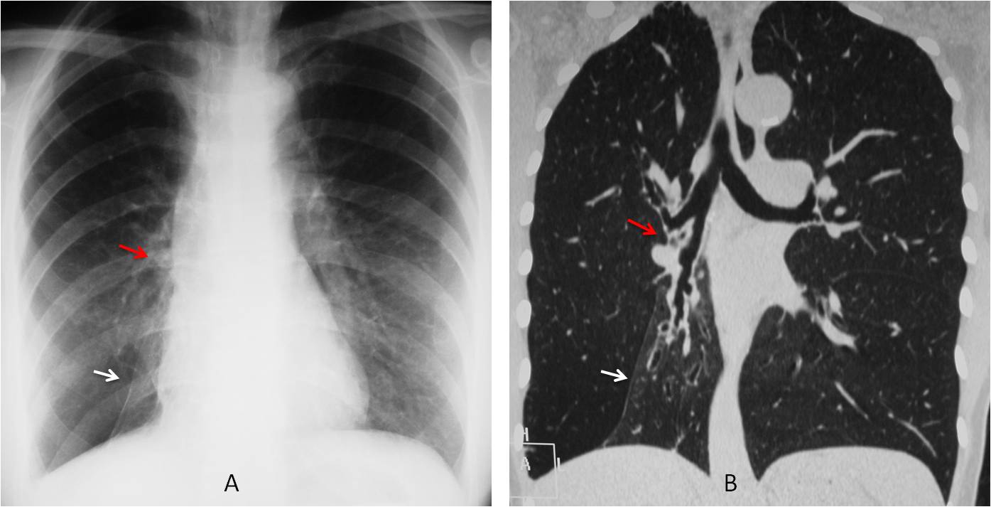
Fig. 5
Fig. 5: Aerated RLL collapse secondary to bronchiectasis. PA radiograph shows a markedly displaced major fissure simulating an inferior accessory fissure (A, white arrow). There is marked downward displacement of the right hilum (A, B, red arrows). Coronal CT shows the collapsed lobe (B, white arrow) with bronchiectasis and open RLL bronchus.
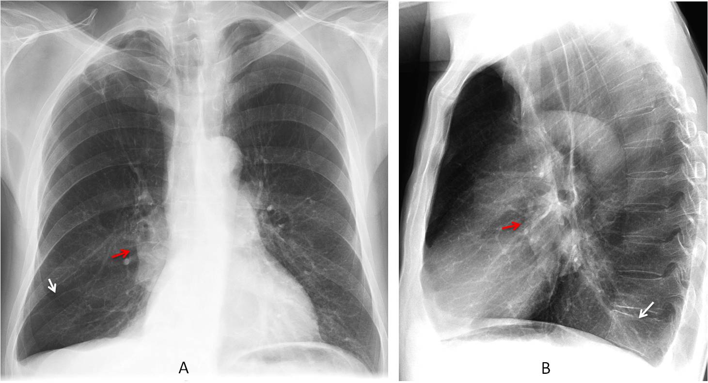
Fig. 6
Fig. 6: Aerated RLL collapse in carcinoma. PA chest film depicts a right hilar mass (A, B, red arrows), with descended hilum. The lowered major fissure is barely visible (A, white arrow). In the lateral view, the collapsed lobe is seen as a faint opacity projected over the spine (B, white arrow).
In marked RLL collapse the opaque lobe hides behind the right side of the heart and may be difficult to detect. The increased lucency of the expanded RUL may be misleading. The descended hilum and upper triangle sign, when present, are clues leading to the correct diagnosis (Figs. 7 and 8). The upper triangle is due to shifting of the superior mediastinum, secondary to RLL volume loss.
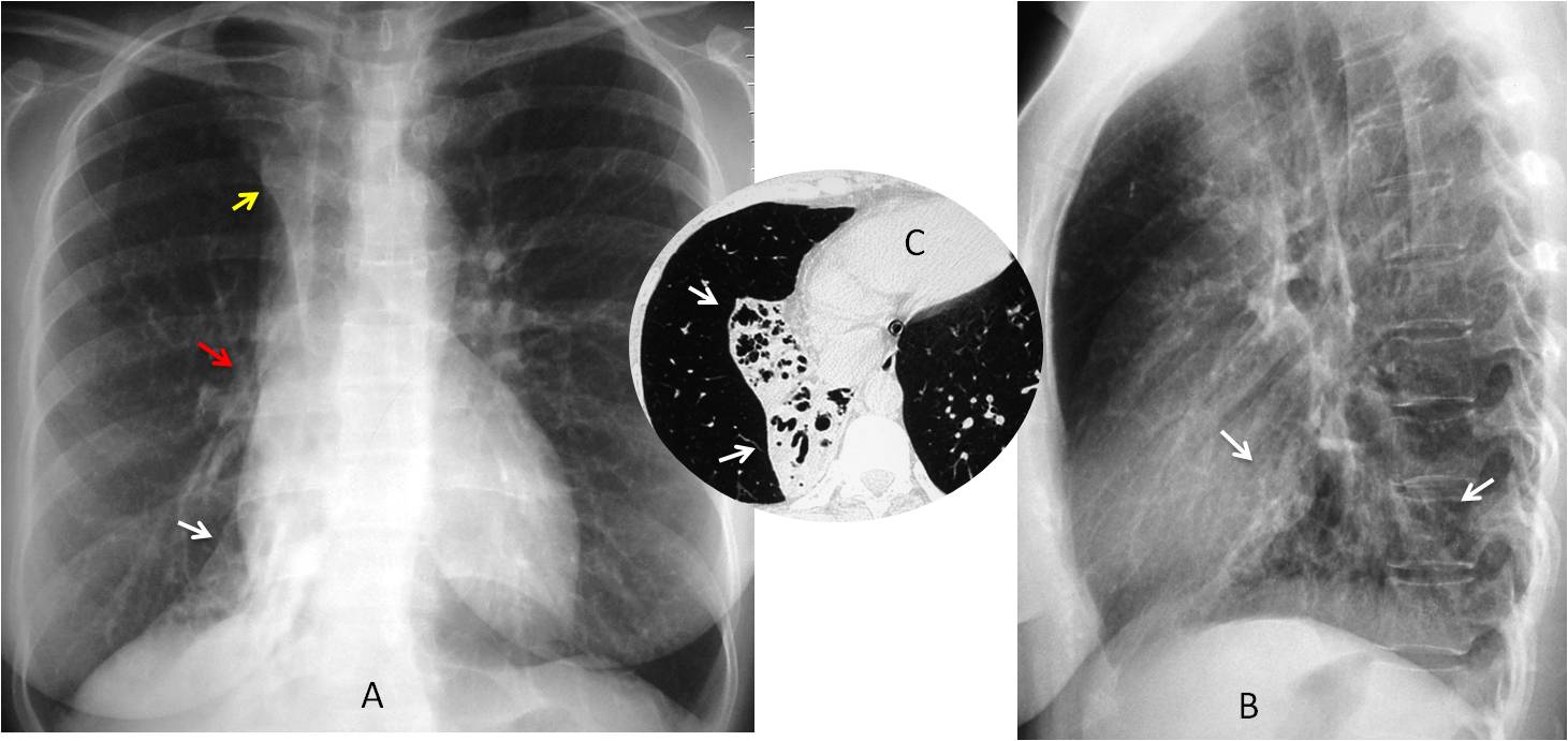
Fig. 7
Fig. 7: PA radiograph shows marked RLL collapse partially hidden behind the heart (A, white arrow), hilum displaced downward (A, red arrow), and upper triangle sign (A, yellow arrow). In the lateral view the changes appear as a triangular RLL opacity blurring the left hemidiaphragm (B, arrows). Diagnosis: inflammatory bronchiectasis (C, arrows).
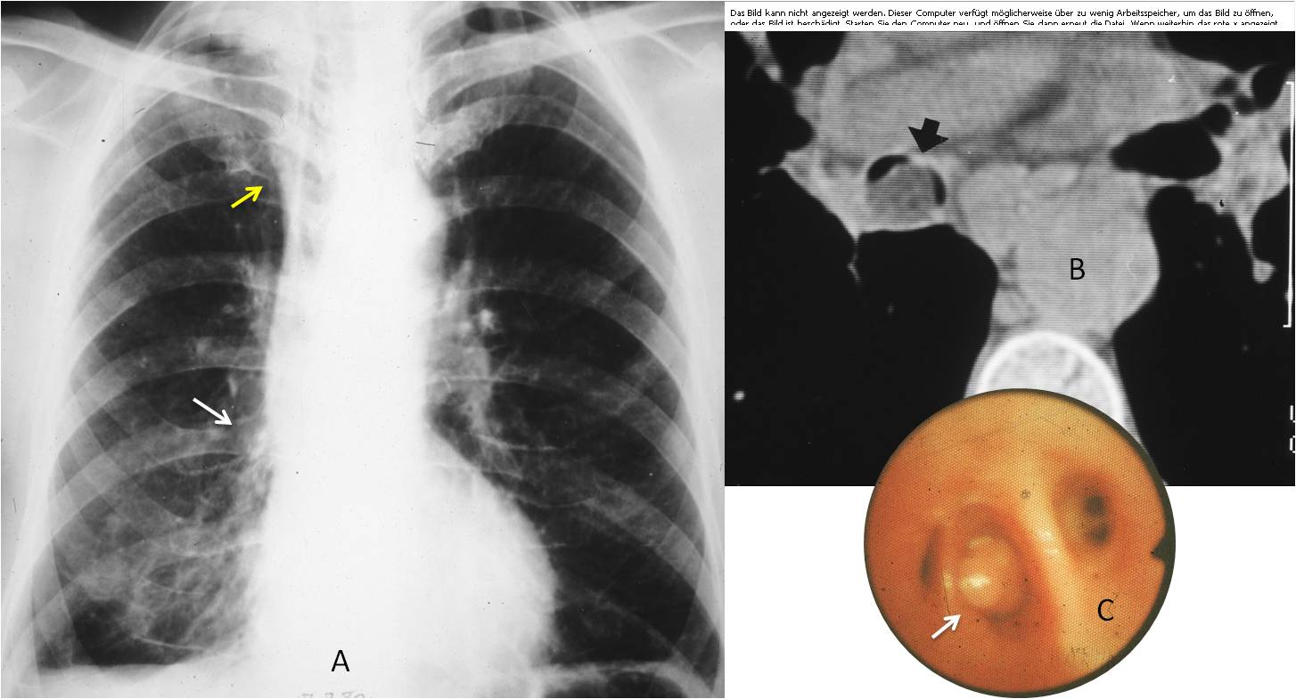
Fig. 8
Fig. 8: 65-year-old man treated in the outpatient clinic for non-specific respiratory complaints. The chest remained unchanged for several years, until somebody noticed the descended hilum (A, arrow) and the upper triangle sign (A, yellow arrow). Axial CT shows a fatty tumour inside the RLL bronchus (B, arrow), confirmed with bronchoscopy (C, arrow). Diagnosis: endobronchial lipoma.

Follow Dr.Pepe’s Advice:
1. In the PA view, typical RLL collapse appears as a triangular opacity at the base of the lung with downward displacement of the hilum.
2. RLL collapse may simulate pleural effusion. Look for a descended hilum.
3. Aerated RLL collapse may be overlooked. It is suspected by visualization of a descended hilum and major fissure.
4. Marked RLL collapse may be hidden behind the right heart. Look for a descended hilum and upper triangle sign.


