ESR Rising Stars Speakers at ECR 2012
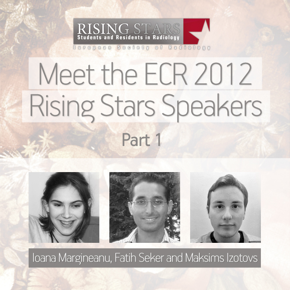
The ESR is very proud of its youth and we try to encourage as much student participation as possible at our annual meeting, the ECR. This year, we again asked for students to submit abstracts on a range of subjects, with the best 20 being given the opportunity to present their work at ECR 2012, in front of a real congress audience. Each of the student speakers will be announced individually on the Rising Stars Facebook page, with a more detailed look, including their full abstracts, here on the ESR Blog. Here are the first three; Ioana Margineanu, Fatih Seker and Maksims Izotovs:
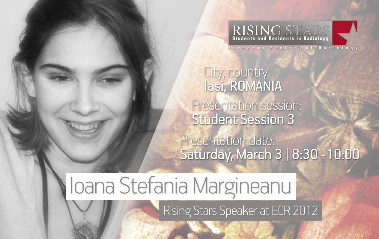
ECR 2012:
Student Session 3 | March 3, 8:30 – 10:00
CV: (excerpt)
Education/Profession:
Since 2006 Studying General Medicine at Gr. T. Popa. University of Lasi, Romania
2009 – 2011 Creative and Publishing Consultant eCongres Medical Events
2009 – 2010 Deputy Head of National Department of Human Rights, National Medical Students’ Association
Extracurricular activities:
Oversea experiences in US, UK and Taiwan (Chinese Taipei)
Abstract:
Background: The author’s experience as a medical student and radiology professor has given him a unique ability to develop questions and search for solutions as to how students view the radiological specialties and curriculum in Romania. Purposes: To determine the impact of exposing medical students to medical imaging, during the second year of their clinical rotation, on their perception of this specialty. To take the results and improve radiology in Romania in order to offer medical students the best possible assistance for the future and also a comprehensive experience of what it means to be a radiologist. Materials and Methods: The study was divided into two phases. In phase one, a group of 65 students were given an essay-based questionnaire with 14 items concerning their knowledge of the field of radiology and the radiology module’s impact on their clinical years and their future profession. The responses underwent qualitative analysis in order to develop the multiple-choice questions needed for phase two. This questionnaire will be given to all fifth and sixth year students and all radiology residents in Iasi, Romania. The results will be quantitatively analysed using SPSS. Phase one results: Out of a total of 65 questionnaires, nine were incomplete. We performed a frequency analysis on the remaining 56 and eliminated all answers with a prevalence of less than three. We qualitatively analysed the answers and constructed a 23 multiple-choice item questionnaire. Phase Two: The questionnaires will be distributed to all fifth and sixth year students and all radiology residents in Iasi, Romania. The goal is to have at least 350 completed questionnaires in order to create a useful statistical analysis for developing solutions to the identified problems.
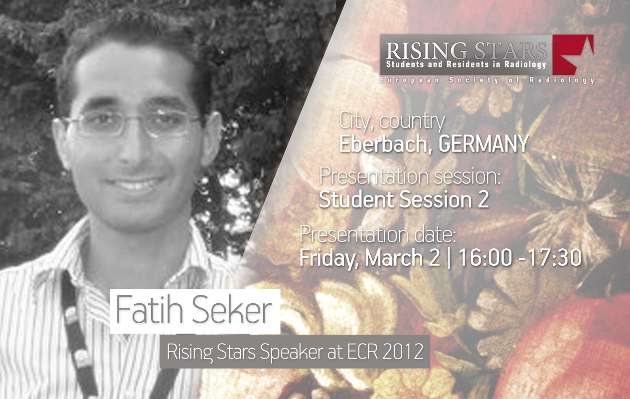
ECR 2012:
Student Session 2 | March2, 16:00 – 17:30
CV: (excerpt)
Education/Profession:
Since 2009 Studying Medicine at Ruperto Carola Univserity of Heidelberg, Germany
Research assistant at Department of Experimental Radiation Oncology, University Medical Center Mannheim,
Ruperto Carola University of Heidelberg, Germany
Extracurricular activities:
08/2010 Participation in 5th World Youth Congress 2010 in Istanbul
Abstract:
For a long time high-tech radiological examination meant morphological imaging such as computer tomography or MRI. However, these technologies mainly allow for a diagnosis based on structural and anatomical information, whereas histopathology is still the gold standard when it comes to diagnosing neoplastic diseases. With the improvement of molecular techniques, pathophysiological processes within cells are now better understood. In molecular imaging these molecular techniques are used for a new kind of imaging procedure. But why is this new technology important for the future of radiology departments in modern hospitals? Molecular imaging, a new radiological discipline, is a product of advances in imaging technology. It uses modern molecular techniques in order to visualise cellular and molecular processes in living tissue. The imaging is based on physiological parameters like proliferative activity, blood flow, oxygen consumption, etc. Molecules like biomarkers or nanoparticles are used to help image the area of interest. For this purpose these molecules connect with delivery agents in vivo and are transported to the pathological area where they accumulate. Thus diseases can be diagnosed at an early stage, before morphological alterations can be seen. These molecular techniques can be used with existing imaging procedures like CT or MRI. Today the successful use of PET-CT in many hospitals has shown the benefits of this new technology. In summary, it can be stated that molecular imaging assists in early detection, especially of neoplastic diseases. This new imaging technology will play a decisive role in the radiologist’s field of work in 2051. It might even replace some histopathological analyses that require invasive procedures.
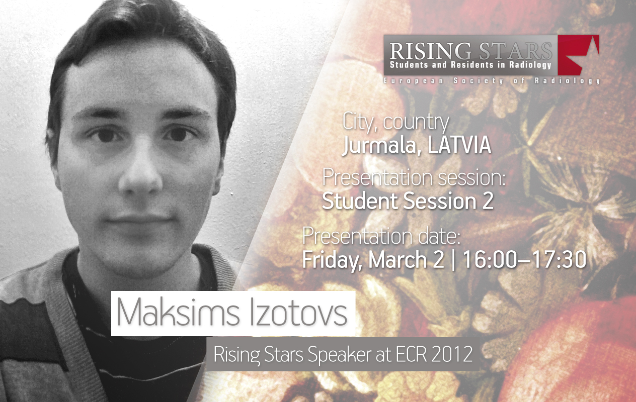
ECR 2012:
Student Session 2 | March 2, 16:00 – 17:30
CV: (excerpt)
Education/Profession:
Since 2009 Studying of Medicine at Faculty of Medicine, Riga Stradins University
Extracurricular activities:
2007 – 2008 PSIA “Jūrmalas trā Palīdzība” | Life-Guard
2011 summer SIA “Saldais Laiks” | Salesman
Abstract:
This topic is very important, because cancer is one of the most feared diseases in today’s society. The initial idea is to predict how cancer treatment will change in the next few decades. Given modern knowledge of the processes occurring in cancer-stricken cells and ongoing research, it may be possible to predict what will happen in the near future. The cause of cancer is found on a cellular level. A cancer cell is a cell that starts to proliferate uncontrollably, a cell which has a malfunctioning cell-division cycle and a damaged reparation system. This uncontrollable growth is caused by both genetic and environmental factors. These days treatment usually involves chemotherapy, radiotherapy, or surgery. Chemotherapy and radiotherapy are based on causing cancer cell apoptosis by blocking DNS synthesis. These methods are not perfect because they lack specificity. Not only are cancer cells destroyed, but healthy cells are also damaged, causing suppression of the immune system, fatigue, hair loss etc. Future methods however will be selective, targeting only cancer cells and leaving healthy cells alone. Developing pharmacogenetics will provide us with drugs that will be able to repair the reparation system by restoring p53 activity. Other drugs could reduce telomerase production in cancer cells causing cells to regain their Hayflick limit and lose immortality. Genetic testing will be widespread, finding people with hereditary cancer and curing them before cancer manifests. It will be possible to use adenoviruses as vectors to transport tumour suppressing genes into rapidly dividing cells. Nanotechnology will also play a role in cancer treatment. The future holds a lot of opportunities for cancer treatment; our duty is to use them. In my opinion, cancer, if not completely defeated, will at least cause less trouble to future generations.

