ESR Rising Stars Speakers at ECR 2012
Here’s the fourth installment of our series introducing our Rising Stars, who submitted the best student abstracts and have been invited to present their work at ECR 2012 in front of a real congress audience. Each speaker will be announced individually on the Rising Stars Facebook page, with a more detailed look, including their full abstracts, here on the ESR Blog. Our next three speakers are Feruza Rozikhodjaeva, Nauris Zdanovskis and Urzula Poznaka:

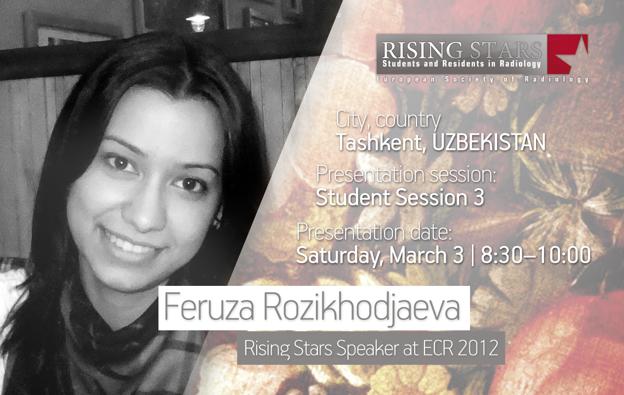
ECR 2012:
Student Session 3 | March 3, 8:30 – 10:00
CV (excerpt):
Education/Profession:
Since 2007 Studying Medicine at the Tashkent Pediatric Medical Institute, Tashkent, Uzbekistan
Extracurricular activities:
August 2011 Particpant in the Summer Academy for Translator” at the Goethe Institute, Tashkent, Uzbekistan
Abstract:
I am a student at the Tashkent Pediatric Medical Institute and in the future I want to devote my life to radiology and to solving various problems in the diagnosis of vascular diseases. Why I am interested with this branch of medical science? I admire the achievements of radiological science over the last few years. The progress made in recent years in the area of medical devices, catheter technology, and contrast application has led to a general improvement in the diagnosis of vascular diseases. Today, through the advancement of other radiological procedures, computer tomography, magnetic resonance and ultrasonic diagnostics are available as alternative technologies for the identification of vascular diseases. Some of these procedures have already successfully been in use for a long time within vascular diagnostics. The progress of non-invasive procedures, above all in the area of magnetic resonance and ultrasound, will lead, in the next few, years to a considerable change in vascular diagnostics. Besides outlining some of the radiological procedures available today this abstract has also aimed to give an account of the developing trends and future perspectives of vascular disease diagnostics.
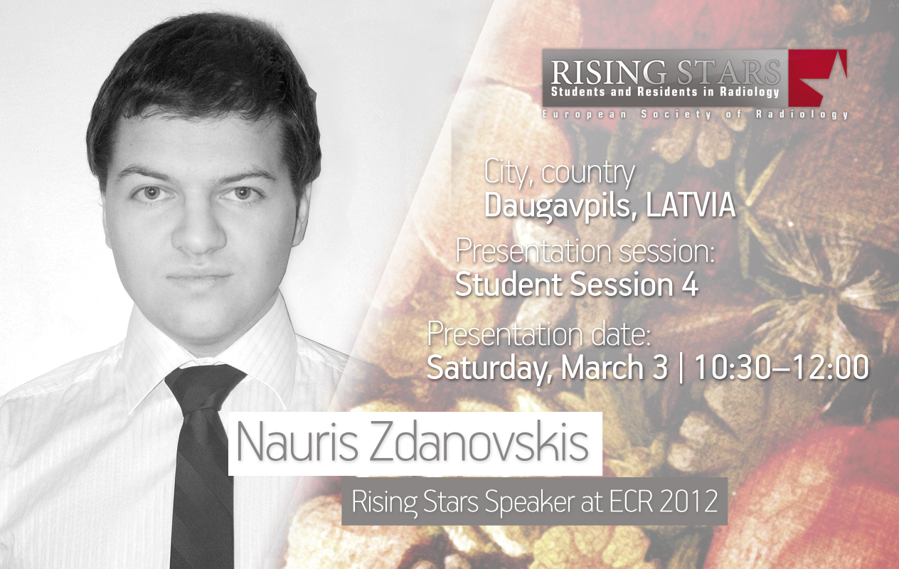
ECR 2012:
Student Session 4 | March 3, 10:30 – 12:00
CV (excerpt):
Education/Profession:
Since 2009 Student at Faculty of Medicine, Riga’s Stradins university, Riga, Latvia
Abstract:
Technology develops so fast that we can’t always keep up with it. Technological innovations enter into our life and it is important that we know how we use them. At present eReaders are becoming more and more popular. The advantages of eReaders are: 1. eReaders vs Books- Students who study medicine have to read a huge volume of books and, usually books take up a large amount of space on a desk. With an eReader it’s not a problem – most only take up the same amount of space as one book. 2. The price of eReaders and eBooks- As the popularity of eReaders grows and competition between manufactures develops the prices should get lower. Now prices are between $69 to $300, depending on the specifications of the eReader. eBooks are 10% – 40% cheaper than conventional books. 4. Convenience- Instead of going to the bookstore or ordering books on the web, it’s possible to get books in a few minutes using Wi-Fi and an online book store. 5. Access to eLearning systems- Many eReaders have internet browsers and thus the ability to access every eLearning system on the web. 6. Additional educational materials- Additional materials for education – presentations, documents, educational videos, and more – can be accessed easily and saved on an eReader. 7. Additional properties: Calendar- Develop sync software with eLearning systems in order to be informed of the latest updates. Dictionary: Problems understanding other languages? It’s not a problem if you have an eReader. 3D atlases. Students can get a better understanding of many disciplines (anatomy, traumatology, radiology, endocrinology etc.) and many others. To sum up then, instead of handing out tonnes of books each year, a university could hand out one eReader to every student. In the long term it would pay off and could be a great educational innovation to improve the quality of education.
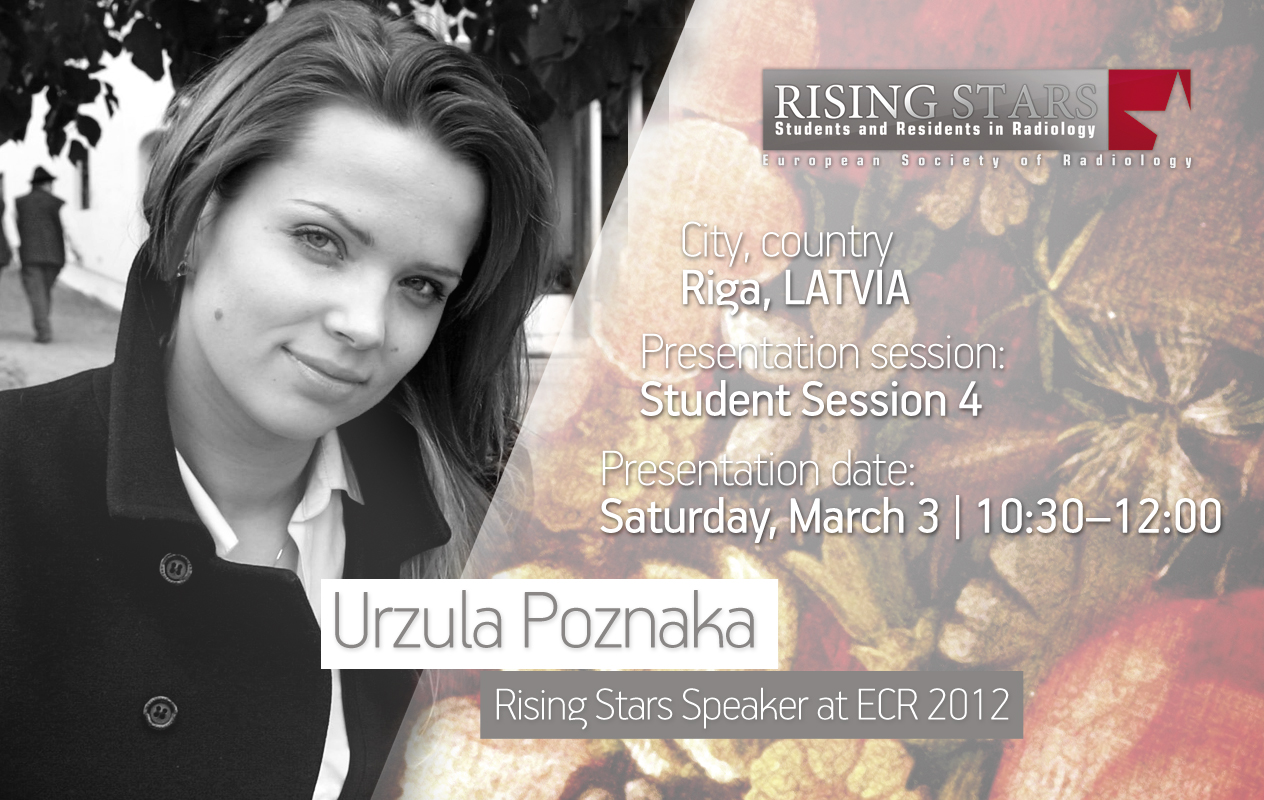
ECR 2012:
Student Session 4 | March 3, 10:30 – 12:00
CV (excerpt):
Education/Profession:
Since September 2005: physician’s programme, Faculty of Medicine, University of Latvia, Riga
Extracurricular activities:
Since 2010 physician’s assistant on the ambulance car unit
Abstract:
Purpose: The primary aims of the research were to identify the incidence of craniosynostosis in Latvia, as well as to compare it with data from other countries, to evaluate performed radiological examinations, and assess surgical strategy. Hypothesis: Use of multi-slice CT with 3D reconstructions for diagnosis of craniosynostosis noticeably improves early detection of this pathology and the possibility of surgical treatment planning. Materials and methods: The study was performed using medical reports from the Children’s Clinical University Hospital within the period from January 2000 to December 2010. During the retrospective study 85 case histories of craniosynostosis patients were analysed, as well as the performed radiological examinations. Results: Overall, craniosynostosis was diagnosed in 85 patients. Incidence in Latvia is 1 per 2800 live births. Since 2005, with the application of multi-slice computed tomography with 3Dreconstructions the number of diagnosed patients increased noticeably from 2.7 to 12.7 patients yearly. Fifty-six nonsyndromic and 11 syndromic cases of craniosynostosis were detected. Nonsyndromic craniosynostoses consisted of sagittal synostosis – 46.6%, multisutural and metopic synostosis both comprised 16.4%, coronal synostosis 11%, lambdoid 6.8% and temporoparietal synostosis 2.7% of cases. Only in 3 of 11 syndromic synostosis cases was the diagnosis proven genetically. There was not a single case of craniosynostosis diagnosed prenatally. The most common age of patients’ age at diagnosis was 4 months. Surgery was performed in 51 cases. Conclusion: The radiological diagnosis of craniosynostosis in Latvia is not comprehensive but rather qualitative. In all patients the diagnosis was based on the results of CT imaging. Since 2005, with the beginning of the application of multi-slice 3DCT, the number of diagnosed patients increased noticeably. The surgical management of craniostenosis in Latvia corresponds to the common concept.

