ESR Rising Stars Speakers at ECR 2012

Here we continue our series introducing our Rising Stars, who submitted the best student abstracts and have been invited to present their work at ECR 2012 in front of a real congress audience. Each speaker will be announced individually on the Rising Stars Facebook page, with a more detailed look, including their full abstracts, here on the ESR Blog. Our next three speakers are József Lackó, Katia Khouri Chalouhi and Ana Jantarada João:

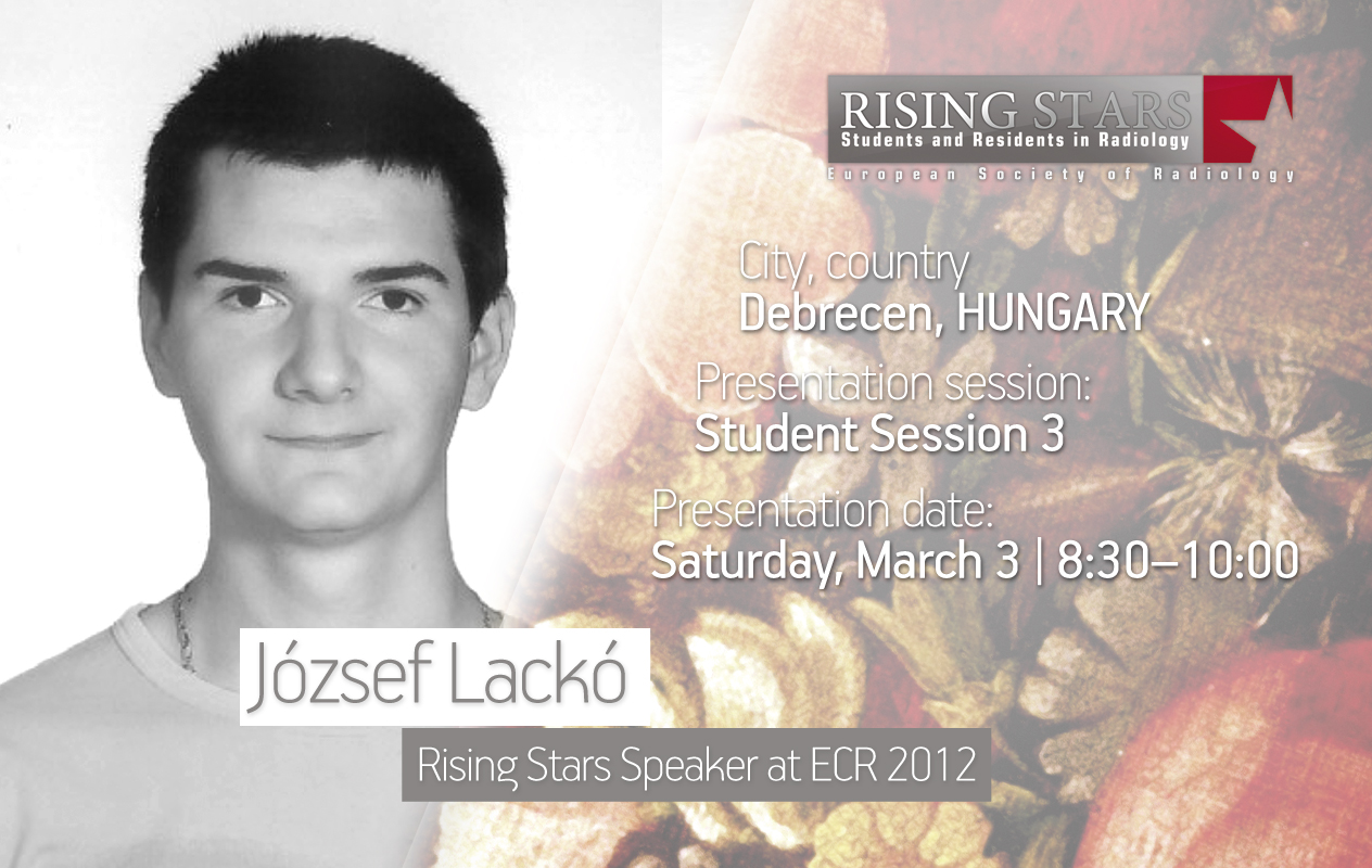
ECR 2012:
Student Session 3 | March 3, 8:30 – 10:00
CV (excerpt):
Education/Profession:
Since 2009 Student at the University of Debrecen Medical and Health Science Center, Faculty of Medicine, Hungary
Extracurricular activities:
Delegate at the University’s Student Centre
Abstract:
Purpose: The appearance of highly differentiated medical imaging devices demands the specialisation of those who work in radiology. Two types of experts are needed for acquiring images and diagnosing diseases properly: radiographers specialising in the operation of the devices and radiologists who evaluate the images. We aimed to compare both professions to each other emphasising, equally, their strengths and weaknesses. Methods and materials: Our research is based on the latest statistics available, studies on the training syllabi at our institution (University of Debrecen, Hungary), the experiences of our current and future colleagues and eventually, our own surveys. We found that radiographers obtain the necessary clinical practice and skills during their bachelor training, while r future radiologists mostly recieve this during their postgraduate specialist training. The theoretical training makes a radiographer capable of understanding, deeply, the procedures of imaging, performing examinations independently, and operating the equipment securely. Radiologists are competent in evaluating the prepared images professionally and making a differential diagnosis. A radiographer’s knowledge includes general physics and radiology, while a radiologist’s includes a wide range of specialised topics in radiology and medicine. However it is important to broaden the radiographer’s expertise in securely recognising pathologies. This could lead radiographers towards a new type of specialised imaging. The process of specialisation should consist of joint radiographer-radiologist image evaluation, unified educational materials, programme packages on computer-assisted learning methods and a standardised exam question bank. Problem-based learning can fit both bachelor and masters training. Conclusion: Training radiographers is a must and is gaining increasing acknowledgement. However their job requires close cooperation with their radiological colleagues and vice versa. These sister professions have a common purpose; treating patients in a specialised, appropriate and professional manner.

ECR 2012:
Student Session 2 | March2, 16:00 – 17:30
CV (excerpt):
Education/Profession:
Student at Faculty of Medicine and Surgery, at University of Milan, Italy
Abstract:
In 1895, W.C. Röntgen discovered x-rays, which gave birth to radiology as a science and medical discipline. For decades, x-rays were the only ‘radiological’ modality. Technological developments during and after World War II allowed for scintigraphy, ultrasound, angiography, CT, PET, and MRI as clinical tools. However, nuclear medicine was separated from radiology and radiology was subdivided according to technique. The radiologist became mainly a human appendix of a machine, a specialist of imaging, not of organs, systems, or body parts. About thirty years ago, radiologists began to understand the need for clinical knowledge across the different imaging modalities. Clinical fields became more important than imaging techniques. Radiological subspecialties such as neuro, pediatric, cardiovascular, breast, musculoskeletal, interventional, thoracic, and so on, were established. However, the human body still remains an entire entity. Thus, the unity of radiology must be conserved, at least when considering two main reasons. First, the borders between the subspecialties are not clearly defined (e.g., who has to study the peripheral nervous system? Neuroradiologists or musculoskeletal radiologists?). Second, when we look for an expected finding but we find more and more incidental findings, a trend increased by population aging, which results in comorbidities. Every radiologist should have a transversal knowledge and a specialised skill in at least two main subspecialties. This is not only needed in order for radiology to survive the fragmentation of knowledge, but it is also the only way to do the best for our patients, and to be prepared for the future merger between radiology and nuclear medicine, which will result from the use of hybrid machines.

ECR 2012:
Student Session 4 | March 3, 10:30 – 12:00
CV (excerpt):
Education/Profession:
Since 2010 PhD in Computational Engineering, CEMAT, Instituto Superior Técnico de Lisboa, Portugal & University of Texas (Austin), USA
Abstract:
Purpose: Cardiovascular diseases are associated with a number of factors that include biochemistry, haemodynamics and genetic predisposition. These factors are specific to each individual and it is important to accurately represent patient-specific information in order to assess patient state at diagnosis and prognosis stages. Here we focus on the effects of uncertainty in clinically acquired medical imaging on variability in the reconstructed vessel geometry, and this error propagation in computed haemodynamics. Methods and materials: The effects of filtering, contrast enhancement and segmentation of medical images are examined with relation to changes in the reconstructed geometry. The images used include MRI and CT data sets, largely focused on the cerebral vasculature. Different image processing methods are chosen and tested, and the most suitable combination of approaches is discussed. Numerical simulations of haemodynamics are performed and the impact of variability in the vessel geometry is considered with respect to flow parameters commonly associated with disease: transport and stresses on the vessel wall. The discussion therefore has a clinical relevance. Results: Results indicate that image pre-processing can substantially alter the quality of the image and improve vessel extraction. This removes a certain level of uncertainty from the segmentation process. Nevertheless, care must be taken to choose appropriate and robust schemes. Results of computational haemodynamics are presented with error bars. Conclusions: Automatic procedures for medical image processing and geometry reconstruction are important for analysing clinical data. Robust schemes are proposed, reducing the effect of errors on subsequent analyses and post-processing.

