This case was provided to me by Dr Edgar Lorente, a smart radiology resident at Hospital Universitario Dr Peset.
This is a 60 year old male with repeated pulmonary infections. This should not be very hard for you…
Well friends, most of you got the exact diagnosis at first glace. Well done!
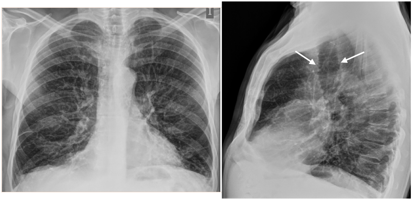
Yes, indeed this is a case of a large trachea and bronchi associated with repeated pulmonary infections: Tracheobronchomegaly or Mounier Kuhn (French physician who described it in 1932).
This is probably a congenital anomaly in which there is an atrophy of the elastic fibers and the smooth muscle of the tracheal and bronchial wall. It usually presents clinically in young adults as repeated pulmonary infections and may mimic COPD.
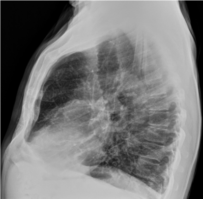
The findings in our case are: Large trachea, seen in the lateral projection (arrows), and some opacities in the lung bases in the PA projection as well as over inflation of the lungs suggesting COPD.
CT images (Lung window settings and Minimum intensity) show the tracheal dilatation and the prominence of the tracheobronchial rings.

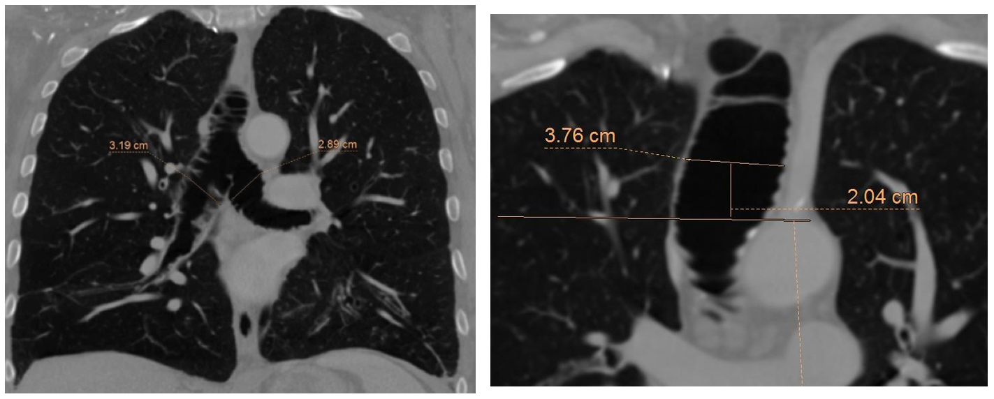
The criterion to define tracheobronchomegaly is to have a tracheal diameter larger than 3 cm measured 2 cm above the aortic knob. Other criteria are the measurement of the diameters of the main bronchi: Left > 23 mm and right > 24 mm.
There are many other causes of tracheal dilatation, some related to diseases such as Ehlers Danlos or Marfan, but also in acquired conditions as in COPD, cystic fibrosis or aging. (https://radiopaedia.org/articles/tracheomalacia-differential?lang=us).
Point to remember:
Remember the lateral chest radiograph is mandatory as some structures are better seen in this projection. The trachea is one of them.


Mounier Kuhn syndrome!
Also congestive changes.
Interstatial lung disease
Tracheobroncomegaly.
Tracheobronquionegaly.
Right lower lobe partial collapse,left lower lobe retrocardiac consolidation and minimal bilateral pleural effusion
Emphysema maybe