José Vilar and Friends Case 23 (Update: Solution!)
Good day Friends,
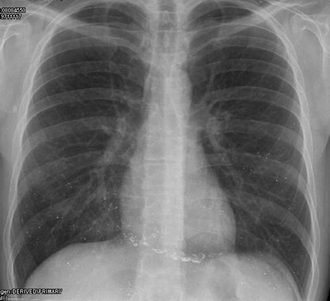
here is a case that I kept in my files. Again, only one image. Let me see what you think!
This was a 38 year old man with psychiatric problems.
Solution:
Remember this is a patient with psychiatric problems, and in this case this may help. Let’s see…
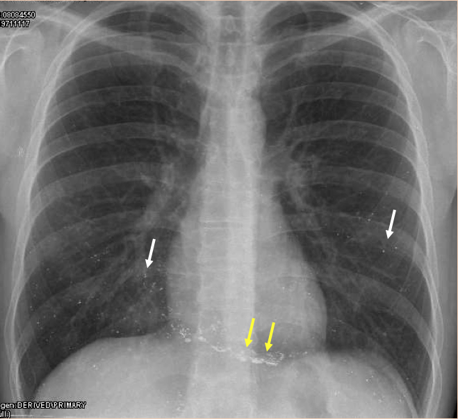
In the PA chest radiograph the obvious finding is the presence of metallic densities in both lower lobes and the cardiac silhouette. The yellow arrows show that these densities seam to adapt to the heart and may show some type of “fluid level “dependency.
Metal can arrive to the lungs either through the bronchi (aspiration) or via the vascular system ( embolization).
In the times of bronchography it was common to see dense lipiodol contrast in the lungs, usually showing the outline of the bronchial walls.
But this is not the case. This is a pathology that I have only seen three times in my life and is easily recognizable if you have seen it before as in my case.
Mercury embolization.
Mercury embolization has been described in patients that intend suicide by injecting themselves with mercury. It also has been described as a potential method to attain muscular strength in athletes.
The three cases I have seen were related to psychiatric problems with suicidal attempts.
The image is always very similar with multiple metallic densities in the lower lobes especially (where there is more flow obviously) and also within the heart in the vicinity of the endocardium.
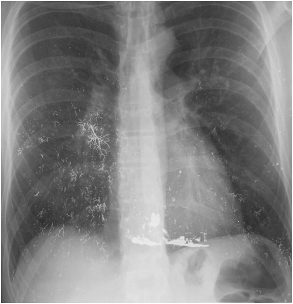
You can see the similar findings in this other case. Branching vascular patterns are seen as well as accumulation of mercury in the right ventricle.
Teaching point: If you ever see a case like this, you will probably make the diagnosis. The aunt Minnie approach often works.
Reference: Celli et al: Mercury embolisation in the lung: N Engl J Med 1976; 295:883-885


Some metallic poisoning
Radiodense material projected over cardiac silhouette and both lungs. I’d check out for history of procedures like vesselplasty, with material migration through paravertebral venous plexus (Batson’s plexus)
Differential diagnosis with aspiration of radioopaque contrast of prior gastrointestinal transit exploration
Could we have a lateral x ray to identify the location of opacities ( intrapulmonary, percardial or pleura )
it could be related to drug induced lung disease , like lithium salt
I see that most of you are in the right direction but: Where are te abnormal opaque densities located?
faint calcification at pericardium ? constricitive pericarditis
IMAGENES RADIOOPACAS PUNTIFORMES EN AMBOS PULMONES ( BASE) ASI COMO INTERDIAFRAGMATICOS Y EN ESTOMAGO : ASPIRACION DE MATERIAL EXTRAÑO ( QUE CONTIENEN MERCURIO , MAS ALEJADO UN BREBAJE O POCIMA) CON SIGNOS DE ASPIRACION
Differential diagnoses of symptoms using machine learning
Diagnosio democratizes healthcare for everyone, everywhere at anytime by using artificial intelligence for differential diagnoses….!
Click Here:>> diagnosio.com
The universal data recovery application functions