José Vilar and Friends Case 24 (Update: Solution!)
Dear Friends,
Today’s case shows a 35 year old woman with abdominal pain and weight loss.
Solution:
Hello friends, I can see that some of you read the case very well. Many made a correct diagnosis of liver metastases, and at least one (Kannan) got very close to the diagnosis. Well done. Indeed, metastases was the initial diagnosis, and the search for the origin was centered mainly in the abdomen, until someone took a good look at the chest radiograph, especially at the LATERAL…
Let us analyze the findings:
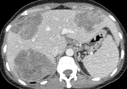
The abdominal CT, and the ultrasound (that I did not include) show multiple solid heterogeneous masses in the liver, with slight enhancement.
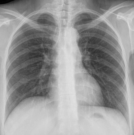
The PA chest radiography showed a convexity in the aorto-pulmonary window (arrows) that was not initially recognized.
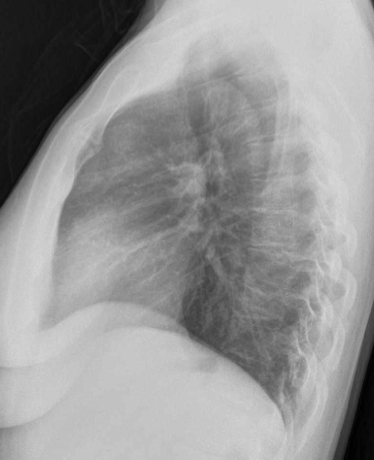
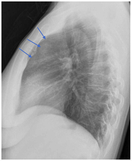
The lateral chest radiograph gives the clue to this case. An abnormal density is seen in the retrosternal space (blue arrows), indicating that the anterior mediastinum is occupied by a probable mass. In this case the finding in the lateral radiograph is subtle, but good enough to make you suspect that “something” is there.
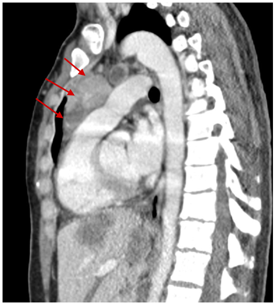
The CT showed a perfect correlation with the chest radiograph, with a solid, partially enhancing mass, in the prevascular or anterior mediastinum. An initial diagnosis of Thymic tumour was then established. And the biopsy revealed an unusual variant of Thymic carcinoma; Thymic Neuroendocrine carcinoma.
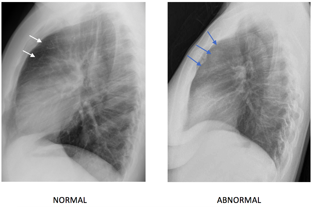
The main teaching point in this case is the importance of the Lateral Chest Radiograph. The anterior clear space, behind the sternum and in front of the heart and main vessels should be dark in normal patients (white arrows in image below). If you see a density in the anterior clear space you should suspect pathology, usually thymic ( Tumours or cysts) although other less common tumours such as Teratomas or ectopic thyroid may give similar findings.
REMEMBER: LOOK AT THE LATERAL, IT’S EASY AND REWARDING!




Colorectal liver metastasis
Metastatic liver lesions.
There’s a lung nodule in anterior segment of right upper lobe (better seen on lateral view). Multiple hipodense liver lesions (segments 8, 7 and 2) consistent with metastasis.
I’d rule out lung primary tumor (including carcinoid, taking care of patient age) with liver metastatic disease.
Could be liver metastases from mediastinal thymoma .
THERE IS AN ANTERIOR MEDIASTINAL MASS LESION-LYMPHNODES WITH MILD INDENTATION OF ANTERIOR TRACHEAL WALL IN LATERAL VIEW
MILD ANTERIOR COMPRESSION OF T 10 VERTEBRA IN LATERAL VIEW
BEAVER TAIL OF LEFT LOBE OF LIVER
MULTIPLE FOCAL LESIONS IN BOTH LOBES OF LIVER
METASTASIS OR LYMPHOMA
OPACIDAD TENUE EN EL MEDIASTINO ANTERIOR :
PERDIDA DE ALTURA DEL CUERPO VERTEBRAL DE D10
MASAS HEPATICAS DE ASPECTO SECUNDARIO
MASA RADIOOPACA EN EL MEDIASTINO ANTERIOR
PERDIDA DE ALTURA DEL CUERPO VERTEBRAL DE D1O
MASAS HEPATICAS ( 3) DE ASPECTO SECUNDARIO