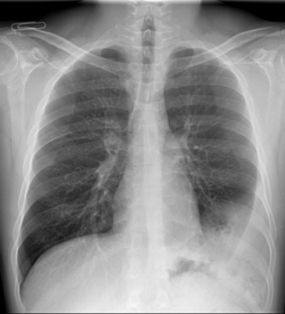
Today’s radiographs belong to a 37-year-old with fever and cough. Rule out pneumonia.
Ok friends, can I see some opinions about this case and what you would recommend?
The findings were interpreted by a pulmonologist as pleural effusion. The attempted pleural puncture was negative.
The patient was interrogated and revealed that he had suffered a car accident some years before. A CT was then performed.

PA radiograph shows a subtle finding: air bubbles (red arrow)- CT , coronal projection demonstrates a large left diaphragmatic hernia containing fat, and bowel loops responsible for the air bubbles in the chest radiograph.
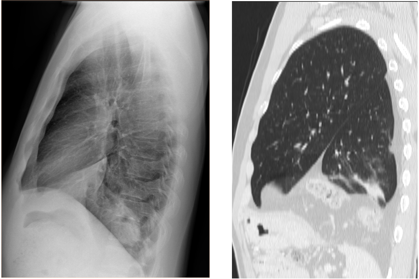
Sagittal CT at the level of the left hemi thorax shows a cleft in the left hemi diaphragm and entrance in the thorax of abdominal contents (fat, vessels and bowel loops).
Diagnosis: Blunt diaphragmatic rupture with herniated content.
Teaching points:
The herniated elements may mimic pleural pathology.
Look for findings inconsistent with pleural disease and other findings such as air (red arrow) from intestinal structures or fractures (not evident in this case).
When suspecting hernias, CT is mandatory to make the diagnosis.
Note: Did you know that Blunt diaphragmatic rupture is more frequent in the left side? (the driver´s side in most countries).


Pneumonia lower lobe to the left. Hydrothorax left
Clearly there is some ill-defined process in the left lower field. I don’t see any relevant pleural effusion. Also, I hardly see the left hemidiaphragm. Regarding the history I’d recommend an ultrasound and/or ce-ct to rule out an empyema.
A pneumonic patch is seen involving the left lower lobe as it is silhoutting the left diaphragmatic copula
Also the inferior lingula may be also involved
Chest X ray PA view shows non homogeneous opacity in LLZ obliterating left cardiophrenic angle,lateral half of left hemidiaphragm with a few lucent areas within.No evidence of air bronchogram /air fluid level/ calcification seen Another homogeneous opacity is seen obliterating left costophrenic angle with superior meniscus margins-may represent pleural effusion. The fundic bubble appears irregular with a focal discontinuity of left hemidiaphragm…
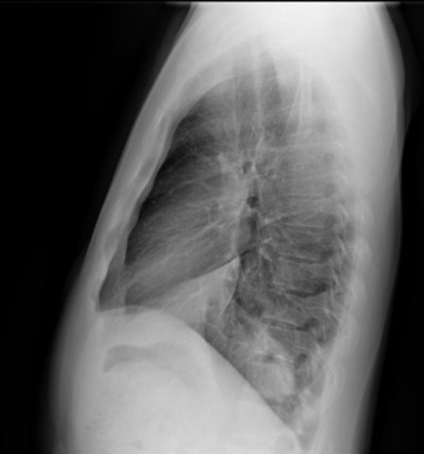
Left lateral X ray shows corresponding opacities in anterior, lateral and posterior basal segment of the left lower lobe with loss of spine lucent of lower dorsal vertebra.the left hemidiaphragm silhouette is also not well delineated . However posterior cp angle is not so dense for effusion.
I would like to do USG chest to rule out effusion.
Also is there history of trauma- diaphragm rupture?
The US shows no effusion.
Pulmonary findings are broad based towards pleura and left oblique fissure- may represent loculated empyema …
There is a extrapulmonary pattern lesion in left lower lobe, with triangular shape and no clear definition of left hemidiafragm. The density is similar to muscle or fat.
In the lateral view it seems that the opacities projected over left lower lobe have some intestinal/gastric pattern, so I’d suspect left diafragmatic herniation with abdominal content and less probably pleural lipomatosis.
I’d recommend CT scan or esophagogram, but I’d try first with ultrasound to confirm if it’s fat or abdominal content.
some of you are doing very well. The solution is coming tomorrow…
I have to congratulate Trinity and ELM because they were just on the spot. Well done.