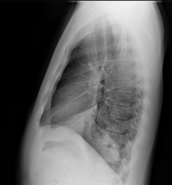
Today’s radiographs belong to a 37-year-old with fever and cough. Rule out pneumonia.
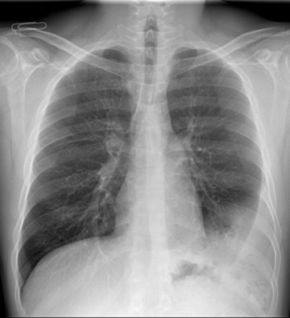
Ok friends, can I see some opinions about this case and what you would recommend?
The findings were interpreted by a pulmonologist as pleural effusion. The attempted pleural puncture was negative.
The patient was interrogated and revealed that he had suffered a car accident some years before. A CT was then performed.

PA radiograph shows a subtle finding: air bubbles (red arrow)- CT , coronal projection demonstrates a large left diaphragmatic hernia containing fat, and bowel loops responsible for the air bubbles in the chest radiograph.
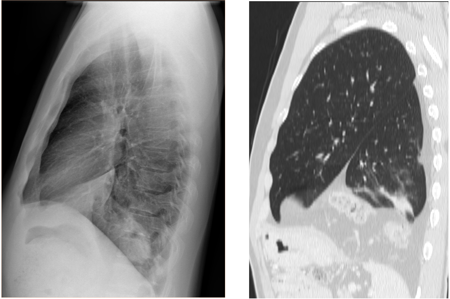
Sagittal CT at the level of the left hemi thorax shows a cleft in the left hemi diaphragm and entrance in the thorax of abdominal contents (fat, vessels and bowel loops).
Diagnosis: Blunt diaphragmatic rupture with herniated content.
Teaching points:
The herniated elements may mimic pleural pathology.
Look for findings inconsistent with pleural disease and other findings such as air (red arrow) from intestinal structures or fractures (not evident in this case).
When suspecting hernias, CT is mandatory to make the diagnosis.
Note: Did you know that Blunt diaphragmatic rupture is more frequent in the left side? (the driver´s side in most countries).