Continuing with the December sale, I am presenting the case of a 25-year-old epileptic male with three weeks of fever and one episode of haemoptysis.
1. Tuberculosis
2. Pulmonary abscess
3. Carcinoma of lung
4. None of the above
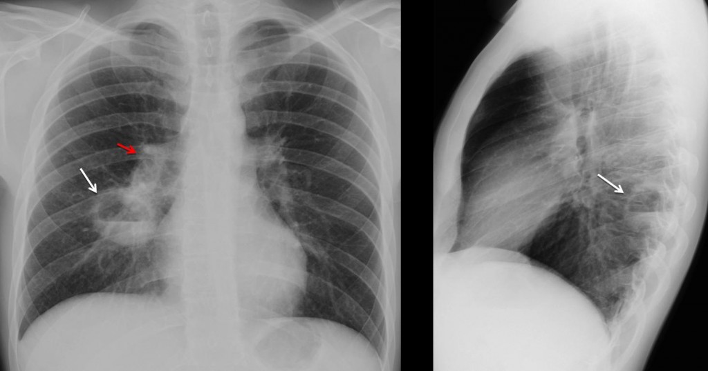
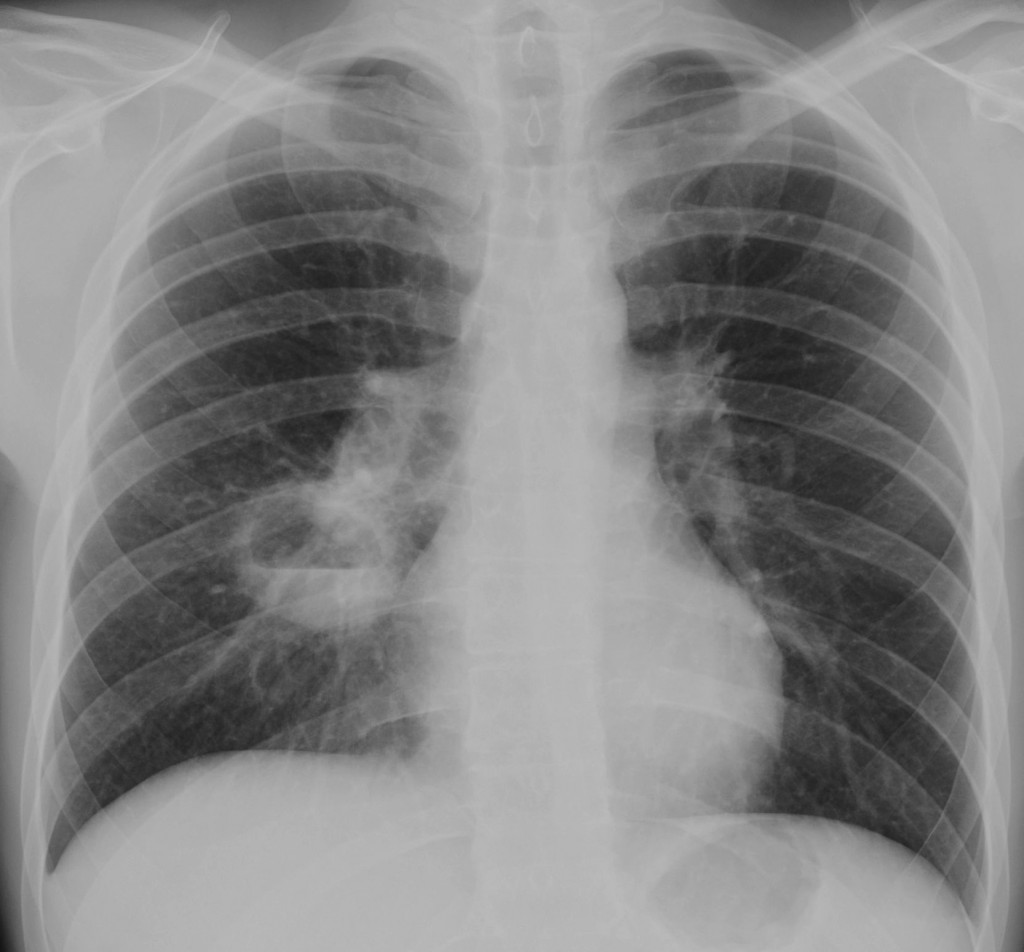
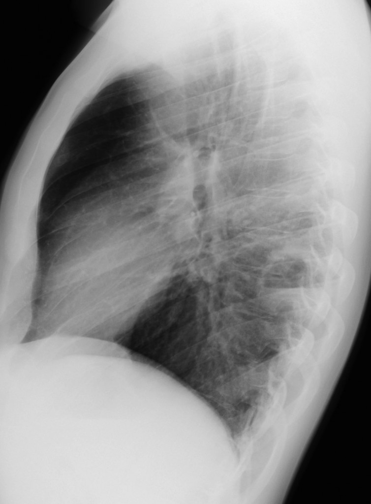
Findings: PA and lateral radiographs show a cavity with an air-fluid level in the apical segment of the RLL (arrows). In addition, there is increased opacity of the right hilum (
red arrow) when compared to the left.

Answer 1
Axial CT confirms the cavitation and enlarged lymph nodes in the right hilum (arrow).

Axial CT
A cavity and unilateral adenopathies suggests either TB or carcinoma. Given the age of the patient (25 years old), Muppet made the diagnosis of TB. However he was wrong (yes, Muppet is fallible and makes mistakes!).
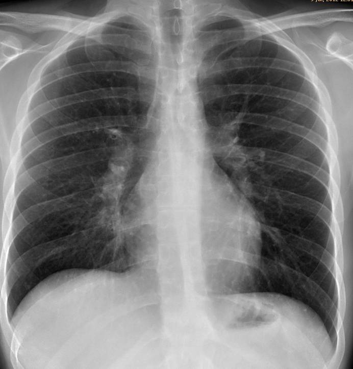
Clinicians suspected aspiration and the patient was treated with antibiotics. A chest radiograph three weeks later shows that the cavity has disappeared. Note that the right hilum has returned to normal.

Answer 3
Final diagnosis: pulmonary abscess secondary to aspiration
Teaching point: all pulmonary cavities have similar imaging appearances and clinical and lab results help in the diagnosis. Don’t forget to look at the hila in every patient. It will offer valuable information, although in this case it was misleading.


lung abscess.
Lung abcess
Pulmonary abscess:
– thin wall;
– air-liquid level;
– mediastinum looks normal;
– history of 3 weeks fever;
– one episode of haemoptysis.
Pulmonary abscess at the apical segment of the right lower lobe – due to the features listed above by DAG, and also due to the h/o epilepsy (increasing the risk of aspiration during altered level of conciousness during epileptic fits) and also due to the young age (making abscess more probably than a necrotic squamous cell carcinoma…which would have a thicker wall in most instances).
Tuberculosis:
– The history of 3 weeks fever is compatible with the diagnosis of Tuberculosis, and the haemoptysis is not specific for any of the above.
– Against it could be the air-liquid level, although not exclusionary, and the location, not typical of TBC.
I disagree about the location of TB. Apical segments of upper and lower lobes are typically affected.
La diagnosi è pronta e non è nessuna di quelle ipotizzate dai colleghi: aspetterò il 90′ minuto per fare goal! Forza Benfica e Forza BARIIIIIII!!!!
Volevo dire Forza BARCELLONA!!!!!
What about the retrosternal extrapulmonary opacity ?
Internal mammary lymph node ?
Costochondral tuberculosis ?
I believe it is a normal costochondral union. There is
another finding, though. It has been overlooked so far.
Right cardiac border is blurred.
The inferior esternum (lateral view) is not well seen and there is that retrosternal extrapulmonary soft tissue…Findings are subtle and equivocal…but anterior chest wall tuberculosis would make such a beautiful case…
Too beautiful to be true…
Addendum: The hila look funny!
On the chest radiograph there are three types of funny hila: increased opacity, increased size and changes in position. Which kind of funny is this case?
These funny hila are at a normal position, left being higher side.
I would say that these funny hila are of the increased opacity type!
🙂
The finding that everybody but Alice overlooked is the increased opacity of right hilum compared to the left one. And now you are on your own!
there seems to be a round to ovoid consoliation in the apical right lower lobe above the cavernous lesion, responsible for the funny looking right hilum.
a malignancy is not very likely given the young age of the patient, although it cannot be ruled out.
another thought: is the horizontal fissure moved upwards or is that just my Imagination?
hmmm… i choose tuberculosis.
I think none of the above…..may be Echinococcus cyst of the lung
Please! are there other small cavitary lesions?
No, there are not.
There is a cavitated lesion in the apical right lower lobe, and an increased right hilum.
Adenopathy + parenchymal lesion.
The patient is too young to have cancer.
Although cavitation is rare, I think it is a primary tuberculosis.
Unilateral hilar lymphadenopathy and pulmonary abscess in the apical segment of a lower lobe (a common site for TB) = TB unless proven otherwise.
I hope that the epilepsy is not a consequence of TB CNS involvement…
Epilepsy was previous. Muppet is a SOB, but fair
😀 @ muppet being a fair SOB!!
Le cause di una lesione cavitaria, con livello idro-aereo,sono molteplici , alcune comuni, altre rare. Una dd con una semplice radiografia deve essere integrata con i dati clinici e di laboratorio, dividendo le patologie in 2 grossi capitoli: infettive e non. La storia clinica nel soggetto parla di febbre da 3 settimane ed emottisi.Non sappiamo altro circa i risultati di laboratorio e l’esame microbiologico dell’espettorato. Il soggetto è giovane( quindi una patologia congenita malformativa) dovrebbe anche essere considerata .E’ vero , vi sono le caratteristiche semeiologiche della RADIOLOGIA che indirizzano verso una cavità e non verso una cisti.Vi è poi un ilo dx , addensato( adenopatico-reattivo?). E la sede , della lesione, inusuale.Si può da questi tre dati estrapolare una diagnosi?Polmonite necrotica ?Naturalmente ho escluso la patologia cerebrale del soggetto in esame, ( semplice associazione o spettro di Sindromi ,es Sclerosi tuberosa di Bourneville, con manifestazioni polomonari di LAM cistica).
Cavitary lesion in apical segment of RLL.
Right hilum is increased in density (adenopathy?)
Primary TB
[(Also seems to be a moderate fine nodular pattern) miliary? what do you think about? from imaging to imagination?]
CT showed some spillage from the cavity , but I don’t think it is visible in the radiograph
IMAGINE: cyst with milk of calcium on CT scans?
IMAGINE: cyst with milk of calcium around on CT scans?
Too much to imagine!
Auguri “galactico” collega, per le festività, da Bari!!!!
Thank you. Hoping Bari plays the European cup next year!
Lung abcess