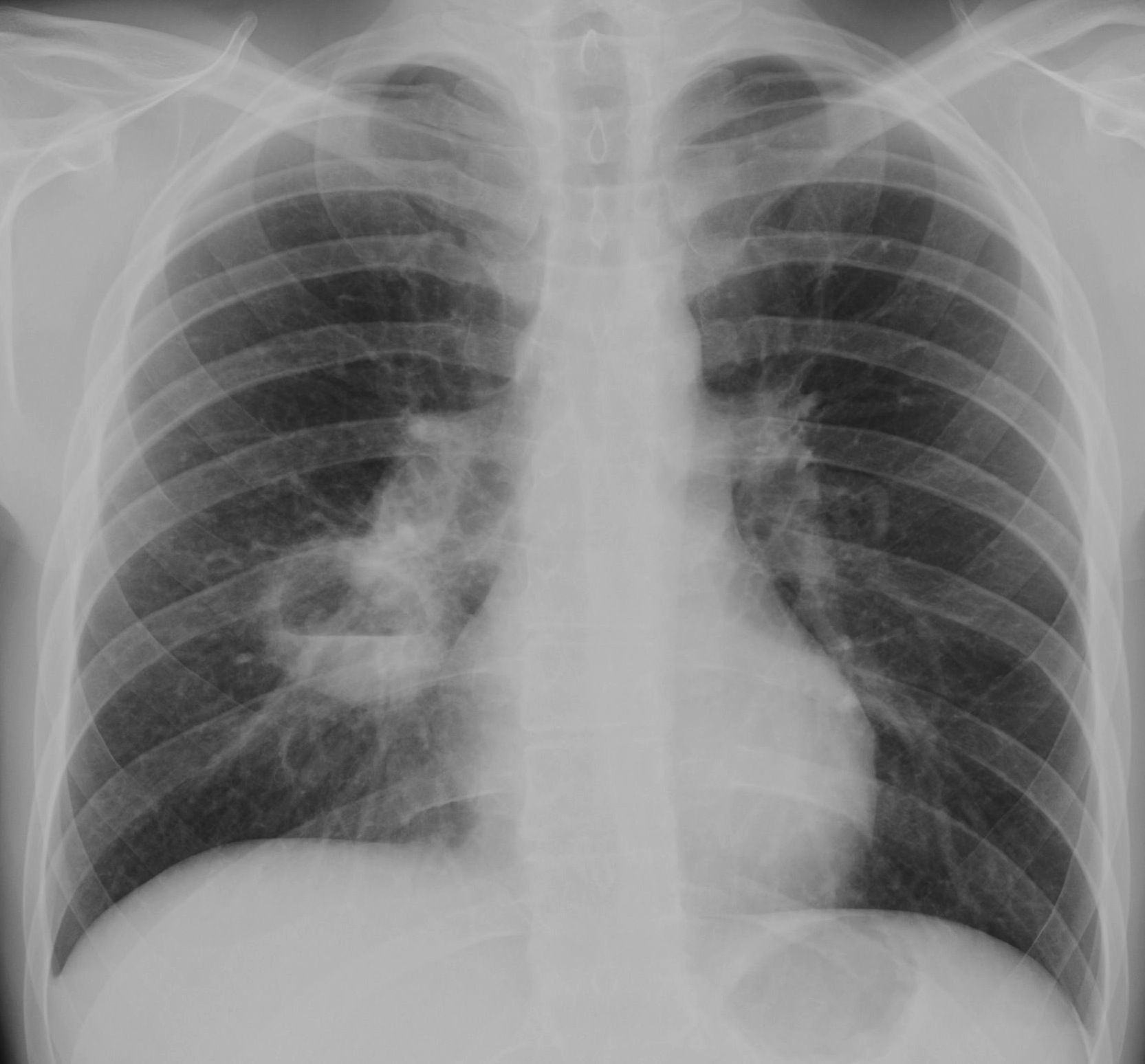
Continuing with the December sale, I am presenting the case of a 25-year-old epileptic male with three weeks of fever and one episode of haemoptysis.
1. Tuberculosis
2. Pulmonary abscess
3. Carcinoma of lung
4. None of the above
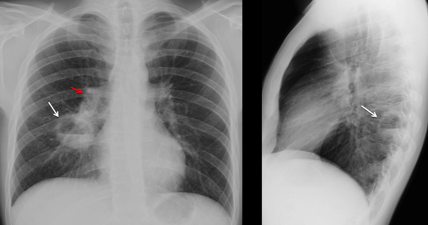
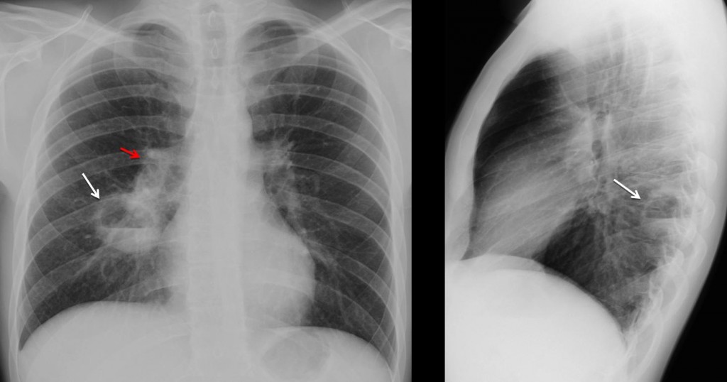
Findings: PA and lateral radiographs show a cavity with an air-fluid level in the apical segment of the RLL (arrows). In addition, there is increased opacity of the right hilum (
red arrow) when compared to the left.
Answer 1
Axial CT confirms the cavitation and enlarged lymph nodes in the right hilum (arrow).

Axial CT
A cavity and unilateral adenopathies suggests either TB or carcinoma. Given the age of the patient (25 years old), Muppet made the diagnosis of TB. However he was wrong (yes, Muppet is fallible and makes mistakes!).
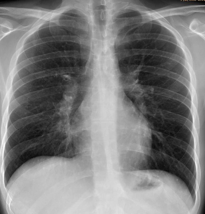
Clinicians suspected aspiration and the patient was treated with antibiotics. A chest radiograph three weeks later shows that the cavity has disappeared. Note that the right hilum has returned to normal.
Answer 3
Final diagnosis: pulmonary abscess secondary to aspiration
Teaching point: all pulmonary cavities have similar imaging appearances and clinical and lab results help in the diagnosis. Don’t forget to look at the hila in every patient. It will offer valuable information, although in this case it was misleading.