
Dear Friends,
Muppet and I just returned from the RSNA meeting in Chicago (Muppet very happy after spending the week with Miss Piggy!). This week we’re presenting a straightforward case to make you happy as well. Images belong to a 63-year-old man with vague chest complaints.
Where is the abnormality and what do you think it is? Leave yout thoughts and diagnosis in the comments section and come back on Friday for the answer.
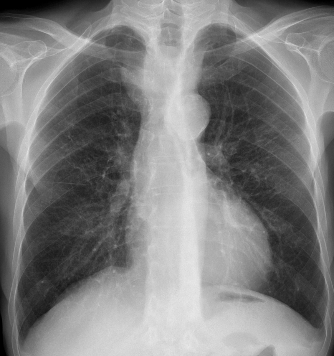
63-year-old man, PA chest
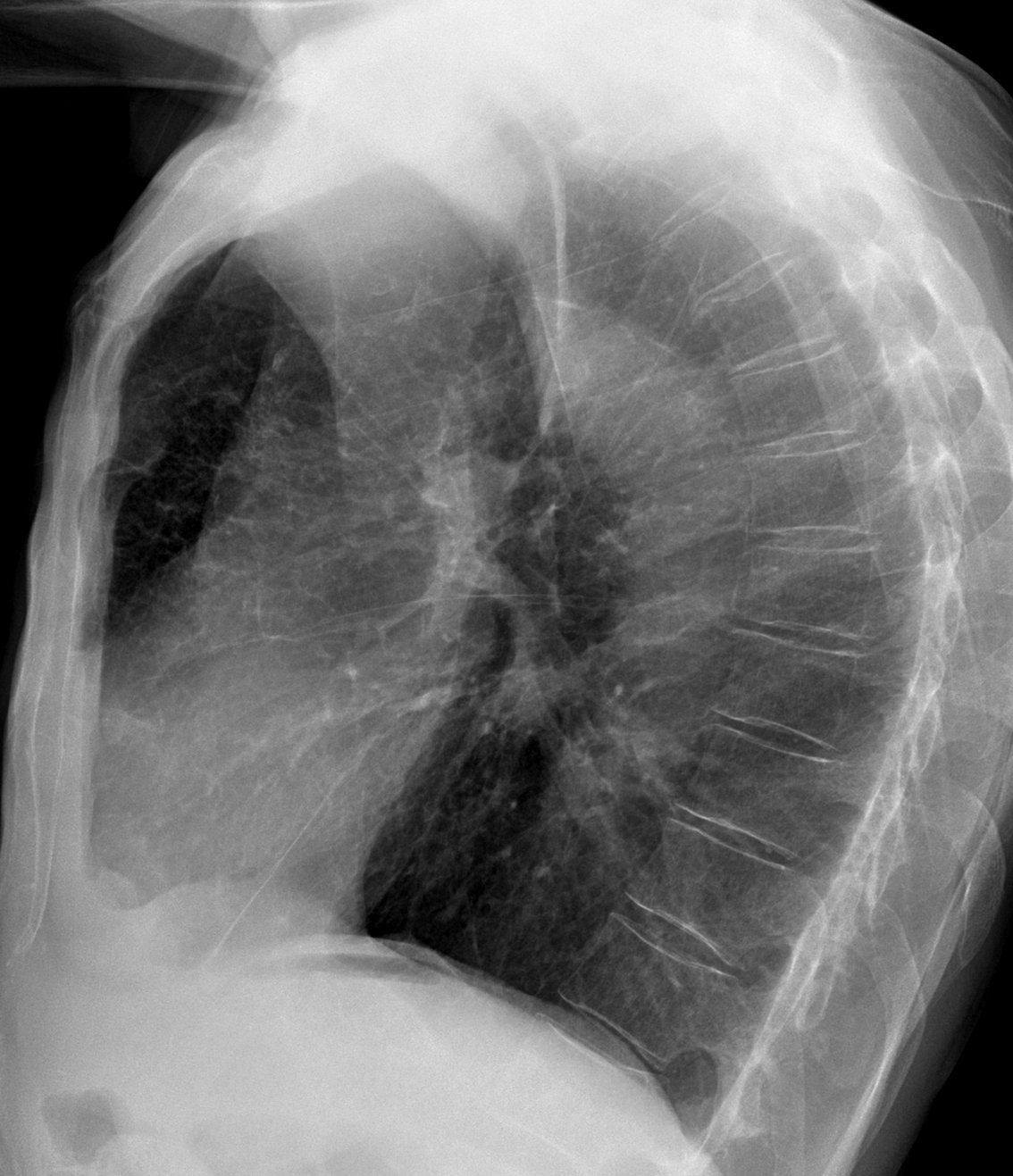
63-year-old man, lateral chest
Click here for the answer to case #81
Findings: PA chest shows an ill-defined increased density in the superior mediastinum (arrows). The lateral view shows narrowing and posterior displacement of the trachea (arrow), indicative of an anterior mediastinal mass.
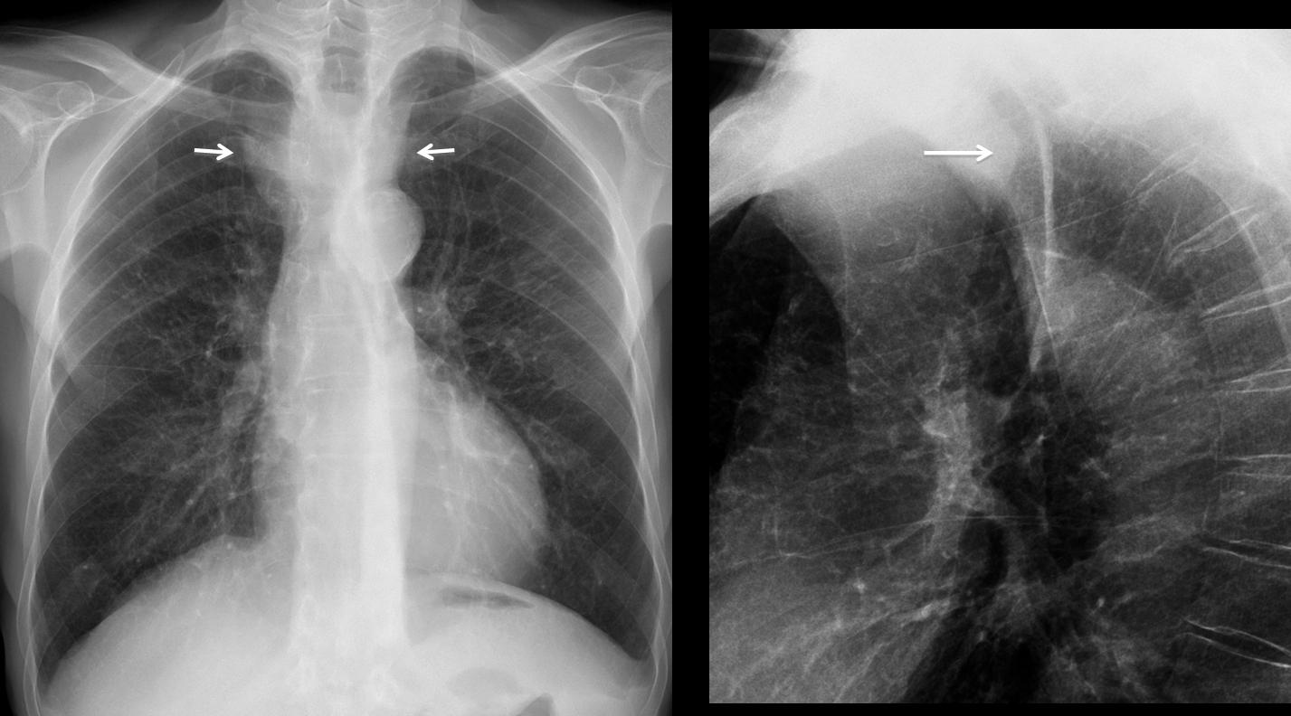
Because of the tracheal displacement, intrathoracic goiter would be my first choice. A midline cystic lesion should also be considered. Thymoma and teratoma are less likely. As always, a vascular lesion cannot be ruled out.
Coronal and sagittal enhanced CT demonstrate an aneurysm of the innominate artery, displacing the trachea backwards (arrows).
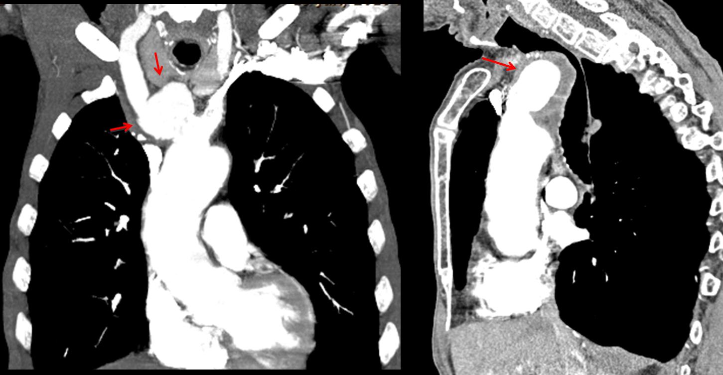
Final diagnosis: arteriosclerotic aneurysm of the innominate artery.
Congratulations to Dr. Sameh Khodair who discovered the tracheal narrowing and Dr. Maria S. who suggested the vascular origin of the mass.
Teaching points (repetition):
1. Remember to look at the trachea on the lateral view.
2. Always consider a vascular origin of any mediastinal mass (see Diploma Casebook case no. 41)


