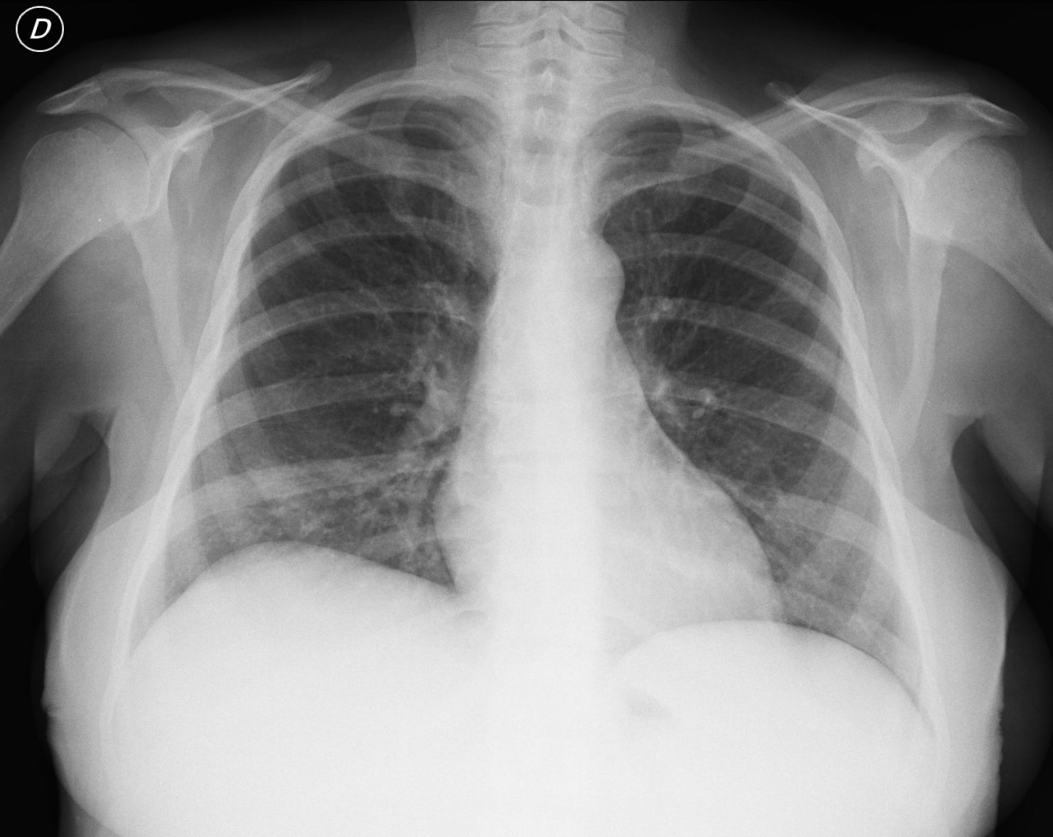
Check the image below, leave your thoughts in the comments section and come back for the answer on Friday.
Findings: notice the uniform density of the abdominal left upper quadrant with medial displacement of the gastric air (Fig 1, red arrow), suggesting an enlarged spleen.
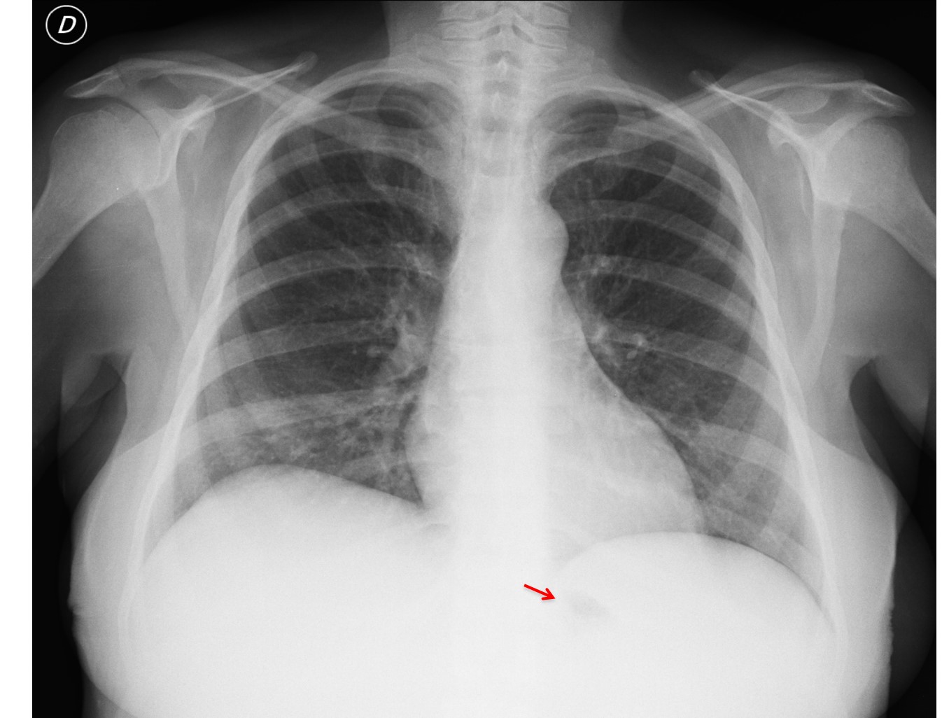
Fig. 1
In addition there is increased density of both humeral heads with lack of trabeculation (compare with normal appearance, Fig 2, arrows). When adding splenomegaly to bone sclerosis, the first diagnosis that comes to mind is myelofibrosis.
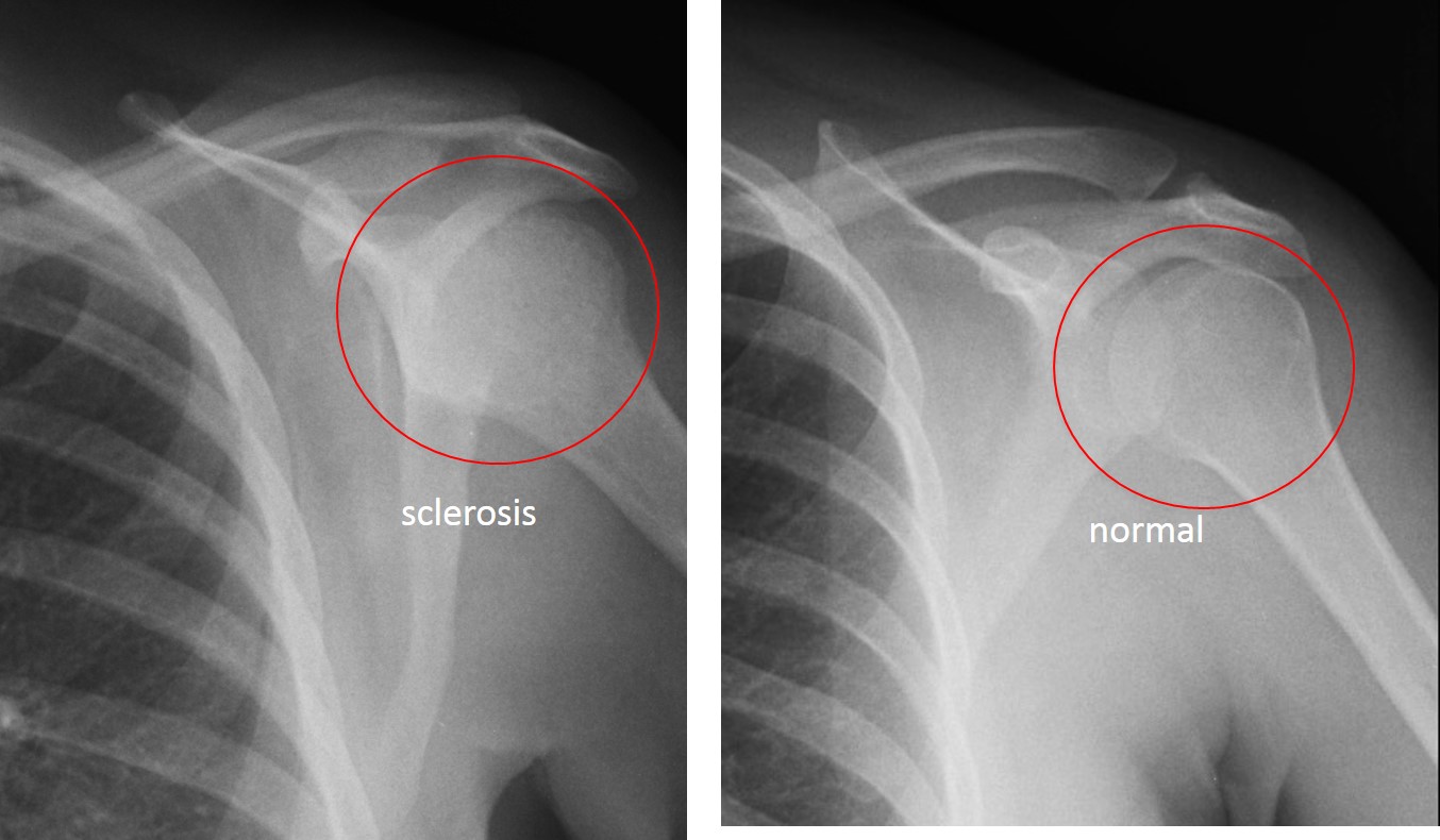
Fig. 2
The patient’s physician was contacted and it turned out that he already knew the diagnosis, which had been made a few years earlier. Images of lumbar spine after surgery confirm the sclerotic bone changes (Fig 3) and the large spleen compressing the left kidney (Fig 4, arrows).
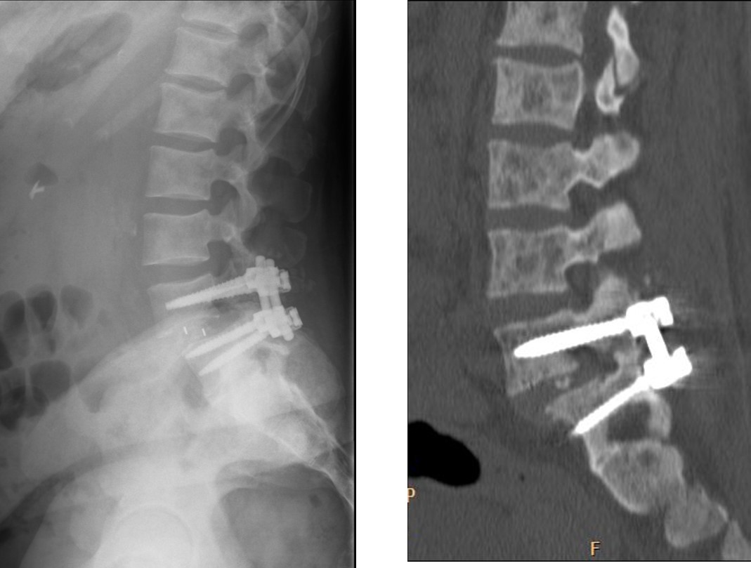
Fig. 3
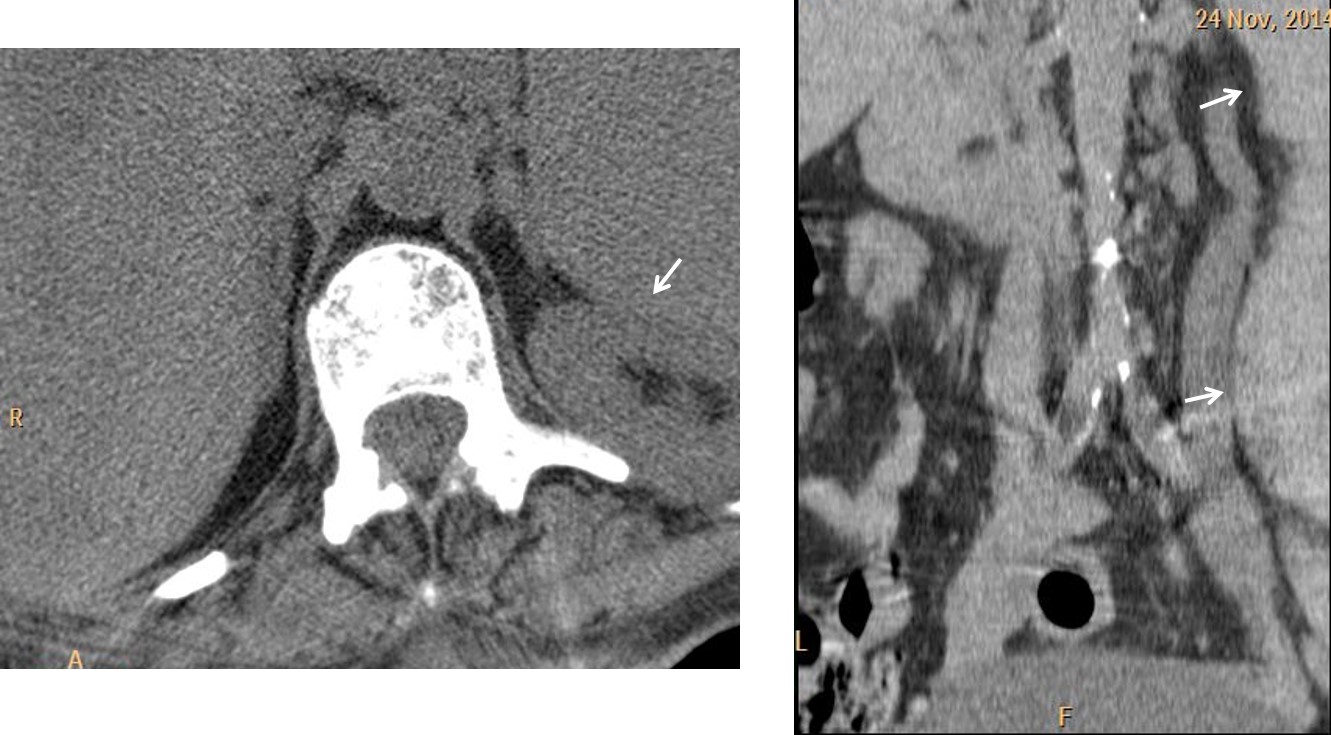
Fig. 4
The enlarged anterior right rib is a normal variant.
Final diagnosis: myelofibrosis
Congratulations to Lola la Piconera and Genchi Bari, who detected the bone changes. However, they missed the splenomegaly, which qualifies them as part-time old timers.
Comment: being an old timer has its advantages; occasionally you can make discoveries such as this and see (again) the gleam of reverence in the eyes of underlings!