Dr. Pepe’s Diploma Casebook: Case 73 – SOLVED!

Dear Friends,
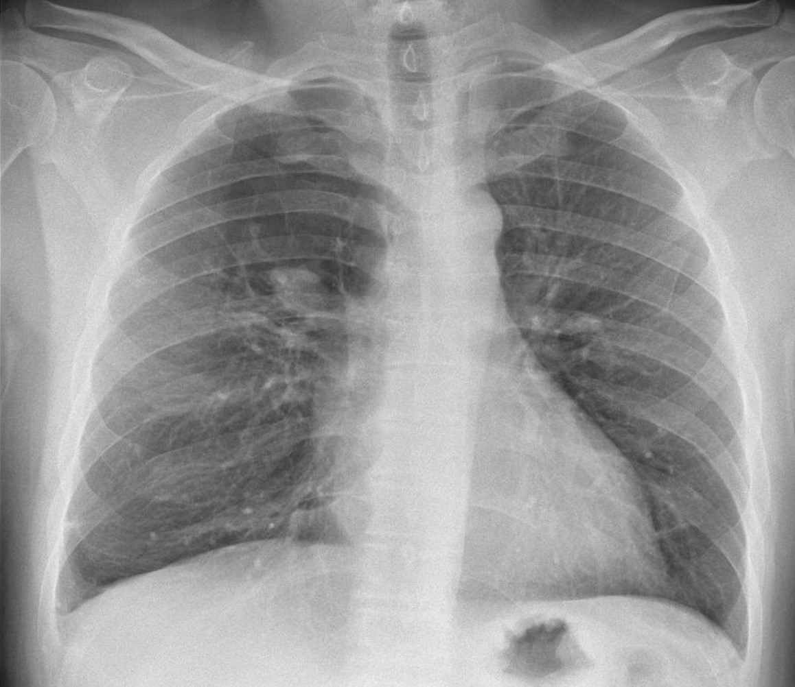
After mid-term vacation, I return with renewed energy! Presenting images of a 54-year-old man with a moderate cough.
Check the image below, leave your thoughts in the comments section and come back on Friday for the answer.
Diagnosis:
1. Apical bulla with fungus ball
2. Carcinoma of the lung
3. TB and granuloma
4. None of the above
Findings: chest radiograph shows a hyperlucent area in the upper third of the right lung, with a lowermost round opacity (A, arrow). Coronal CT shows a large mucus plug at the origin of the apical bronchus of RUL (B, arrow) and no distal bronchi. The appearance is highly suggestive of segmental bronchial atresia.
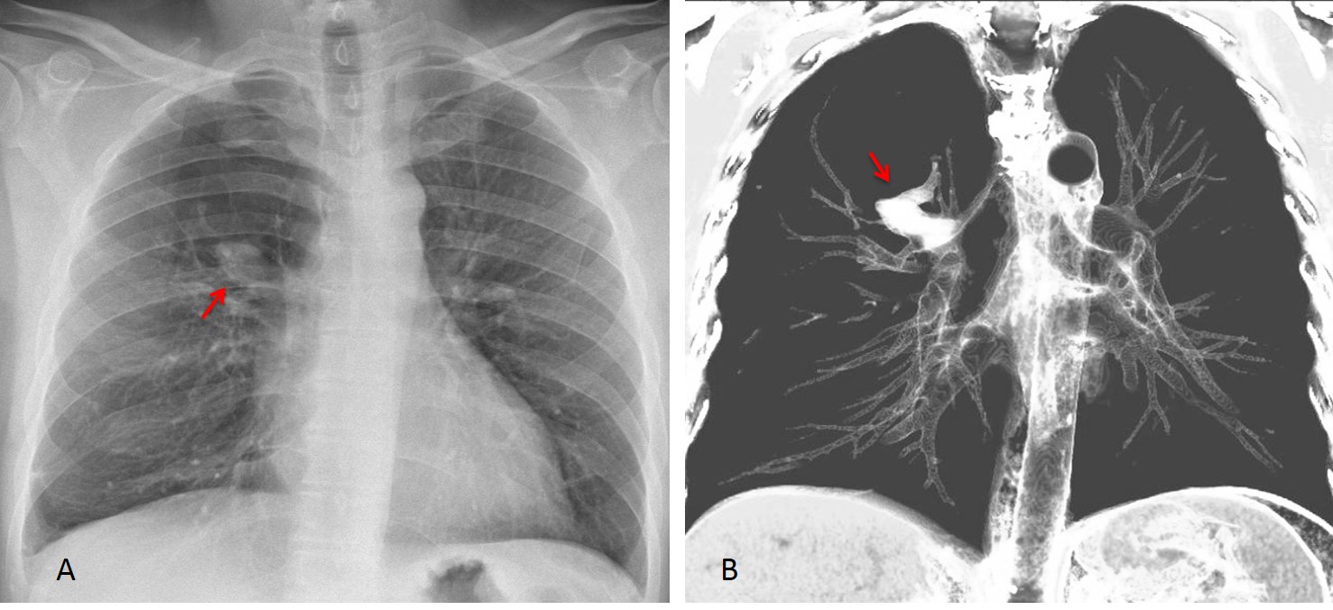
Final diagnosis: congenital bronchial atresia
The objective of this presentation is to discuss common congenital malformations of the lung. Contrary to what may be believed, congenital diseases are not rare in adults. My intention is to present the imaging features of the most common ones so that they will be recognised and not mistaken for more serious conditions. Congenital malformations can be focal (bronchial atresia and pulmonary sequestration) or affect one lung (agenesis of the lung and hypogenetic lung syndrome).
In my experience, congenital bronchial atresia is the most common lung malformation seen in adults. Congenital bronchial atresia results from proximal interruption of a segmental bronchus, which causes overinflation of the affected segment and secondary mucus impaction. Chest radiography shows a focal hyperlucent area with internal mucus impaction. CT depicts these findings better (Fig. 1). Inspiration and expiration films help to confirm air-trapping (Fig. 2). In case of doubt, bronchoscopy excludes other causes of bronchial obstruction.

Fig 1, A+B
Fig 1: PA chest film shows numerous serpiginous shadows in the LUL (A, arrows) with associated hyperlucency of the underlying lung. Serpiginous shadows bring to mind two possibilities: arteriovenous malformations and mucus plugs. The hyperlucent lung and the LUL location suggest congenital bronchial atresia as the first option.
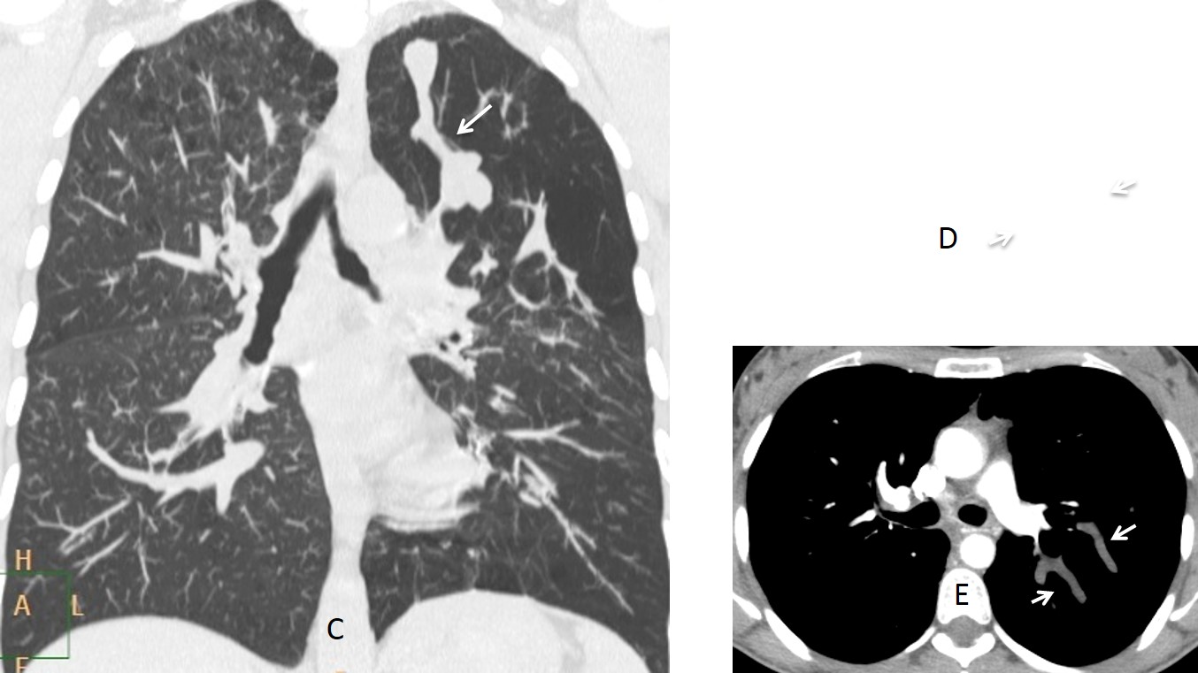
Fig. 1, C,D,E
Coronal and axial CT show the segmental hyperlucency and mucus plugs (C and D, arrows). Enhanced axial CT confirms the non-enhancing mucus plugs (E, arrows).
Diagnosis: congenital bronchial atresia
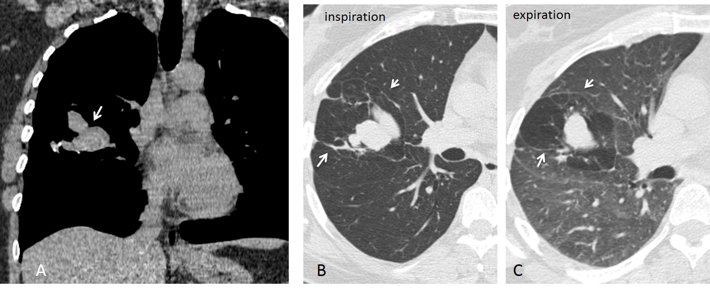
Fig. 2
Fig. 2: 48-year-old woman with bronchial atresia. Unenhanced coronal CT demonstrates the typical finger-in-glove appearance of mucus impaction (A, arrow). Axial CT with pulmonary window shows areas of emphysema around the mucus impaction (B, arrows), with air-trapping on expiration (C, arrows). Bronchoscopy was negative. This case emphasises the value of expiratory films to demonstrate air trapping.
Intralobar pulmonary sequestration consists of abnormal lung tissue that is not connected with the bronchial tree and is irrigated from systemic vessels. The chest radiograph shows a lower lobe opacity with recurrent infections that do not heal completely. CT confirms the diagnosis by demonstrating the systemic artery arising from the aorta (Figs. 3 and 4).

Fig. 3, A,B
Fig. 3: 41-year-old man with recurrent pneumonia in the RLL. PA and lateral radiographs depict an opacity in the RLL with cystic areas showing air-fluid levels (A and B, arrows). The appearance is non-specific, but the recurrent pneumonia and cysts suggest pulmonary sequestration. CT should be performed to investigate this possibility.
Enhanced axial CT shows cystic areas (C, arrows) within a large basal infiltrate. A caudal view shows a systemic artery arising from the abdominal aorta (D, arrow). The systemic artery is better depicted in the volume reconstruction (E, arrow). Surgical diagnosis: pulmonary sequestration.
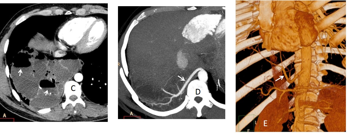
Fig. 3, C,D,E
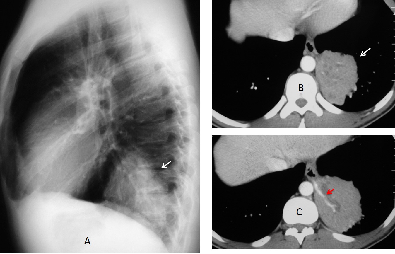
Fig. 4
Fig. 4: 25-year-old man with a solid-looking mass in the LLL (A, arrow). Enhanced axial CT shows a mass with minute cysts (B, arrow) with a systemic artery arising from the aorta (C, arrow). Surgical diagnosis: sequestration.
Hypogenetic lung (scimitar syndrome, venolobar syndrome) is a relatively common malformation that occurs secondary to agenesis of one or two lobes, with abnormal venous drainage of the lower lung in 80% of cases. The malformation usually affects the right lung. On plain films it appears as a small right hemithorax with mediastinal shift to the right. The scimitar vein is visible in about 50% of chest radiographs and is helpful in cases of mild hypoplasia. CT confirms the findings by demonstrating the absent lobe(s) and the abnormal vein, when there is one (Figs. 5 and 6).
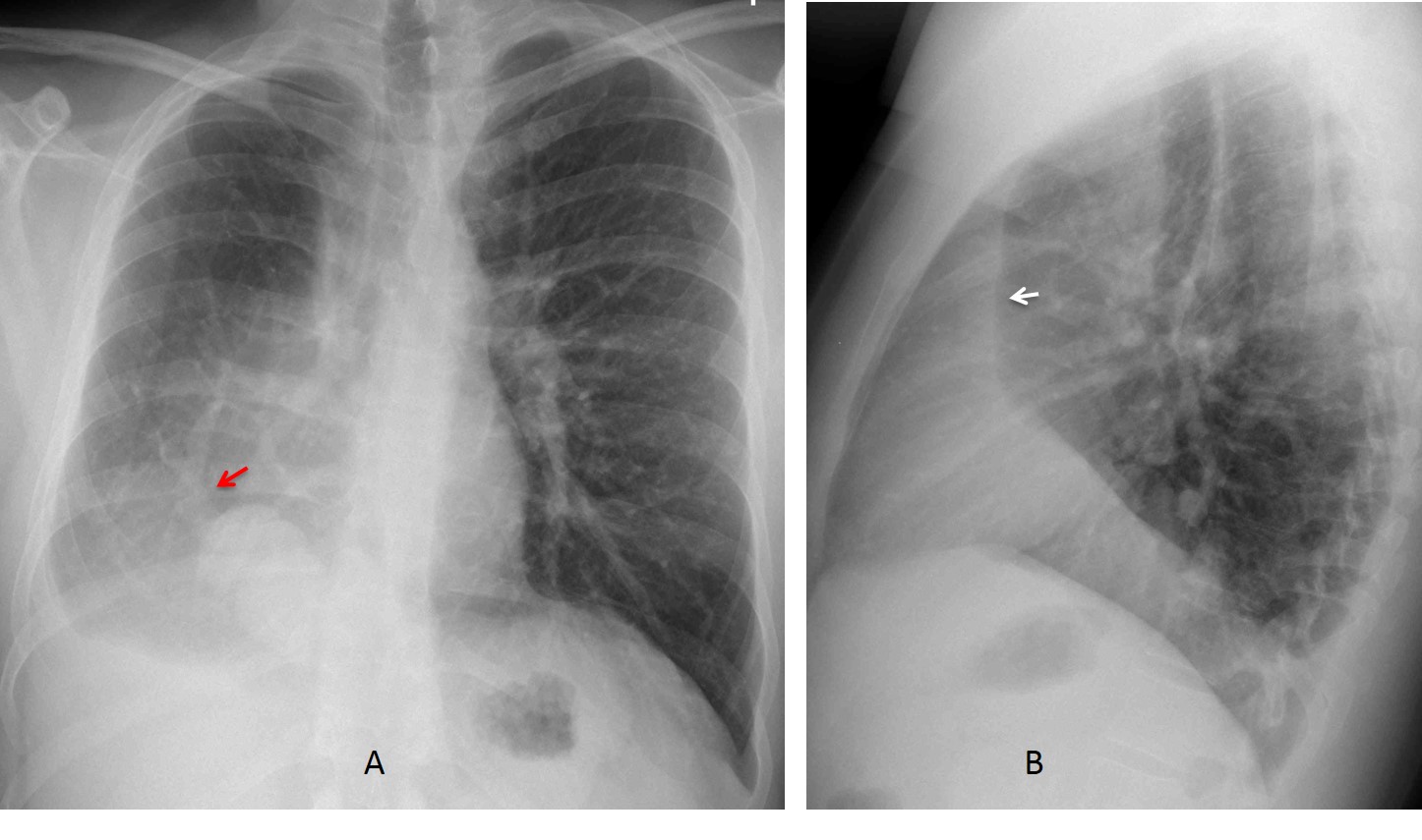
Fig. 5, A,B
Fig. 5: 22-year-old man, asymptomatic. PA radiograph shows typical findings of hypogenetic lung: small right hemithorax with right mediastinal displacement and blurring of the right heart border. A scimitar vein is visible (A, arrow). The vertical retrosternal line (B, arrow), is present in most cases.
Enhanced coronal reconstruction confirms the scimitar vein (C, arrow) Axial CT shows the abnormal bronchial branching (D, arrow) and the smaller right lung. The shortened A-P diameter of the lung is the cause of the retrosternal line in the lateral view. Diagnosis: hypogenetic lung.

Fig. 5, C,D
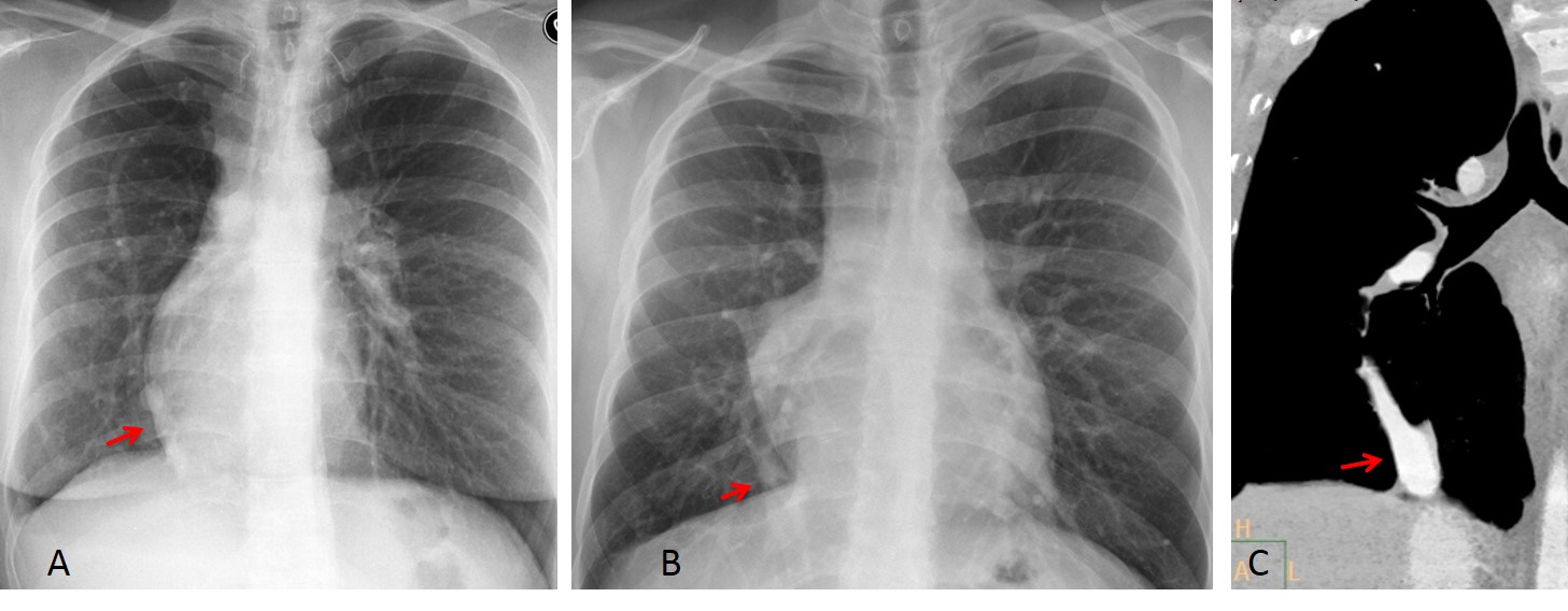
Fig. 6
Fig. 6: two examples of mild hypogenetic lung. In both cases, there is slight displacement of the mediastinum and heart towards the right, simulating dextrocardia. The scimitar vein helps to suggest the correct diagnosis (A and B, arrows). Enhanced CT confirms the vein in the second case (C, arrow).
Unilateral pulmonary agenesis is due to arrested development of one lung. It occurs equally on both sides. It is asymptomatic and usually discovered accidentally. Plain radiography shows opacity of one hemithorax with marked mediastinal shift, simulating a pneumonectomy. The lateral view shows en enlarged anterior clear space secondary to herniation of the opposite lung. CT confirms absent or minimal lung tissue.
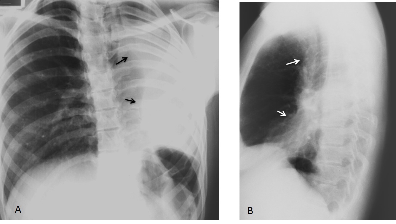
Fig. 7, A,B
Fig. 7: 40-year-old man with left lung agenesis. The PA radiograph shows a small airless left hemithorax with crowding of the ribs and marked herniation of the contralateral lung (A, arrows). The lateral film confirms herniation across the anterior clear space (B, arrows).
Axial CT depicts a small remnant of hepatised lung tissue (C and D, black arrows) and the absent left bronchus (D, red arrow). Note herniation of the right lung behind the sternum. An interesting finding is that the homolateral pulmonary artery is usually of normal size (C, blue arrow).
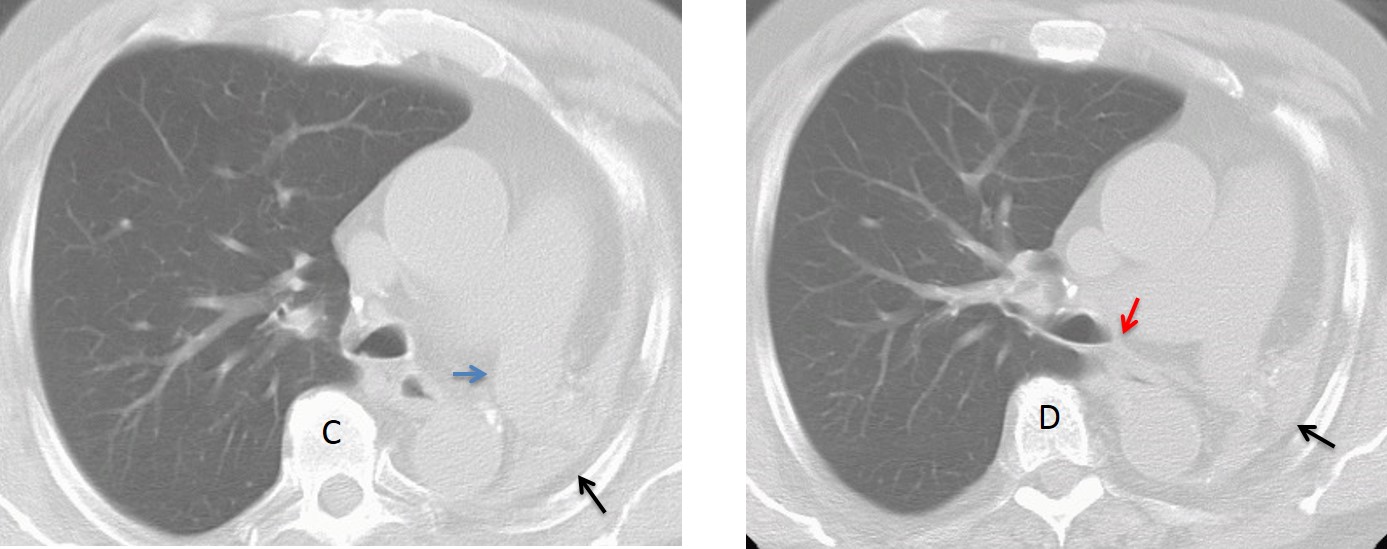
Fig. 7, C,D
In lung agenesis, the normal lung sometimes herniates markedly into the contralateral hemithorax. In the chest radiograph, this may produce the deceiving appearance of aerated lung with chronic pleural disease. The diagnosis is obvious on CT.

Fig. 8
Fig. 8: marked right lung herniation in left pulmonary agenesis. The PA view simulates chronic pleural disease of left side (A, arrows). CT shows marked herniation of the normal lung, reaching the opposite chest wall (B and C). There is considerable posterior displacement of the heart and mediastinal structures.

Follow Dr.Pepe’s Advice:
1. Congenital lung malformations are not unusual in adults. Most are asymptomatic and are discovered when a chest radiograph is taken.
2. Bronchial atresia is characterized by focal hyperlucency and mucus plug.
3. In intralobar sequestration there are recurrent lower lobe infections.
4. Hypogenetic lung is accompanied by a scimitar vein in 80% of cases.
5. Congenital agenesis simulates a previous pneumonectomy.


