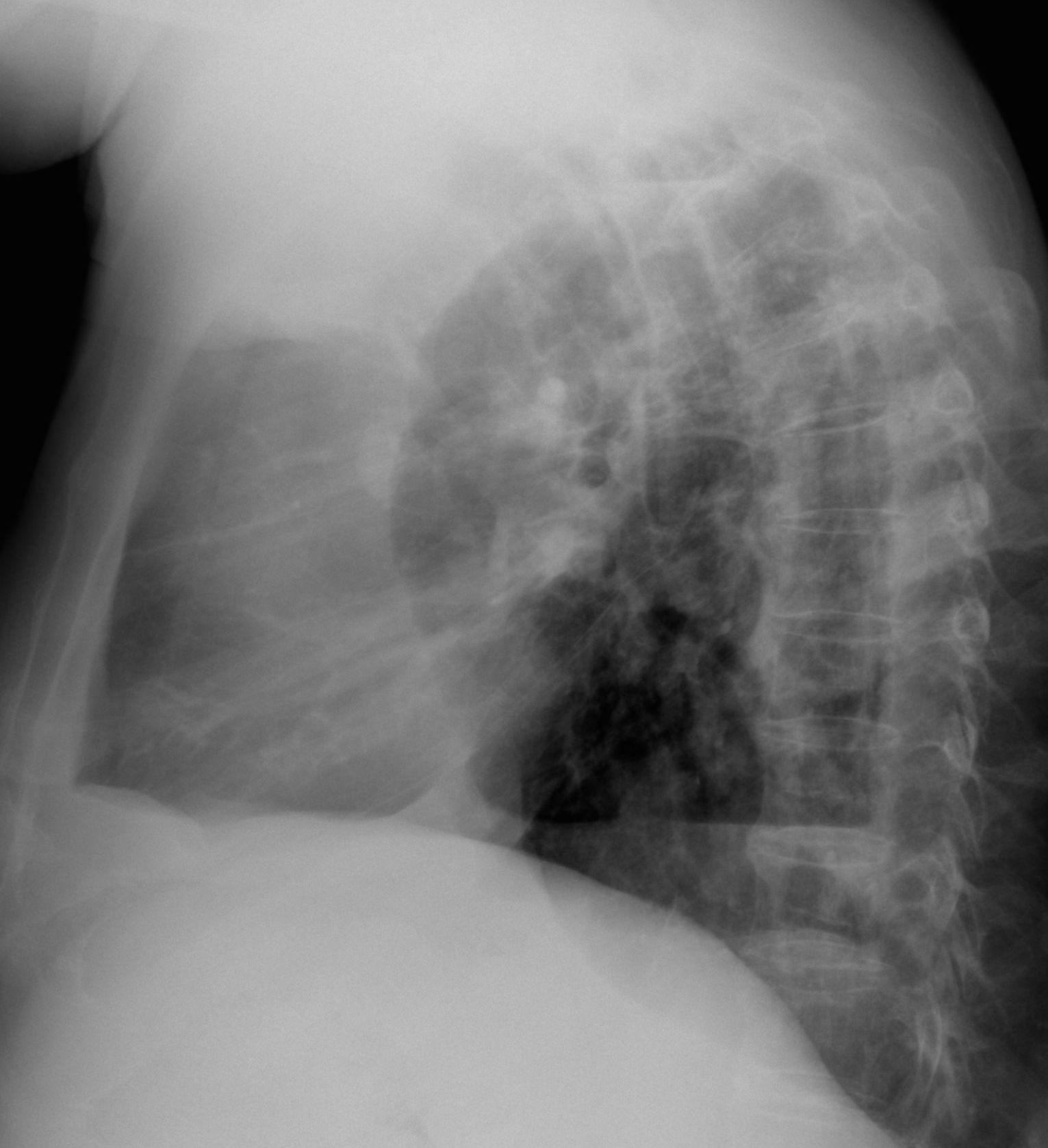
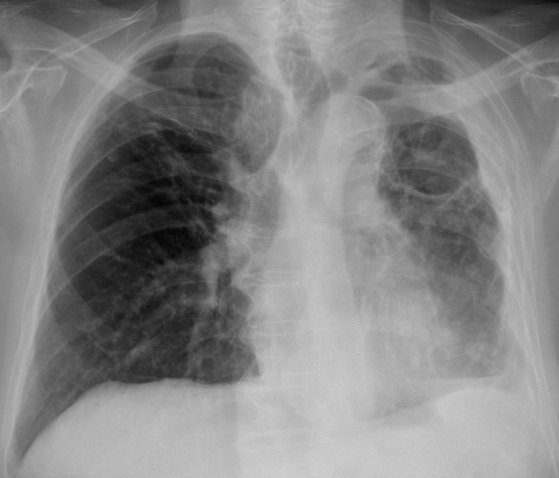
Today I am showing a case provided by my former resident and good friend Dr. Nadine Romera. Images belong to an 87-year-old man with TB in the past. He presents now with loss of weight and low-grade fever.
What do you see?
Check the images below, leave your thoughts in the comments section, and come back on Friday for the answer.
Findings: PA radiograph shows a small left hemithorax with pleural thickening (A, arrows). Chronic changes in both lungs are visible. In addition, two air-fluid levels are seen in the pleural space (A-B, red arrows). With these findings, the first diagnosis that comes to mind is chronic pleural disease with broncho-pleural fistula.

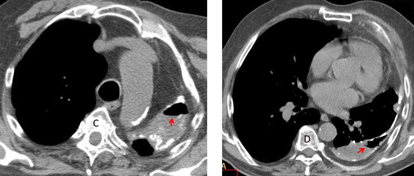
Axial CT shows the calcified pleura with loculated fluid and air-fluid levels (C-D, arrows), confirming the suspicion of broncho-pleural fistula.
CT also demonstrates a small pleural mass that was overlooked in the initial examination, made in October 2014 (E, arrow). A follow-up CT three months later shows an increase in size of the mass (F, arrow) with a rim of increased uptake in PET-CT (G, arrow).
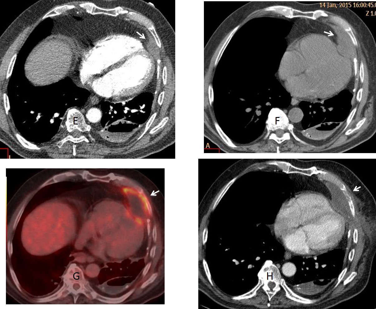
Percutaneous biopsy came back as lymphoma. While in treatment, CT one month later shows invasion of the overlying rib (H. arrow).
Final diagnosis: Chronic pleural changes with broncho-pleural fistula and secondary lymphoma.
Congratulations to all of you who made the correct diagnosis. Cannot single out anyone. The lymphoma is obviously not seen in the chest radiograph and, as a matter of fact, was missed in the first CT.
Teaching point: the presence of an air-fluid level in chronic pleural disease is practically pathognomonic of broncho-pleural fistula. In this particular case, the associated lymphoma is a serendipitous finding. It is well know that malignant tumours are a complication of long-standing inflammatory pleural disease (see Caceres’ Corner case 34)