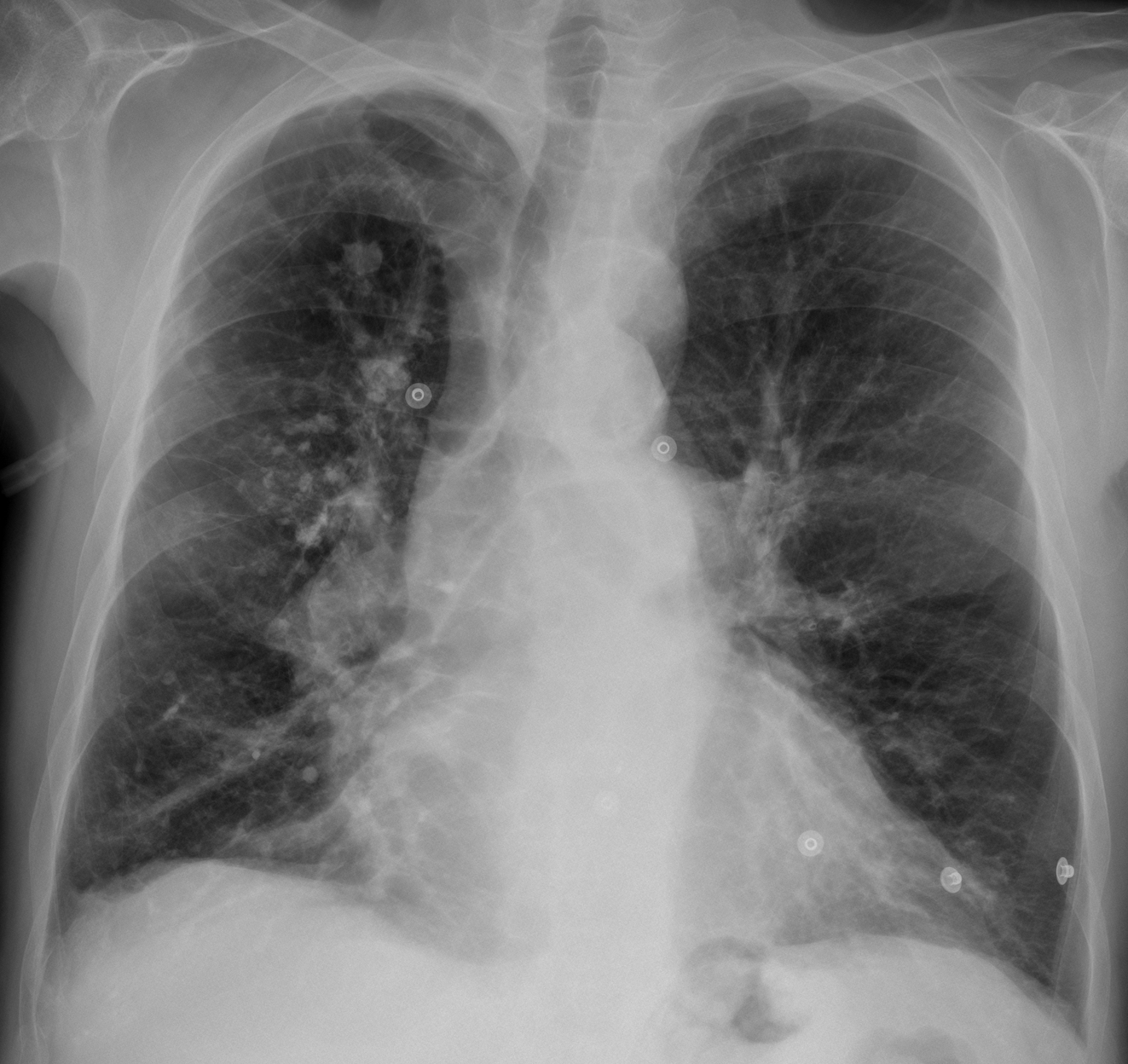
Today’s images belong to a 57-year-old male who is going to undergo a lung transplant for chronic lung disease.
What do you see?
Check the images below, leave your thoughts in the comments section and come back on Friday for the answer.
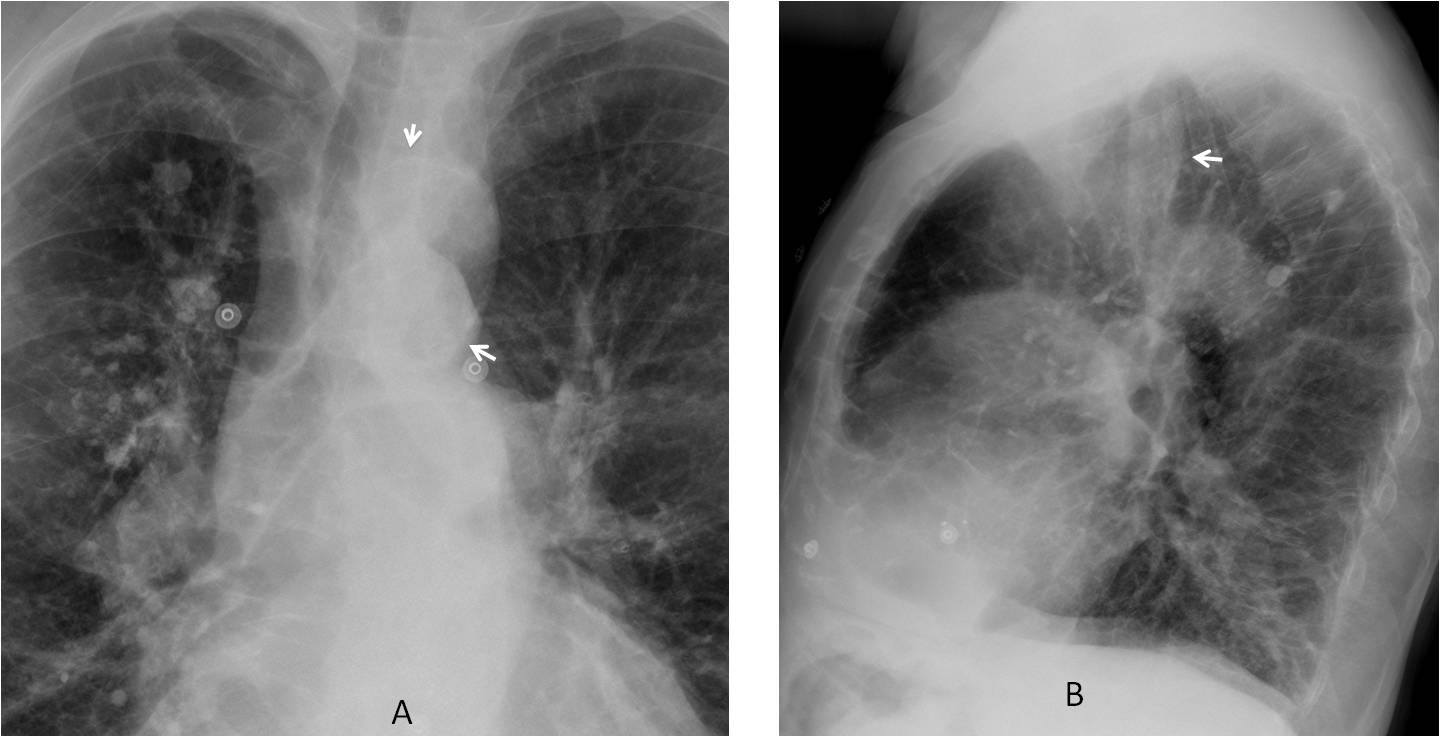
Findings: chest radiographs show emphysematous lungs with calcified granulomas and signs of pulmonary hypertension. The PA film shows an apparent mass with circular calcification (A, arrows) above the aortic knob. It is visible behind the trachea in the lateral view (B, arrow).
Unenhanced sagittal CT shows that the apparent mass represents a high aortic arch (C, red arrow), which angles when joining the descending aorta (C-D, red arrows).
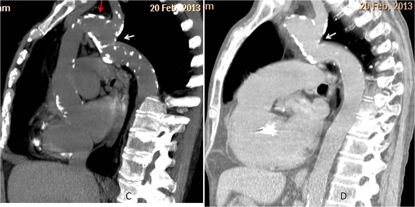
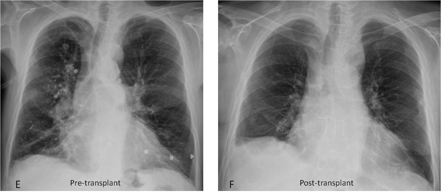
This appearance is typical of aortic pseudocoartation, which should be suspected in the plain film (I have seen several with similar appearance. See Cáceres’ Corner Case 42). The patient had normal blood pressure and underwent a bilateral lung transplant without complications (E-F).
Final diagnosis: unexpected aortic pseudocoarctation in a patient with severe emphysema pending lung transplant.
Congratulations to Pasquale and sht, who were the first to suggest a vascular anomaly of the mediastinum.
Teaching points:
1. When seeing circular calcification in the mediastinum always think of a cyst or a vascular structure.
2. Avoid satisfaction of search. Do not focus on the obvious.