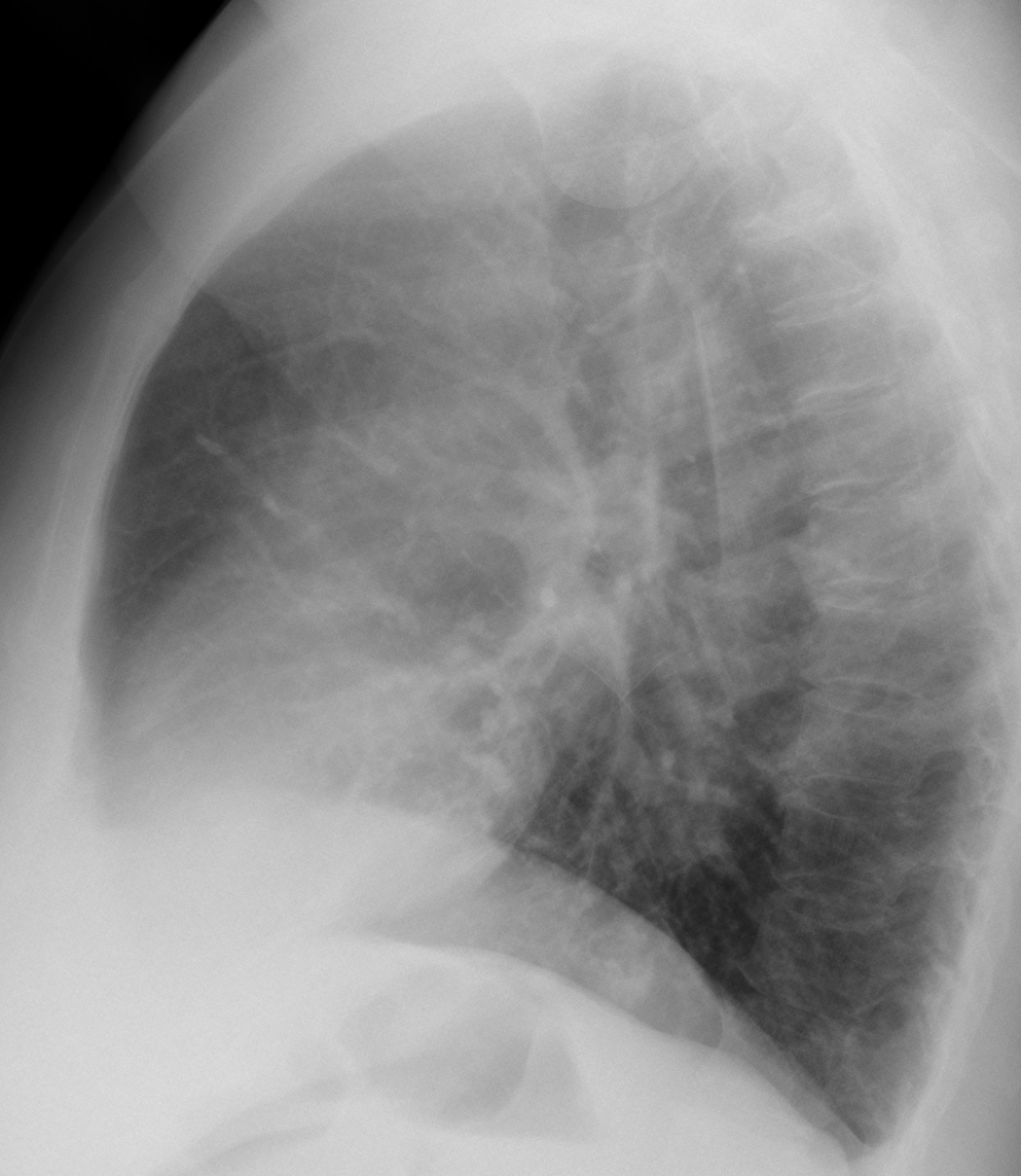
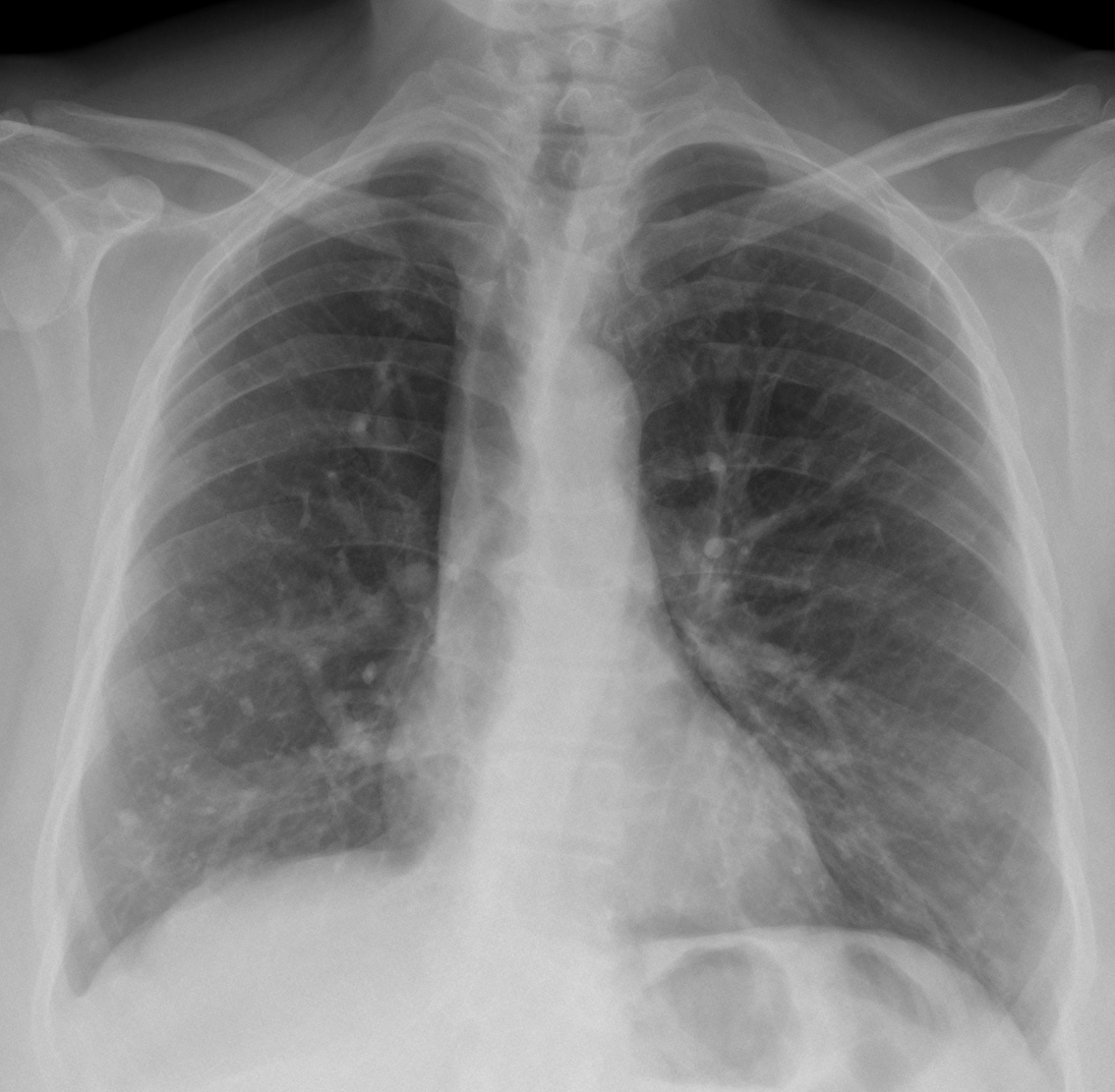
Today we are presenting chest images of a 47-year-old woman with a cough. What do you see?
Check the images below, leave your thoughts in the comments section and come back on Friday for the answer.
Findings: the PA radiograph shows an obvious downward displacement of the right hilum (A, red arrow). The intermediate bronchus has a vertical course (A, arrow), practically following the path of the trachea.
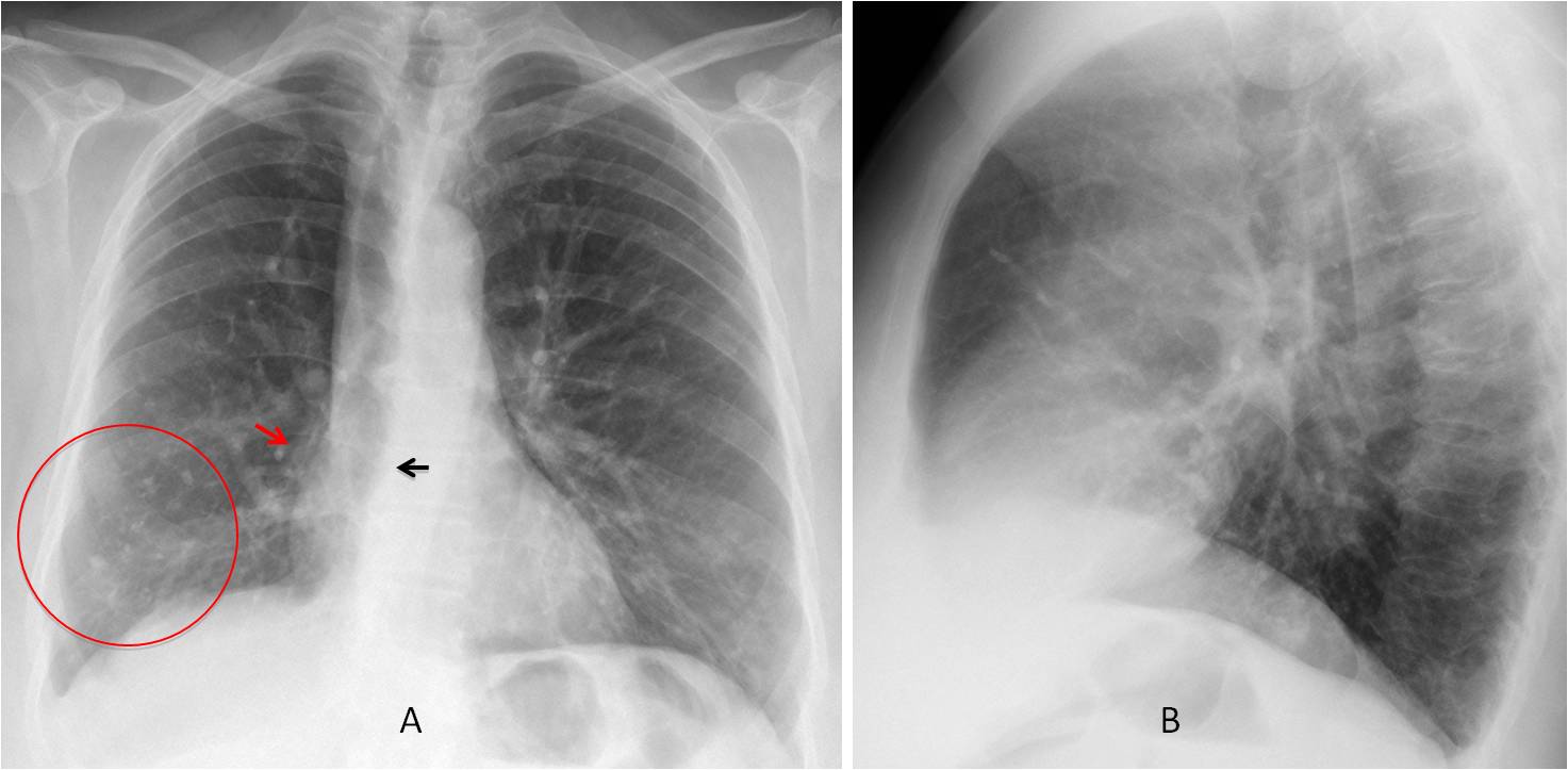
These findings are highly suggestive of marked RLL collapse (see Dr. Pepe’s Diploma Casebook case #79). There are calcified granulomas at the right lung base (A, circle). The lateral view does not offer any additional information.
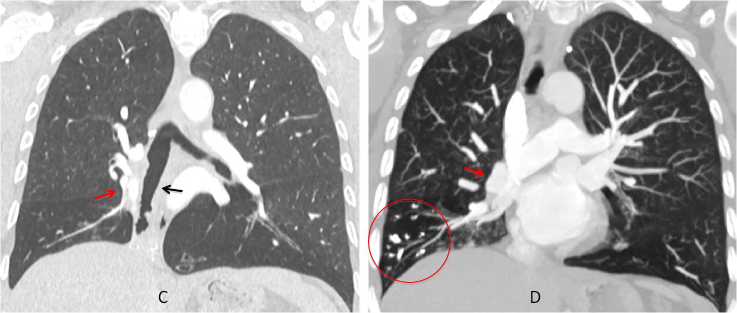
Coronal CT confirms the vertical path of the intermediate bronchus (C, arrow) and the downward displacement of the right hilum (C-D, red arrows). Calcified basal granulomas are seen (D, circle).
Axial and coronal CT show a small triangular shadow with coarse calcium (E-F, arrows), which represents a markedly collapsed RLL.
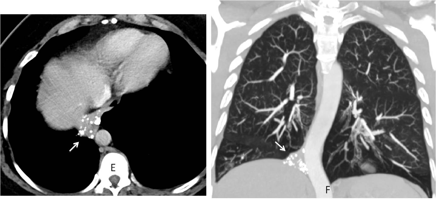
Final diagnosis: marked RLL collapse secondary to previous TB.
Teaching point: remember to look at the hila! A displaced hilum is an excellent sign to suspect lobar collapse.