Dear Friends,
April is the cruelest month. Dr. Pepe has eloped with Miss Piggy and has not prepared any Diploma cases. To cover for him I have selected five cases to be shown during April. Paraphrasing the title of a film by Jack Nicholson, I am calling them “Five easy pieces”. All of them show subtle findings, easily seen if you look carefully.
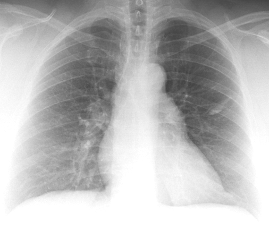
The first case is a PA radiograph of a 53-year-old woman, preo-op for bariatric surgery. Check the image below, leave me your thoughts in the comments section, and come back on Friday for the answer.
Click here for the answer to case #134
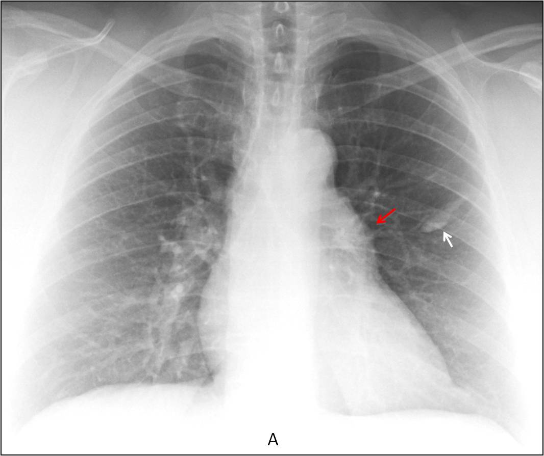
Findings: PA radiograph shows a partially calcified elongated image in the left middle lung field (A, arrow). Milk of calcium, as suggested by Borsuk, cannot be excluded. There is convexity of the pulmonary arch (A, red arrow) that was overlooked in the initial reading. This finding is compatible with enlargement of the main pulmonary artery or a mediastinal mass (See
Dr. Pepe’s Diploma Casebook case #52).
Unenhanced sagittal and axial CT shows a partially calcified mucous impaction (B-C, arrows) and an unexpected solid anterior mediastinal mass (C, red arrow).
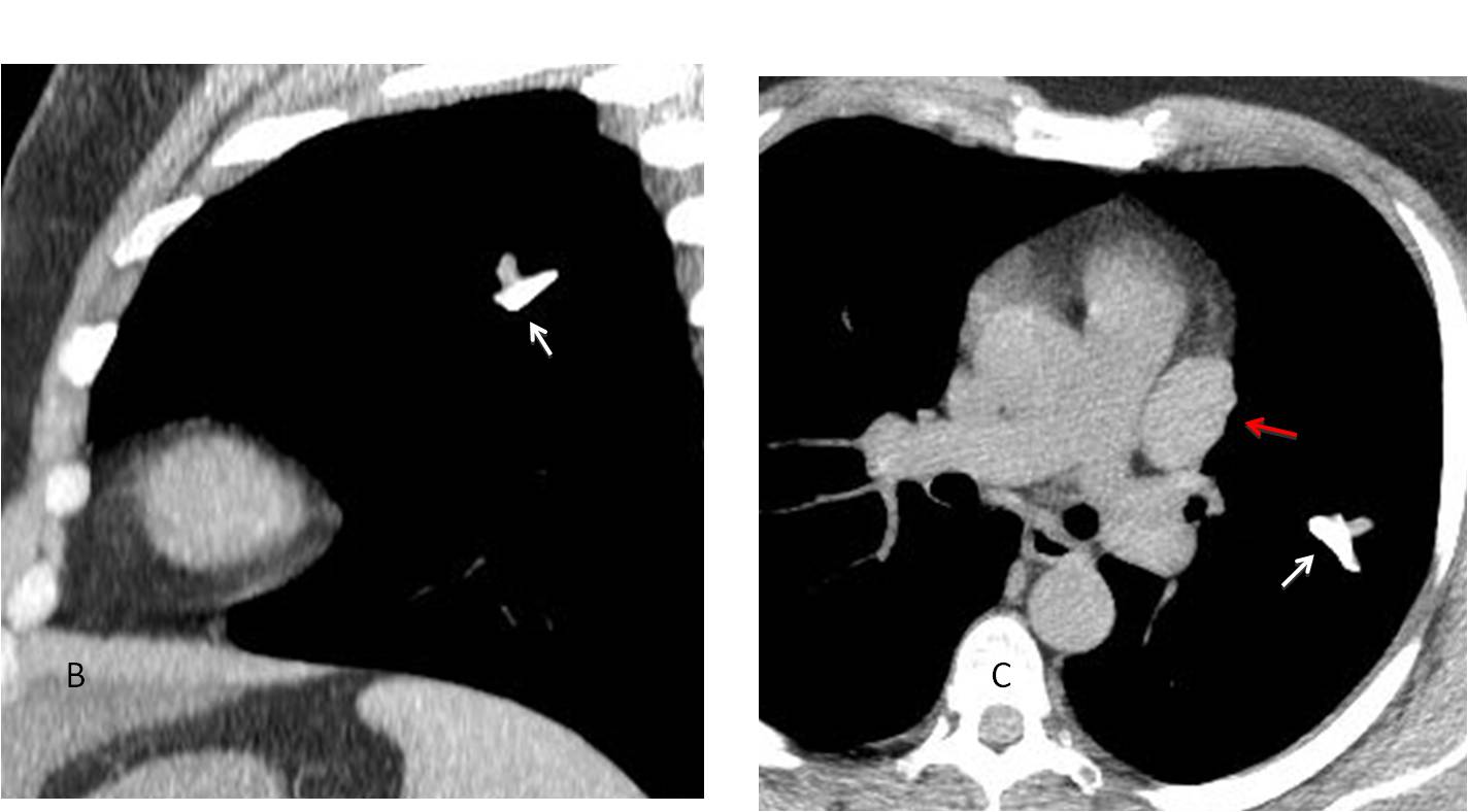
Final diagnosis: calcified mucous impaction and unrelated thymoma.
Congratulations to Aleksandar, who was the first to recognise the mediastinal abnormality.
Teaching point: Look carefully at pre-op films. Important pathology may be discovered.


