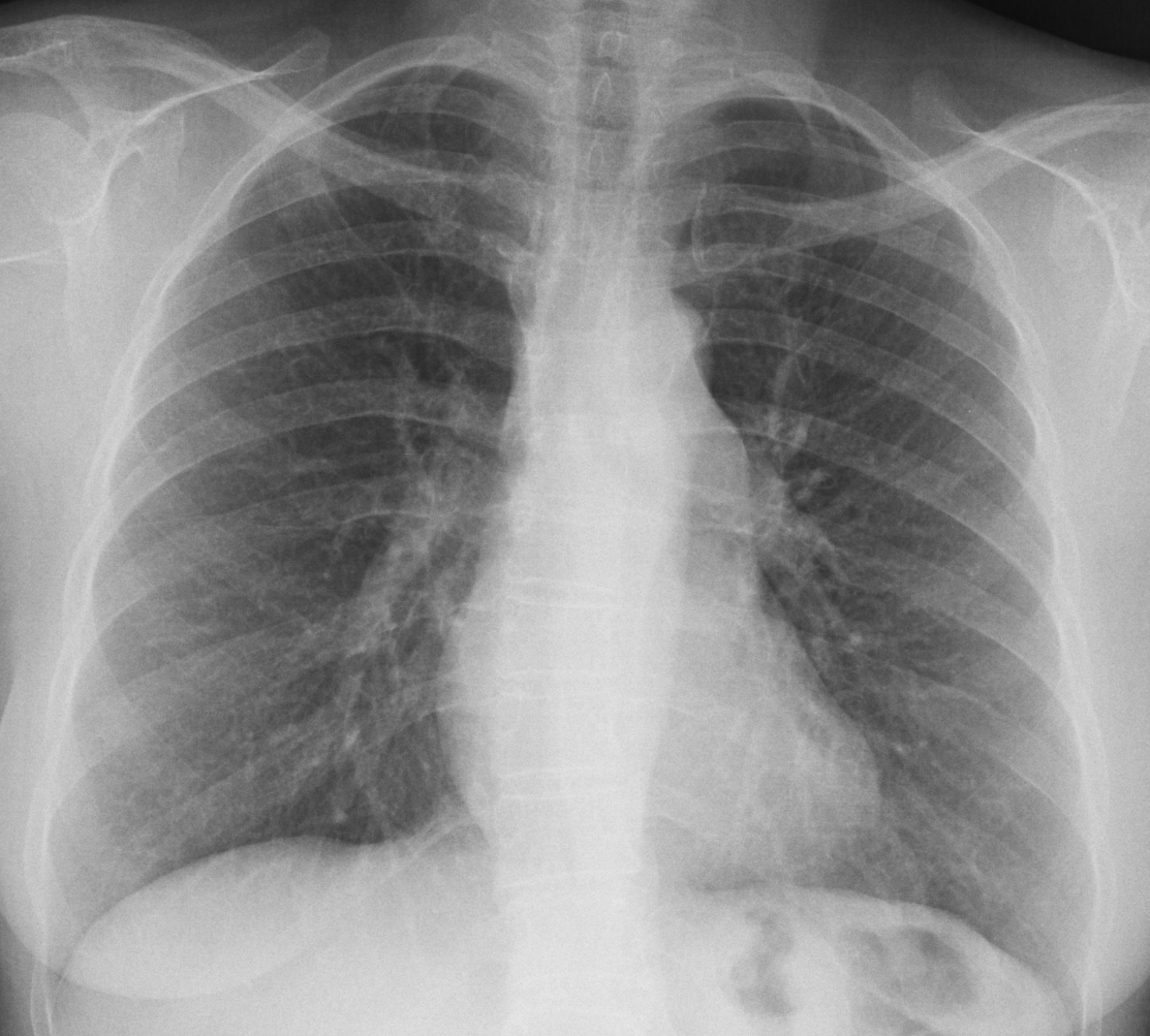
The case of this week is a pre-op radiograph for a knee prosthesis in a 57-year-old woman. What do you see?
Check the image below, leave your thoughts in the comments section and come back on Friday for the answer.
Findings: the PA chest radiograph shows a localised bulging of the left paraspinal line, better seen in the cone down view (A-B, red arrows). In addition, there is an obvious prominence (A-B, arrows) of the pulmonary artery arch (See
Dr. Pepe’s Diploma Caesbook case #52).
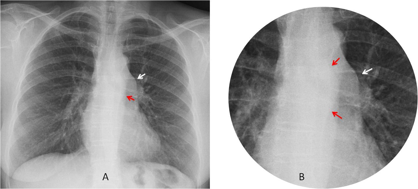
The location of abnormalities in both posterior and middle mediastinum suggests enlarged lymph nodes and I considered lymphoma a good possibility (and I was wrong).
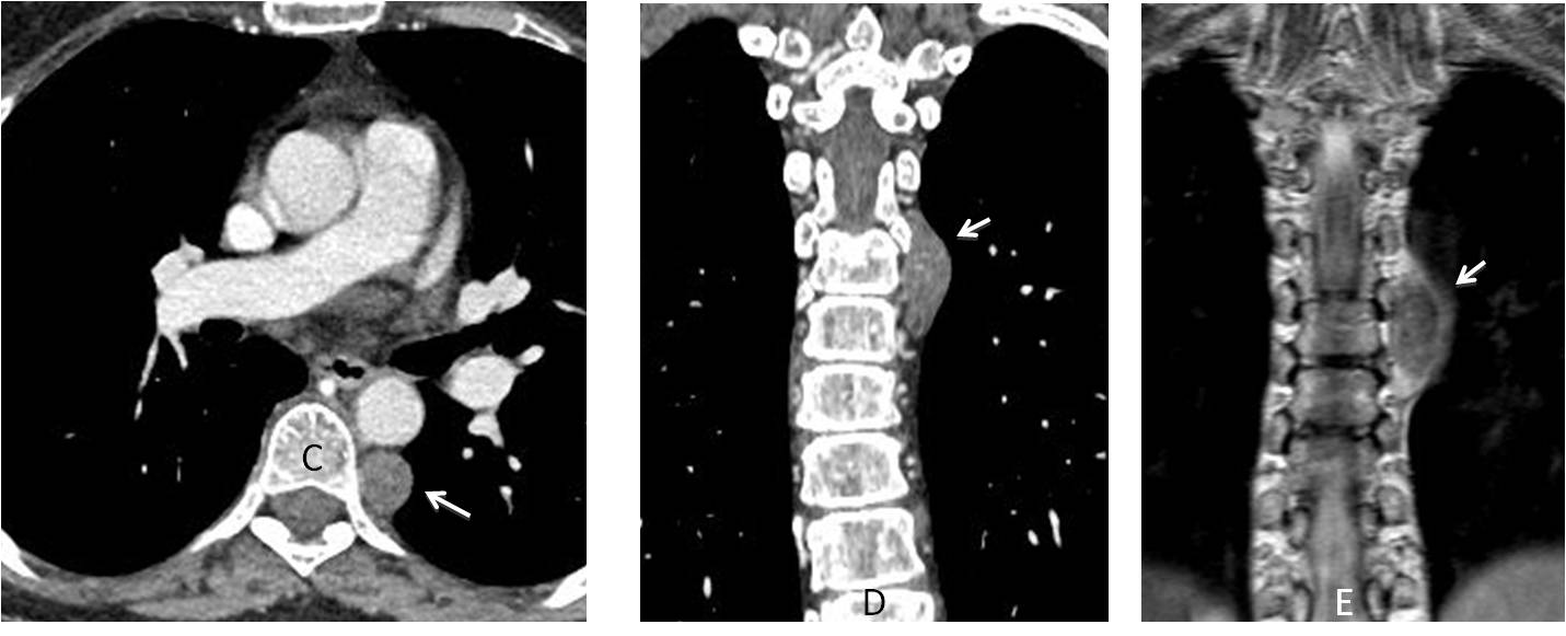
Enhanced CT and MRI confirm a solid posterior mediastinal mass, compatible with neurogenic tumour (C-E, arrows).
Enhanced axial and sagittal CT show a dilated main pulmonary artery (F-G, arrows). Further studies failed to demonstrate a gradient across the pulmonary valve.
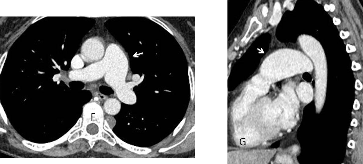
Final diagnosis: unsuspected schwannoma and idiopathic dilatation of the pulmonary artery.
Congratulations to Diogo Pinheiro and M.A. Fahmy who teamed up to make the correct diagnosis
Teaching point: this is another case of satisfaction of search. Most of you saw only one abnormality, either the posterior mass or the prominent artery. I saw two, but still I did not fare any better!