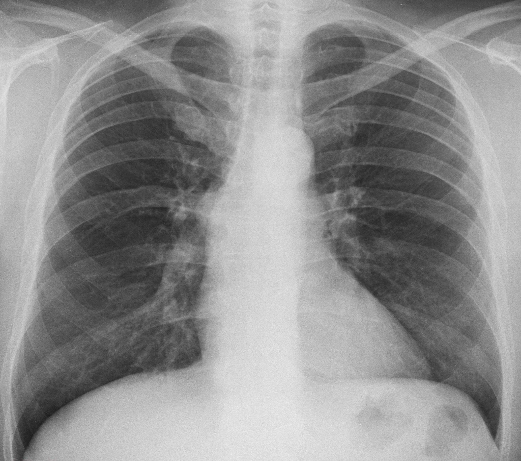
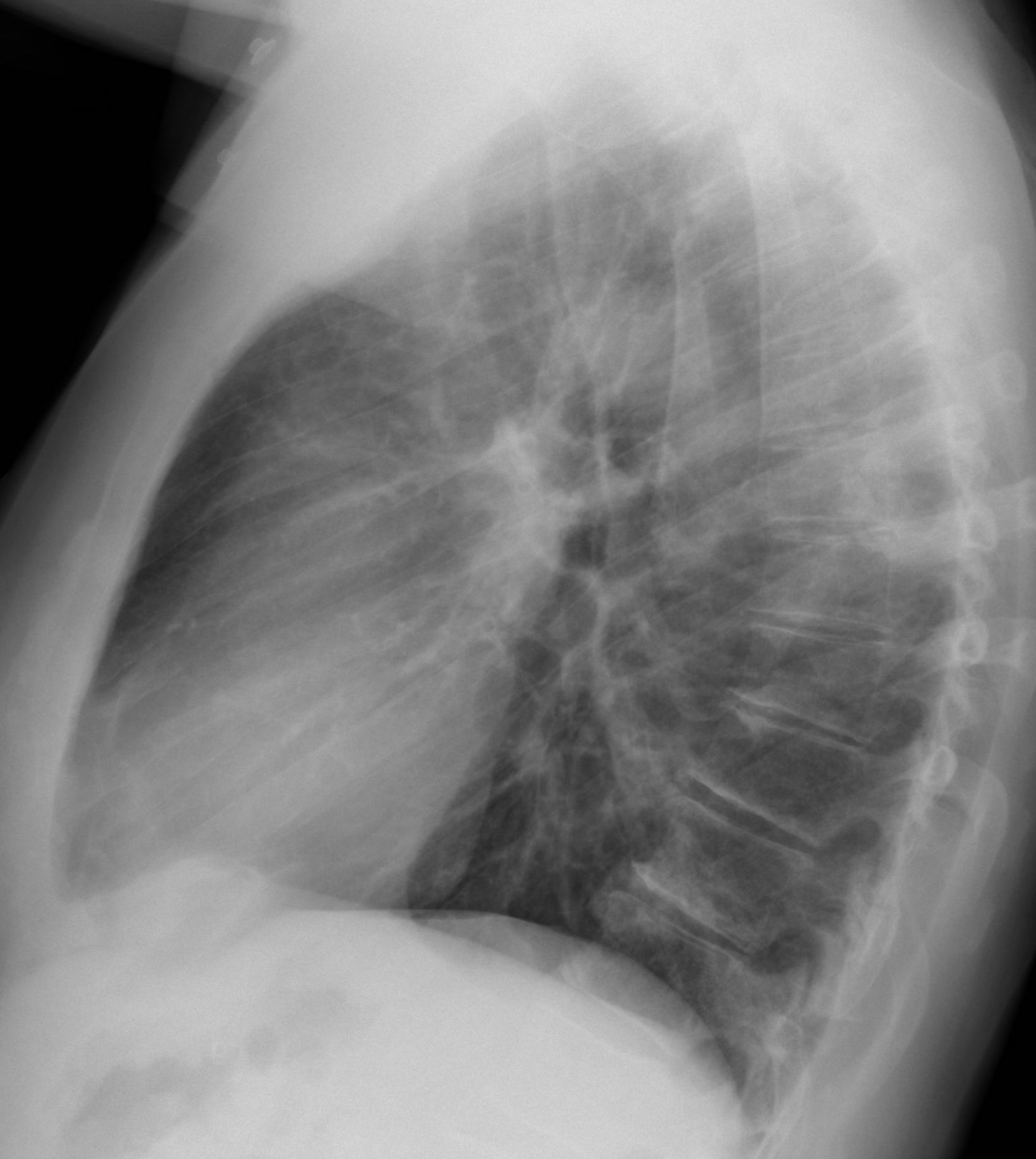
Showing today pre-op radiographs for a hip prosthesis in a 62-year-old man.
What do you see?
Check the images below, leave your thoughts in the comments section, and come back on Friday for the answer.
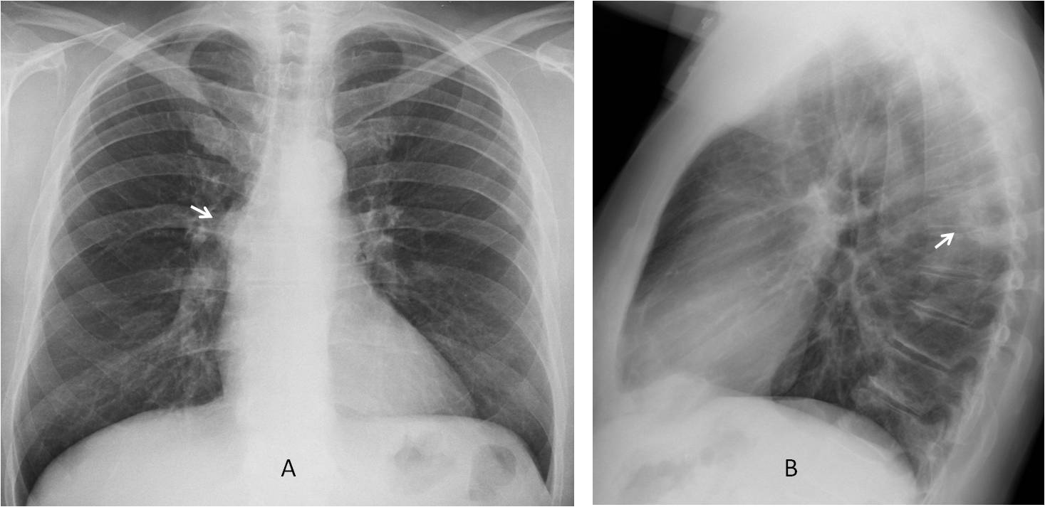
Findings: PA radiograph shows an extrapulmonary lesion projected over the right hilum (A, arrow), located in the posterior mediastinum in the lateral view (B, arrow).
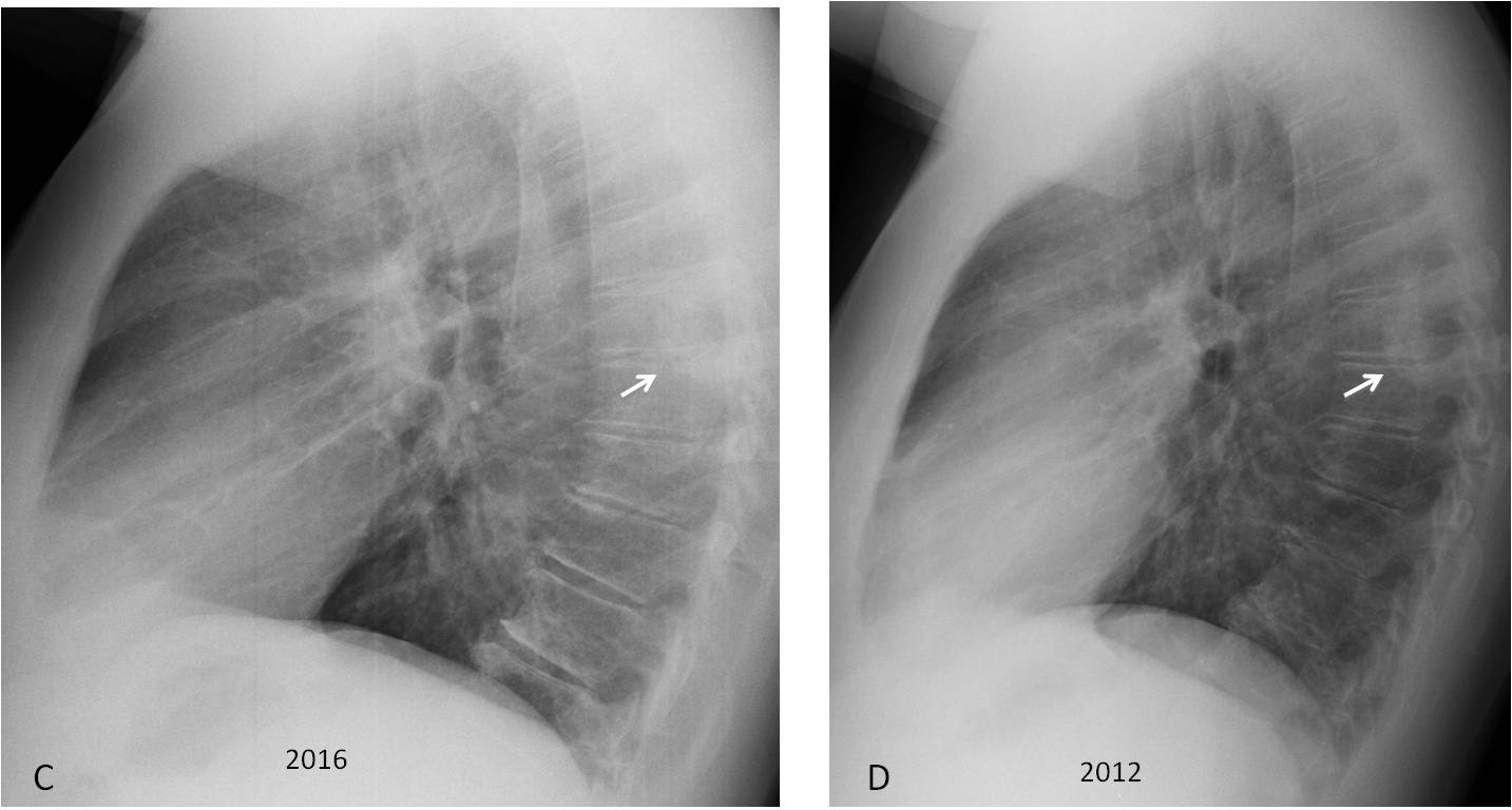
The lesion is very similar to the one shown in case 135 and neurogenic tumour is the first diagnosis that comes to mind. Comparison with previous film shows that the lesion has not changed in a four-year interval (C-D, arrows).
Coronal and sagittal CT demonstrates that the lesion corresponds to a large osteophyte (E-F, arrows).
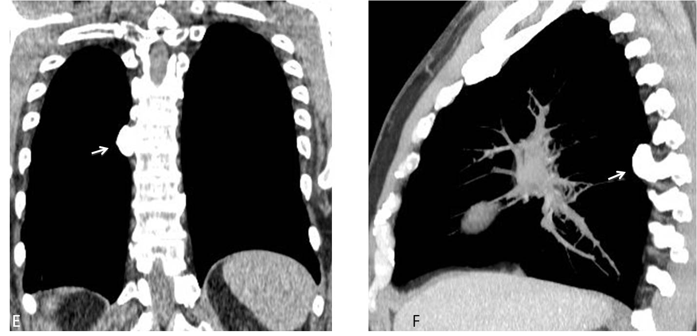
Final diagnosis: osteophyte simulating a posterior mediastinal mass.
Congratulations to all of you who saw the mediastinal lesion. It is important to emphasize that high KV makes it difficult to recognise calcium.
Teaching point: remember that not all posterior mediastinal masses represent neurogenic tumours. Processes arising from the spine and mediastinal fat should also be considered (see Dr. Pepe’s Diploma Casebook case 64).