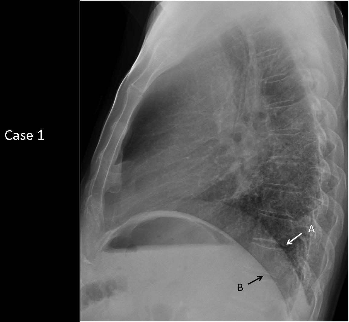
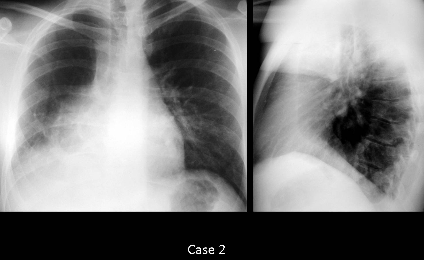
Muppet has the silly notion that he is good at semiology and wants to test your interpretation skills with the following two cases.
1. A
2. B
3. Can´t tell
1. Yes
2. No
3. Can’t tell
Hope you enjoy the challenge. Answer next Monday.
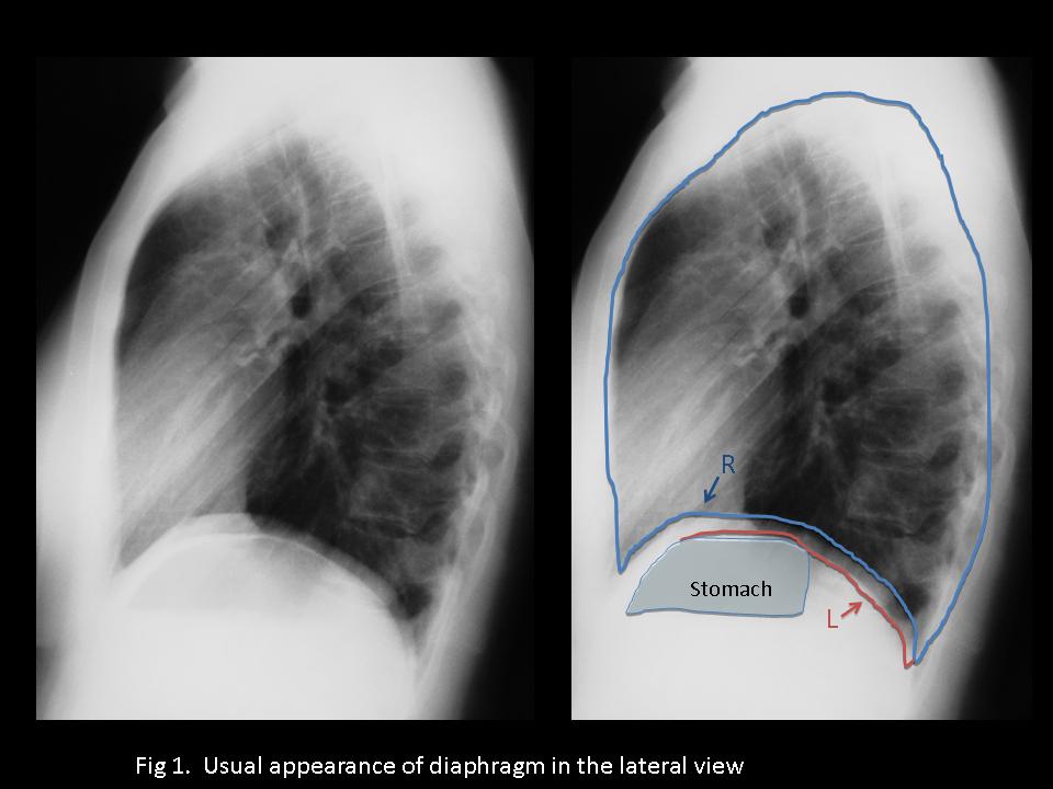
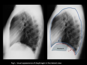
Before giving an answer to case 1, it is necessary to understand the usual appearance of the diaphragm in the lateral view: the right hemidiaphragm is seen all the way and it is slightly higher than the left. The left hemidiaphragm is not seen anteriorly because the heart obscures the anterior portion and has the stomach bubble under it (see Fig 1).
The information in case 1 is misleading: one hemidiaphragm is higher (suggests right), but stops when reaching the heart (suggests left). The other goes all the way to the front (suggests right), but has the gastric bubble under (suggests left).
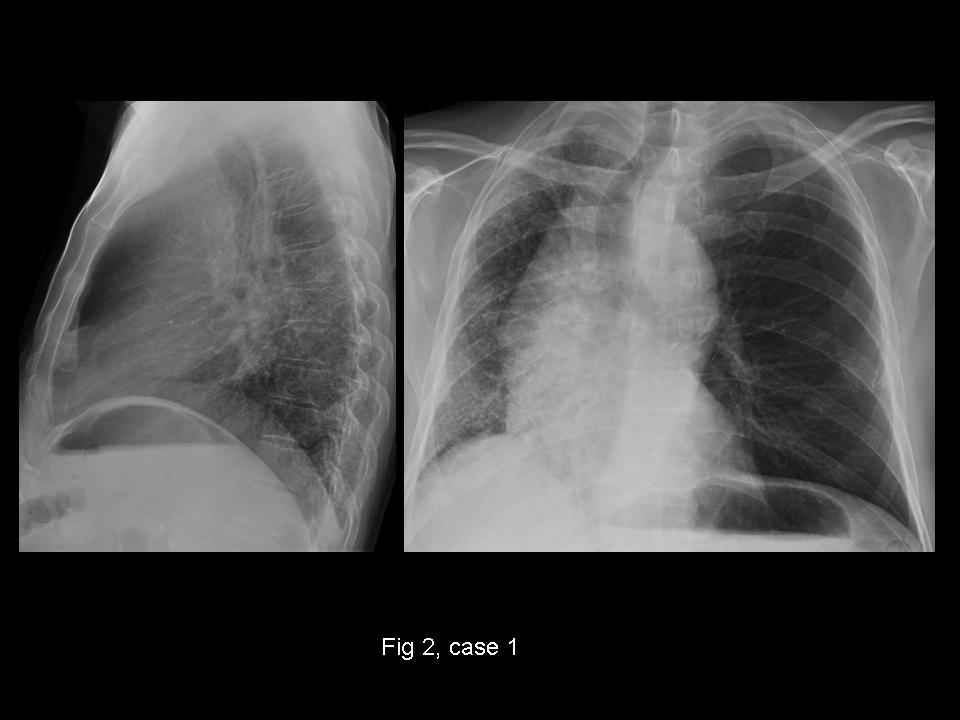
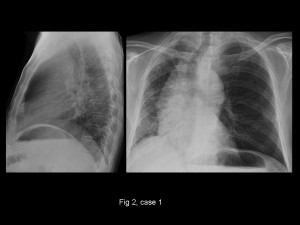
The explanation for this discrepancy is that the heart is in the right hemithorax, explaining the obliteration of the anterior portion of the right hemidiaphragm and the total visibility of the left one. PA confirms the findings: patient had unilateral left lung transplantation, with the expanded left lung pushing the heart into the right hemithorax and accounting for the findings in the lateral view (Fig 2).
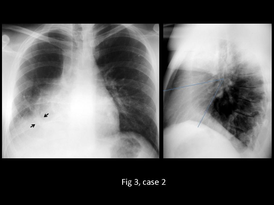
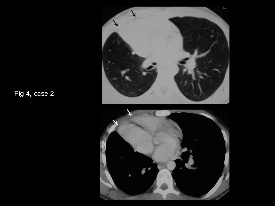
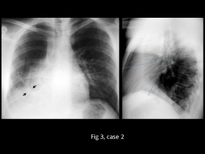
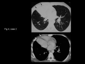
The answer to case 2 is simple: even though there is blurring of the right heart border, it is not due to RML disease, as evidenced by the lateral view, which does not show the typical triangular appearance of RML disease (Fig 3). This case is a good example of hypogenetic lung syndrome (go back to case 6), with the typical findings of small hemithorax, shifting of the heart towards the right, and a scimitar vein (arrows). Blurring of the right heart border is common in this condition and it is secondary to rotation of the heart, which contacts with the chest wall and loses definition (silhouette sign) (Fig 4, arrows).
I’m sorry to say most of you failed this case (Muppet sad). Congratulations to all of you who made the diagnosis, especially Mm, who offered an excellent discussion (Muppet happy).
Incidentally, can you identify the right and left hemidiaphragms in the lateral view of case 2?