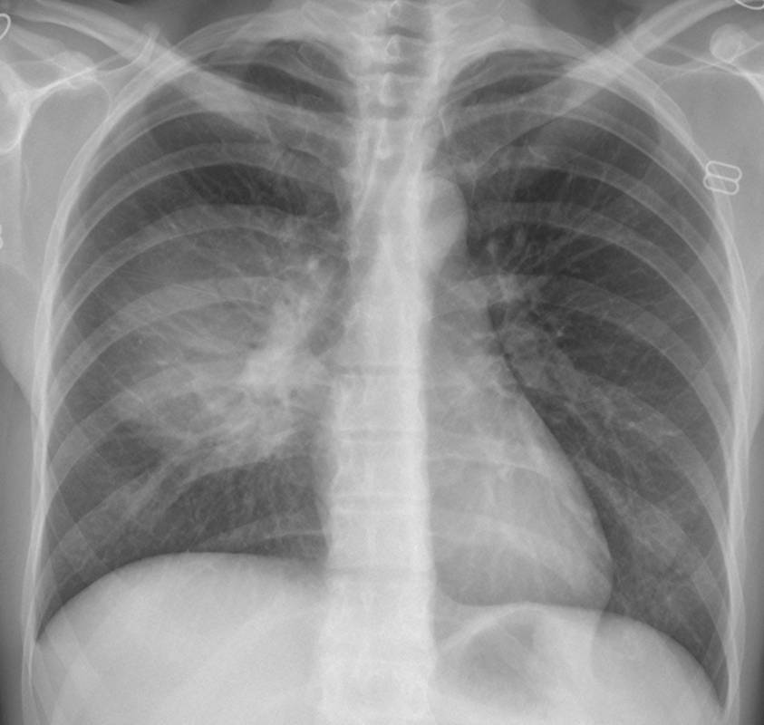
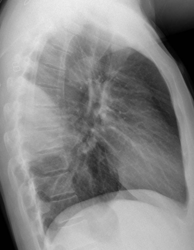
Muppet is becoming soft and wants to show easy cases. The following case is a 44-year-old woman with a cough, low-grade fever, and back pain.
1. Lung mass
2. Loculated pleural fluid
3. Chest wall mass
4. None of the above
Before giving the answer, it is important to understand the semiology of extrapulmonary lesions: they are well defined (because of the pleural layer which surrounds them), and have obtuse angles at the corners (to see an example of extrapulmonary lesion go to
case 1 in Dr. Pepe’s Diploma Casebook).
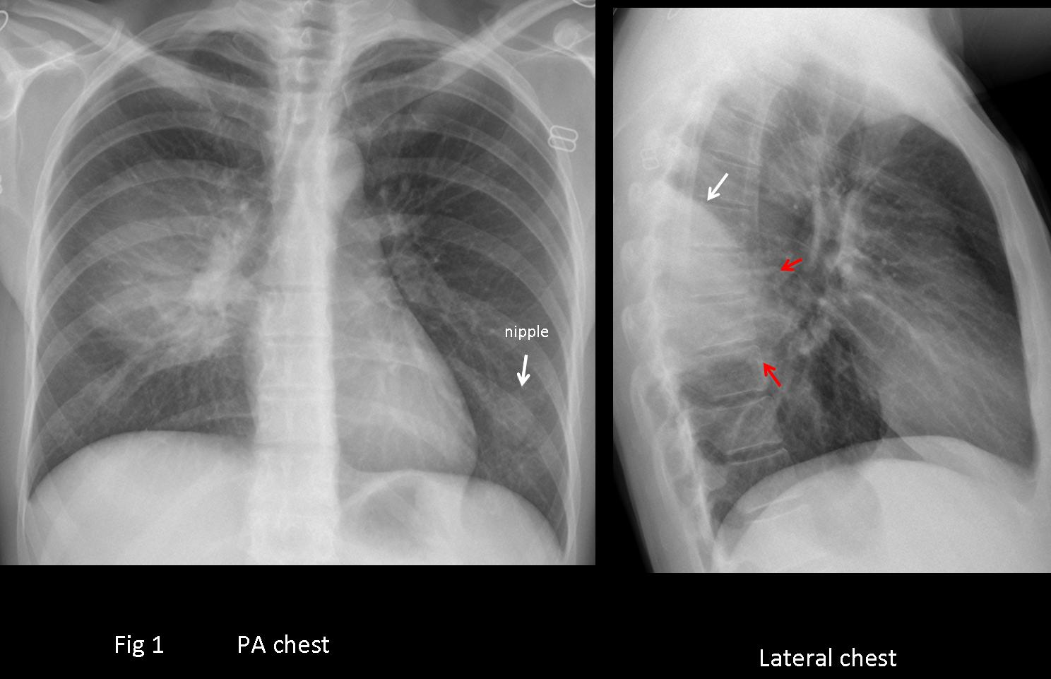
In case 31, the lateral view shows good definition of the lesion in the superior portion when it abuts the major fissure (arrow), whereas two thirds of the contour is ill-defined (red arrows).
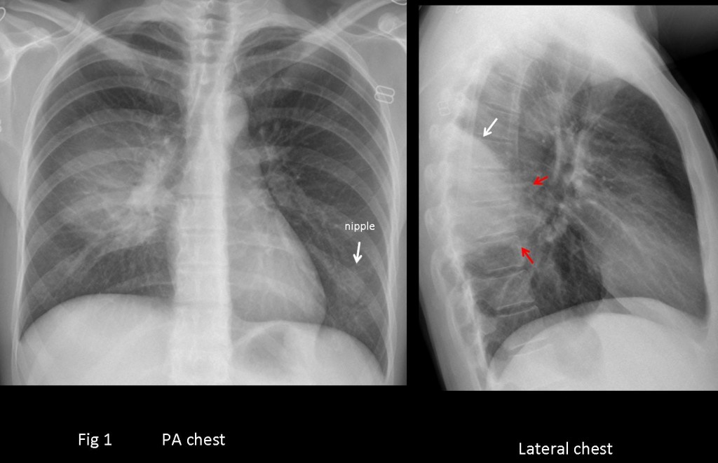
Fig. 1
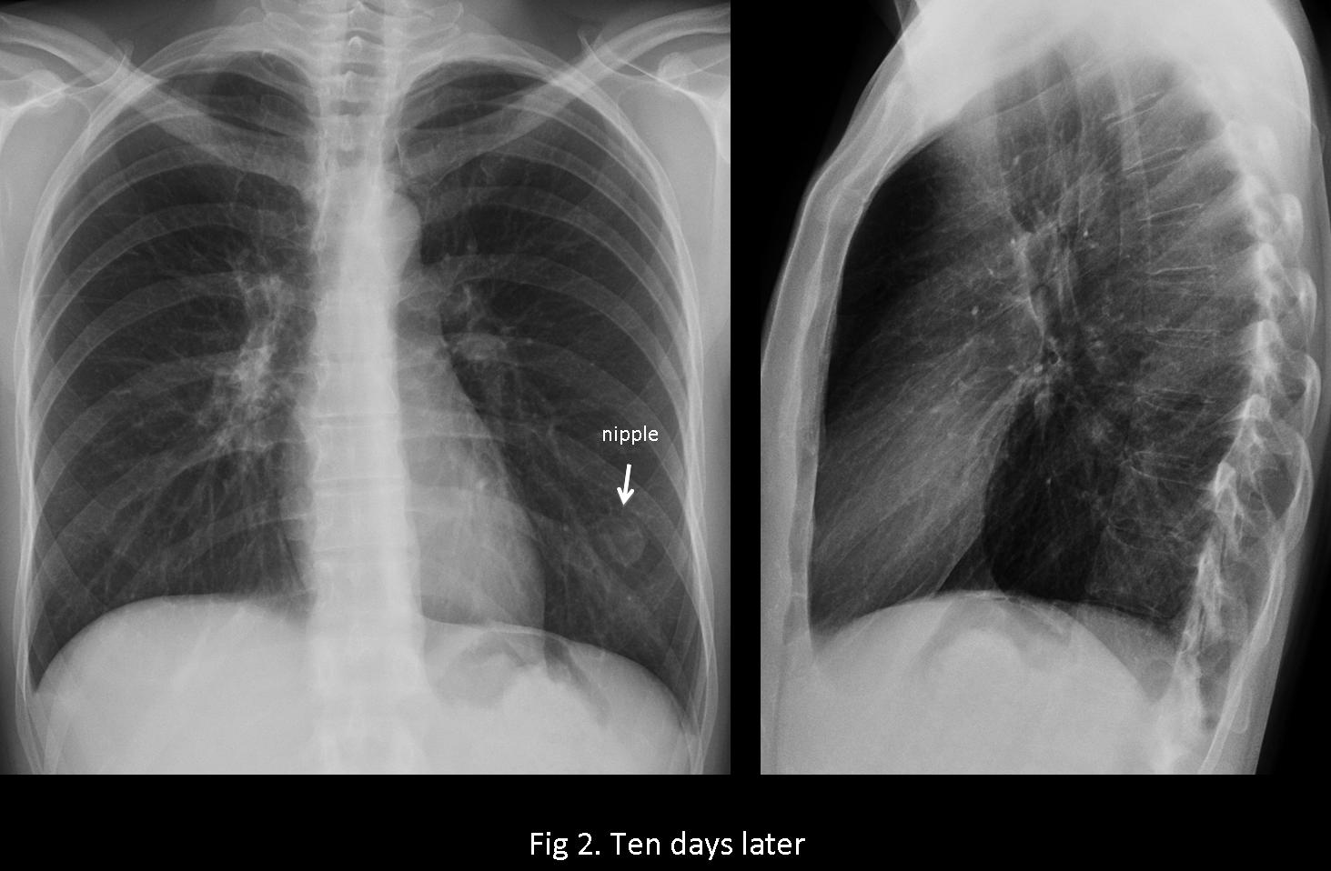
The lack of well-defined borders excludes an extrapulmonary lesion (answer 2 and 3 are out) and places the lesion within the lung. Although a pulmonary mass cannot be ruled out, the symptoms point to a round pneumonia. Correct management is to treat the patient and repeat the examination. It is unnecessary to do a CT. In this case, chest radiographs after ten days show complete resolution of the pneumonia (fig 2). Final diagnosis: round pneumonia in superior segment of right lower lobe.
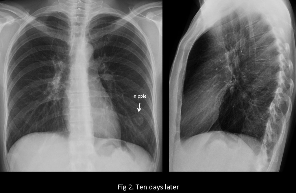
Fig.
Teaching point: when discovering a lesion in a chest radiograph, our first consideration should be to define where such a lesion is located: mediastinum, lung, or pleura/chest wall. Placing the lesion in the wrong compartment will lead to an erroneous diagnosis.