This is just the first of many weekly cases I’m going to show here on the ESR blog, to help you prepare for the European Diploma in Radiology. They are all typical of what you might expect to find in the exam and I will guide you through each one and remind you of the key points to consider.
To start off, I’m going to follow in the footsteps of my good friend Jose Caceres (although I will be more compassionate), and show you a chest case. If you have any comments or questions, please leave them in the comments section under this post. The answer will be posted on Thursday.
1. Myeloma/metastases
2. Fibrous tumour of pleura
3. Extrapleural fat
4. All of the above
Findings:
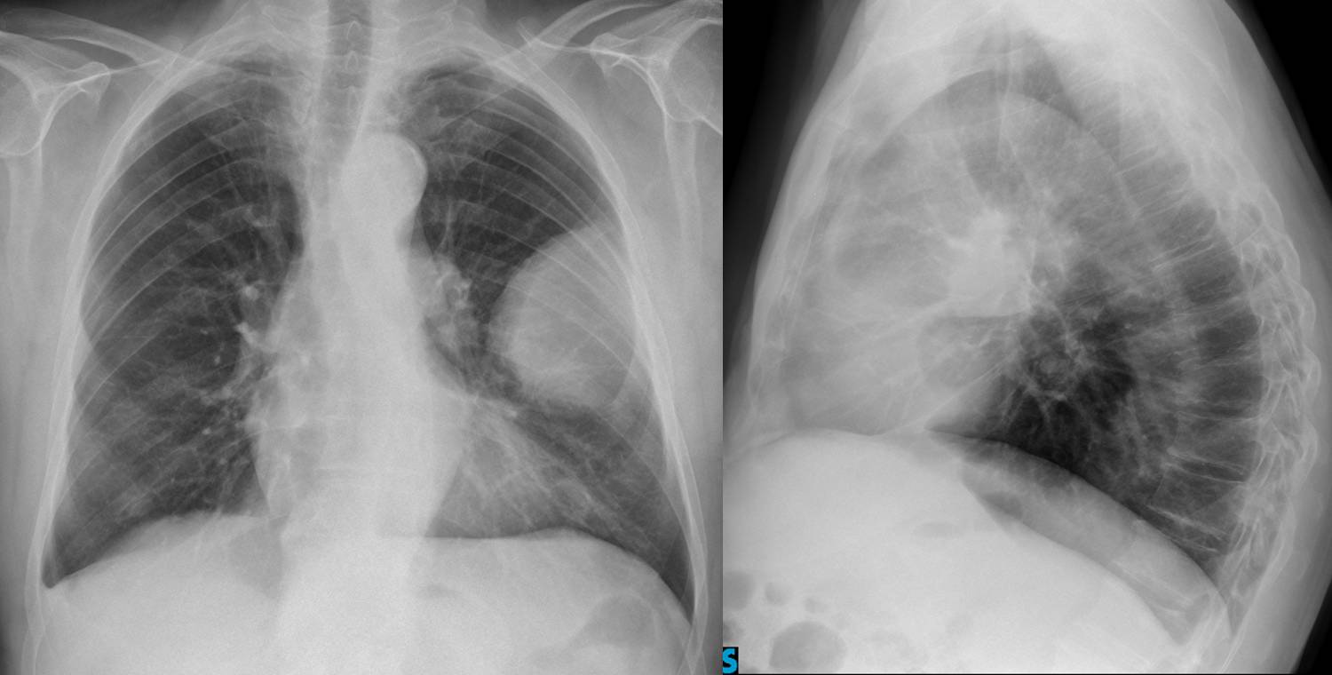
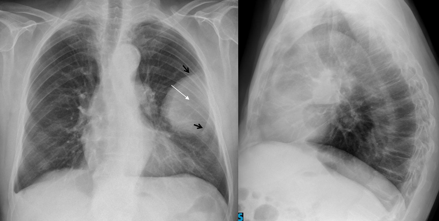
There is an extrapulmonary mass in the left hemithorax with destruction of the anterior aspect of the 4th rib, which is not visible (white arrow), whereas the anterior 3rd and 5th ribs are easily visible (black arrows).
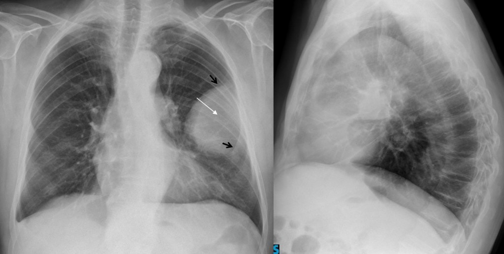
Fig 2
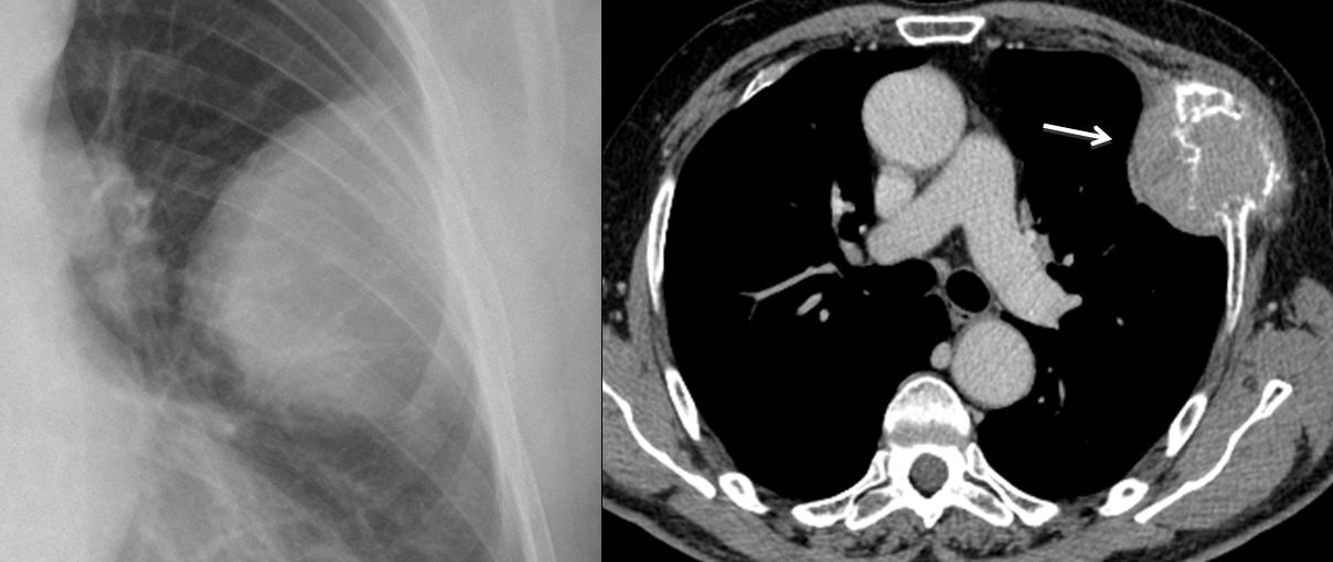
Given that the rib is destroyed, the most likely diagnosis is a malignant costal lesion, either a metastasis or myeloma, neither of which can be distinguished by imaging. CT confirms the rib destruction and the extrapulmonary mass (arrow).
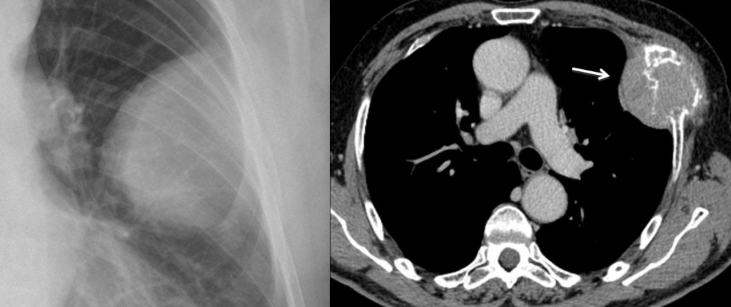
Fig 3
Final diagnosis: metastasis from hepatocarcinoma.
Extrapulmonary masses are easily identified by their typical appearance: location in the periphery of the lung, well-defined outline, and obtuse angles at the corners (pregnancy sign).
The differential diagnosis is simple: they may arise from a rib (metastasis, myeloma), chest wall (extrapleural fat) or pleura (pleural tumour, loculated fluid). Involvement of a rib is usually visible in the chest radiograph and narrows down the diagnosis to a malignant rib lesion. If no rib lesion is visible, the lesion can be of soft tissue density (tumour), water density (loculated fluid) or fat density (extrapleural fat). Differentiation between densities can only be made by CT.
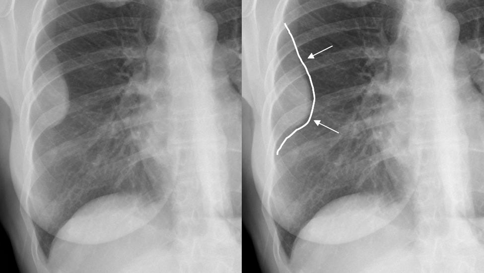
Fig. 4
1. Fat Density
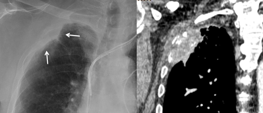
In this 57-year-old man we see carcinoma of the lung and left extrapleural lesion (arrow).
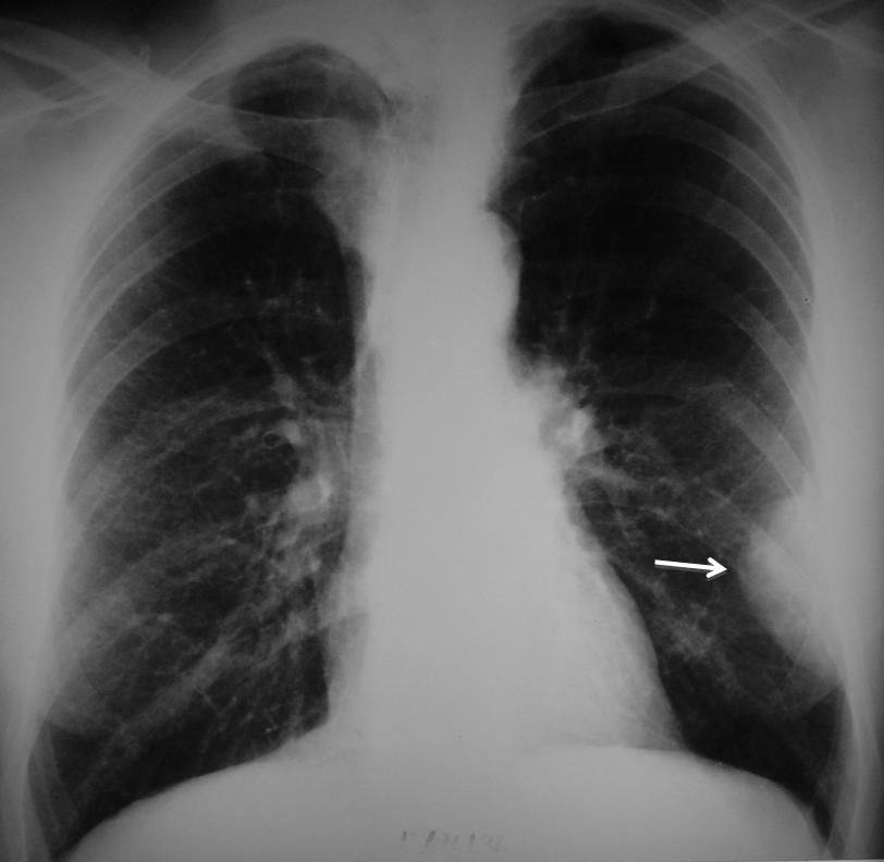
Fig 5
CT demonstrates that the lesion corresponds to extrapleural fat and is, therefore, innocuous.
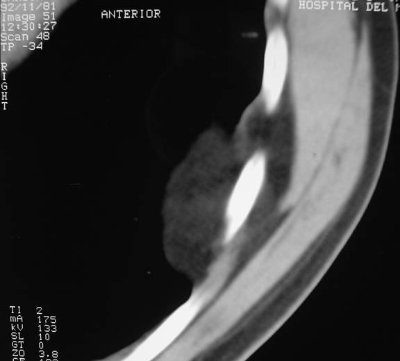
Fig 6
2. Water Density
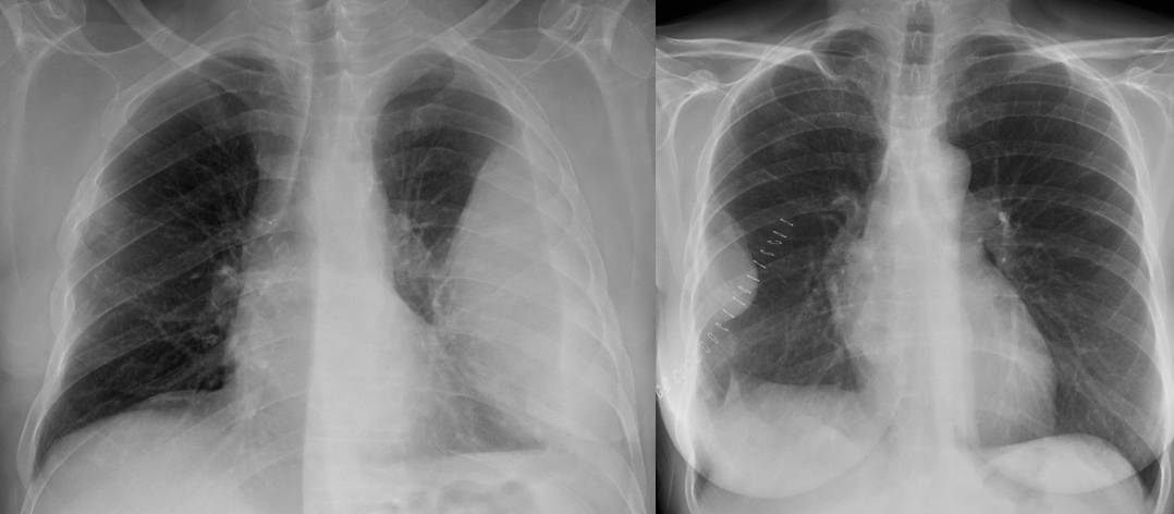
Two patients with loculated fluid and typical ‘pregnancy sign’ in the chest radiograph.
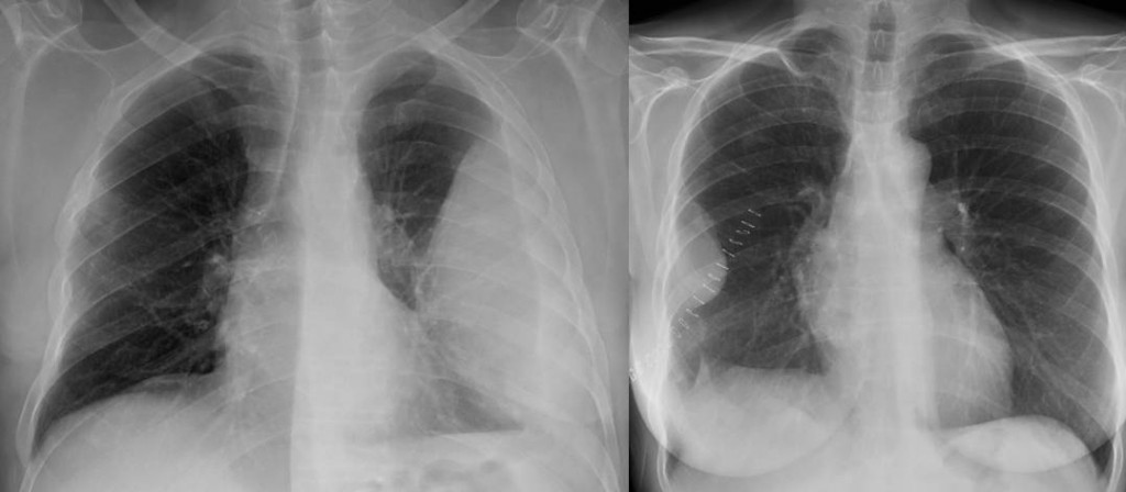
Fig 7
CT in one of them shows that the lesion is of water density, confirming the diagnosis.
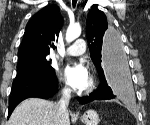
Fig 8
Note that both patients show blunting of the costophrenic angle, which always accompanies loculated fluid.
3. Soft Tissue Density
The radiograph shows an extrapulmonary lesion (arrow). CT shows a soft-tissue density, which likely corresponds to a fibrous tumour of pleura. An alternative would be pleural metastasis.

Fig. 9
Pitfalls
A lesion with indistinct borders, like the one below, shouldn’t be mistaken for an extrapleural lesion. The ragged border in this case indicates a peripheral lung carcinoma invading the chest wall or, less commonly, a chest wall lesion invading the lung.
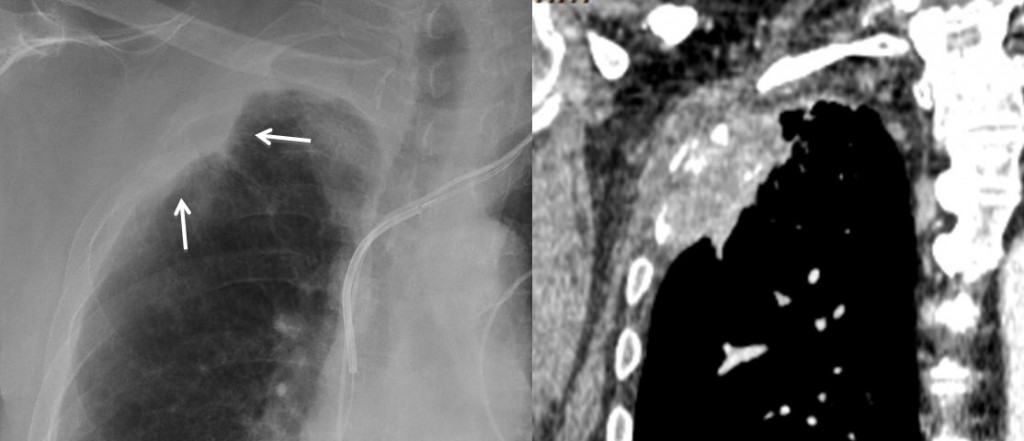
Fig 10
Follow Dr. Pepe’s advice:
- Extrapulmonary lesions are recognised in the chest radiograph by the so-called ‘pregnancy sign’.
- Always look for involvement of the underlying rib (metastases, myeloma).
- All others need a CT.
Suggested reading: chapter in chest wall lesions. JC Reed, Chest radiology. Patterns and differential diagnosis.