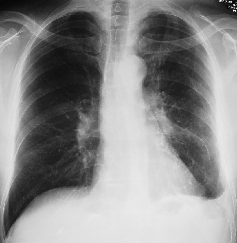
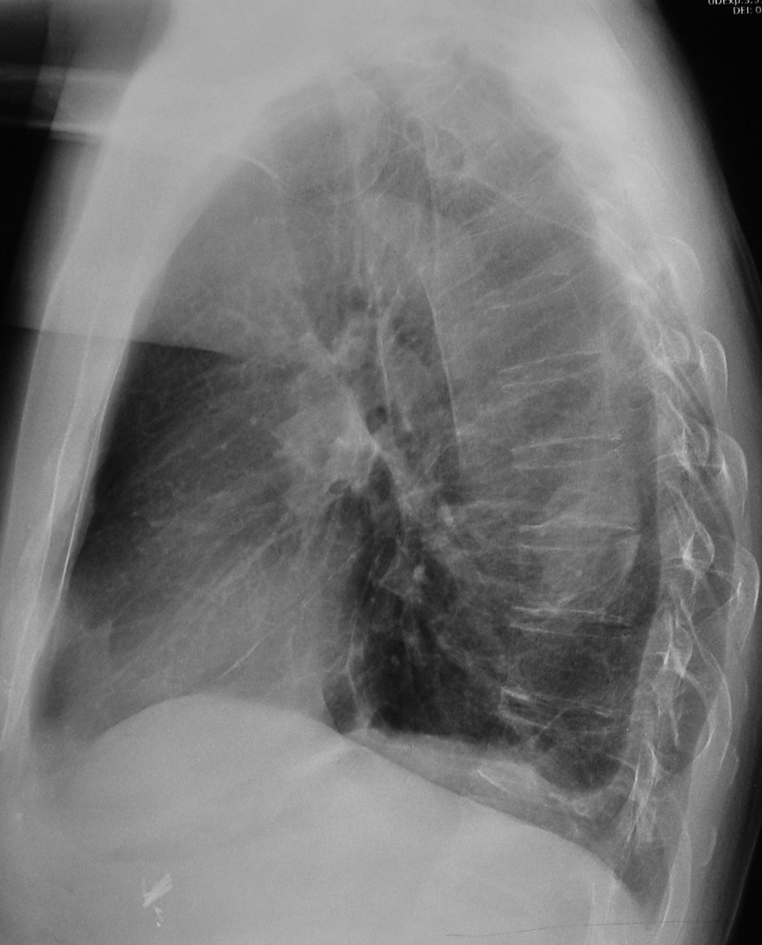
Muppet is so happy to have reached our 50th case, that he wants everybody to get the right answer. Here we have radiographs of a 52-year-old man with mild fever, blood-tinged sputum and left chest pain.
1. Carcinoma of the lung
2. Tuberculosis
3. Pulmonary embolism
4. None of the above
Findings: PA chest radiograph shows blunting of left costophrenic angle. There is medial displacement of left major fissure (arrows), indicating LLL volume loss. The left hilum is denser than the right one, suggesting a hilar mass. However, the left hilum is normal on the lateral view (
red arrow).
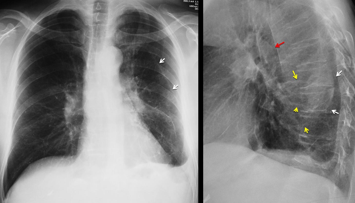
Its apparent increased density is due to a posterior ill-defined opacity (arrows), which is superimposed in the PA view and gives the false impression of increased hilar opacity. The clue to the diagnosis is identifying curving vessels (yellow arrows) leading to the posterior opacity, which should suggest rounded atelectasis. Axial and sagittal CT confirms the diagnosis by showing the atelectasis (A) the curving vessels and the thickened pleura.
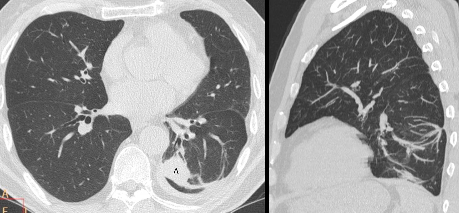
Final diagnosis: rounded atelectasis of LLL.
Congratulations to Dr. Strangelove, who was the first to mention the diagnosis (even though he called Muppet tricky).
This case was chosen as homage to Muppet’s first case, shown here on September 9, 2011. I quote:
“When we put together a peripheral rounded lesion, loss of volume and pleural thickening, rounded atelectasis immediately comes to mind and it is the correct diagnosis in this case. The typical findings of this condition are confirmed with CT.”
Teaching point: rounded atelectasis are not uncommon. Remember the four findings which suggest it: peripheral rounded opacity, loss of volume, curving vessels and pleural thickening.
Quoting again from case #1:
“Rounded atelectasis anchors in thickened pleura. Do not suggest the diagnosis if the pleura looks normal.”