
Dear Friends,
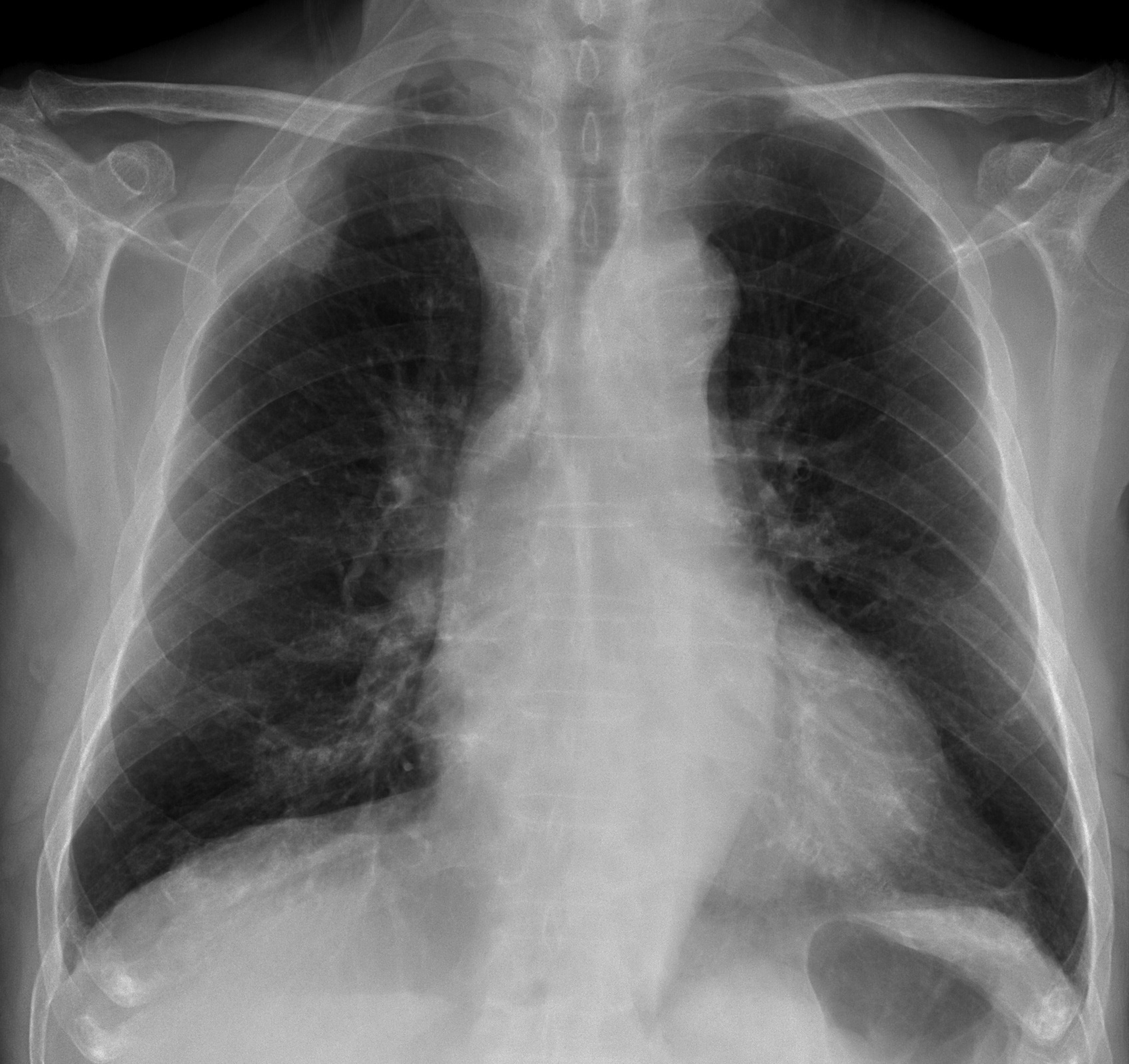
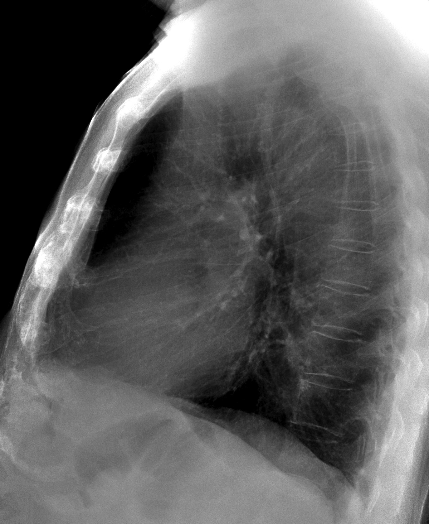
Muppet is traveling abroad on a dangerous mission. If he doesn’t make it back, he hopes you will remember him fondly. For this reason he is showing a clear-cut case of pre-op radiographs in a 81-year-old patient with prostate carcinoma and right shoulder pain. Diagnosis?
1. Lung carcinoma
2. Fibrous tumour of pleura
3. Chest wall metastases
4. None of the above
Click here for the answer to case #53
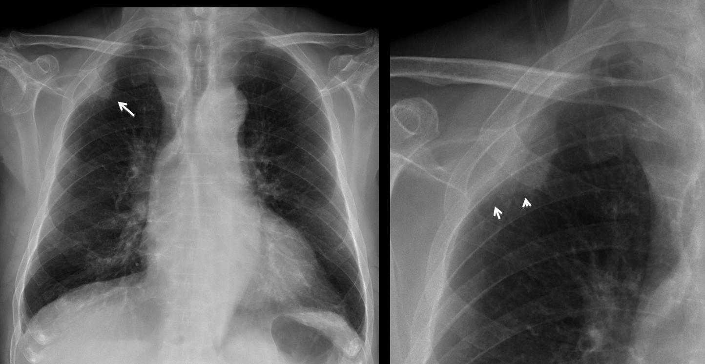
Findings: PA chest radiographs show a peripheral lesion (arrow), better seen in the cone down view. Although the upper limit has an obtuse angle, the border is not sharp and is clearly indistinct in the lower aspect (arrows). These findings are typical of a pulmonary process and exclude an extrapulmonary lesion (options 2 and 3).
CT confirms the pulmonary location of the lesion and the fuzzy border.
Four months later there is obvious invasion on the underlying rib (arrow).
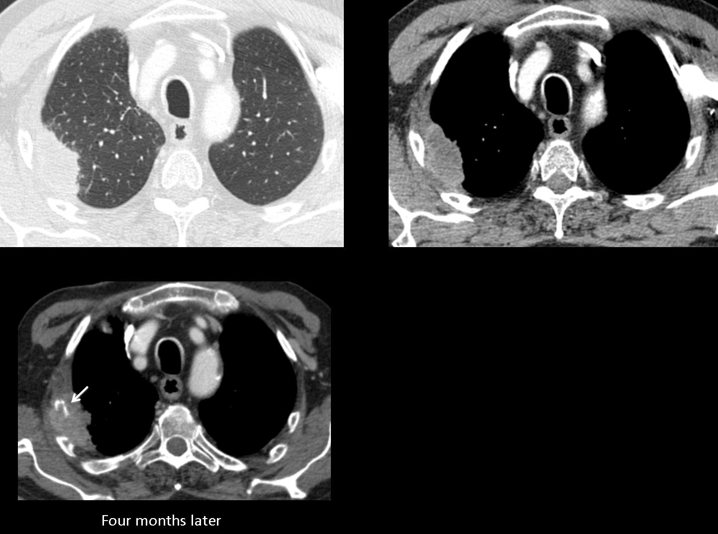
Final diagnosis: peripheral lung carcinoma
Teaching point: extrapulmonary lesions have obtuse angles at the borders and are sharply defined. If one of these characteristics is missing, we should suspect a peripheral pulmonary process (see discussion of case no. 1)


