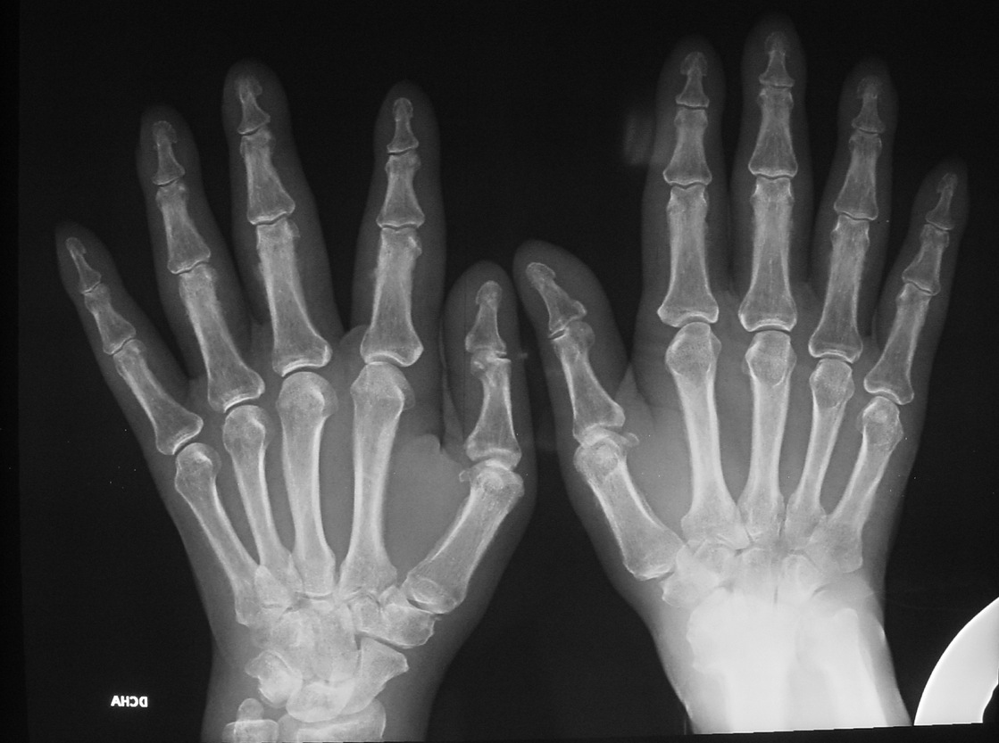
Muppet is running out of chest cases and wishes to show a bone case instead. Presenting an AP radiograph of both hands of a 59-year-old male with pain in the joints.
1. Rheumatoid arthritis
2. Psoriatic arthritis
3. Hyperparathyroidism
4. None of the above
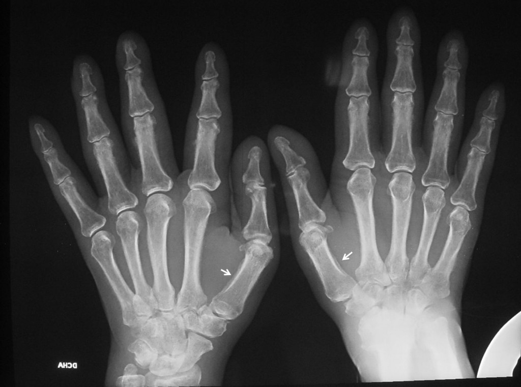
Fig. 1
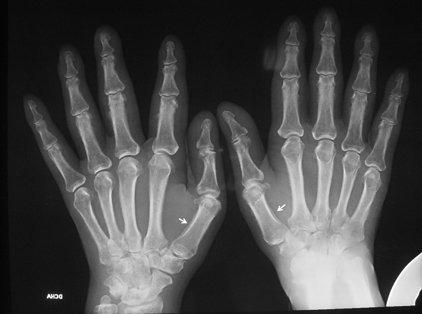
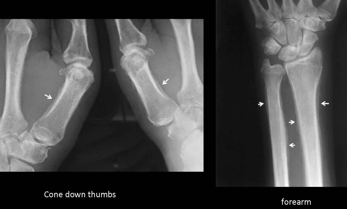
Findings: the AP radiograph of both hands shows periostitis in the proximal phalanges, more obviously in the first metacarpals (arrows), and better seen in the cone down view.
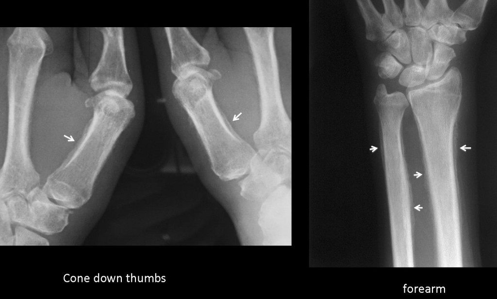
Fig. 2
There is also extensive periostitis in the bones of the forearm (arrows). There are not clear signs of arthritis.
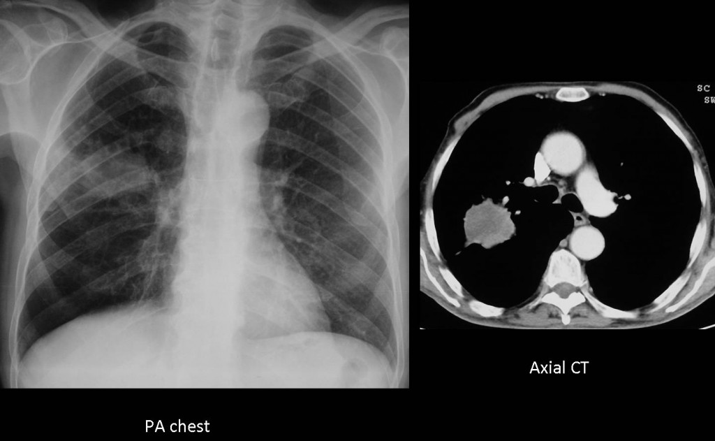
Fig. 3
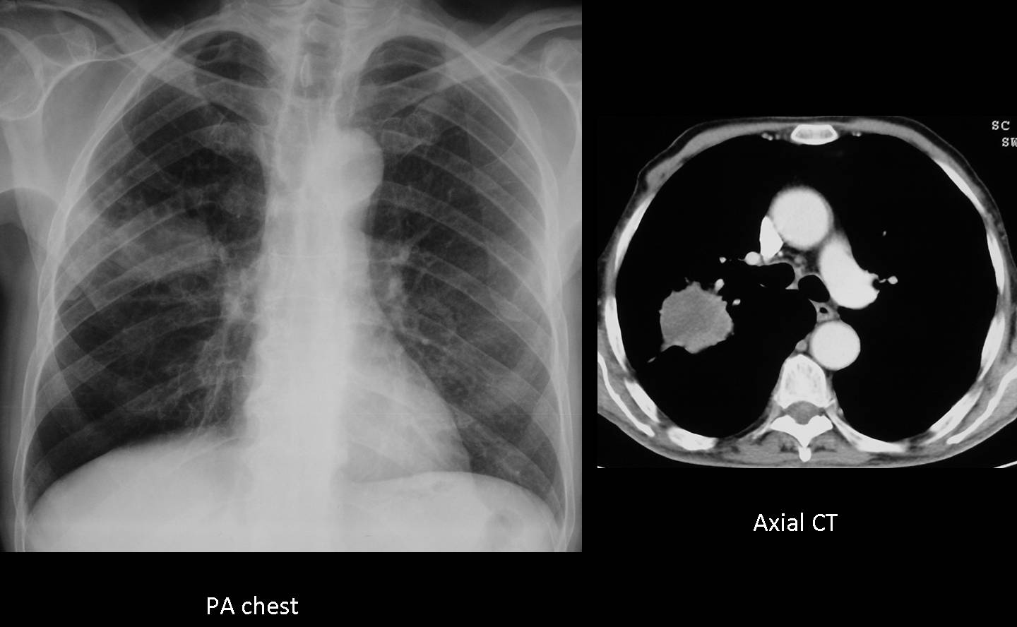
Differential diagnosis includes disease with widespread periostitis (pachydermoperiostosis, thyroid achropachy, vitamin A intoxication, etc.). In an adult, hypertrophic pulmonary osteoarthropathy should be considered and a chest radiograph taken. In this patient, chest film showed a nodular opacity in the right lung, confirmed with CT.
Final diagnosis: hypertrophic pulmonary osteoarthropathy secondary to carcinoma of the lung.
Congratulations to Drs. Yp and Marcolino who made back-to-back diagnosis in an interval of two hours.
Teaching point: sometimes the initial symptoms of a lung carcinoma may be extra-thoracic (bone or brain metastases, hypertrophic osteoarthropathy) and it is important to know them to suspect the correct diagnosis.
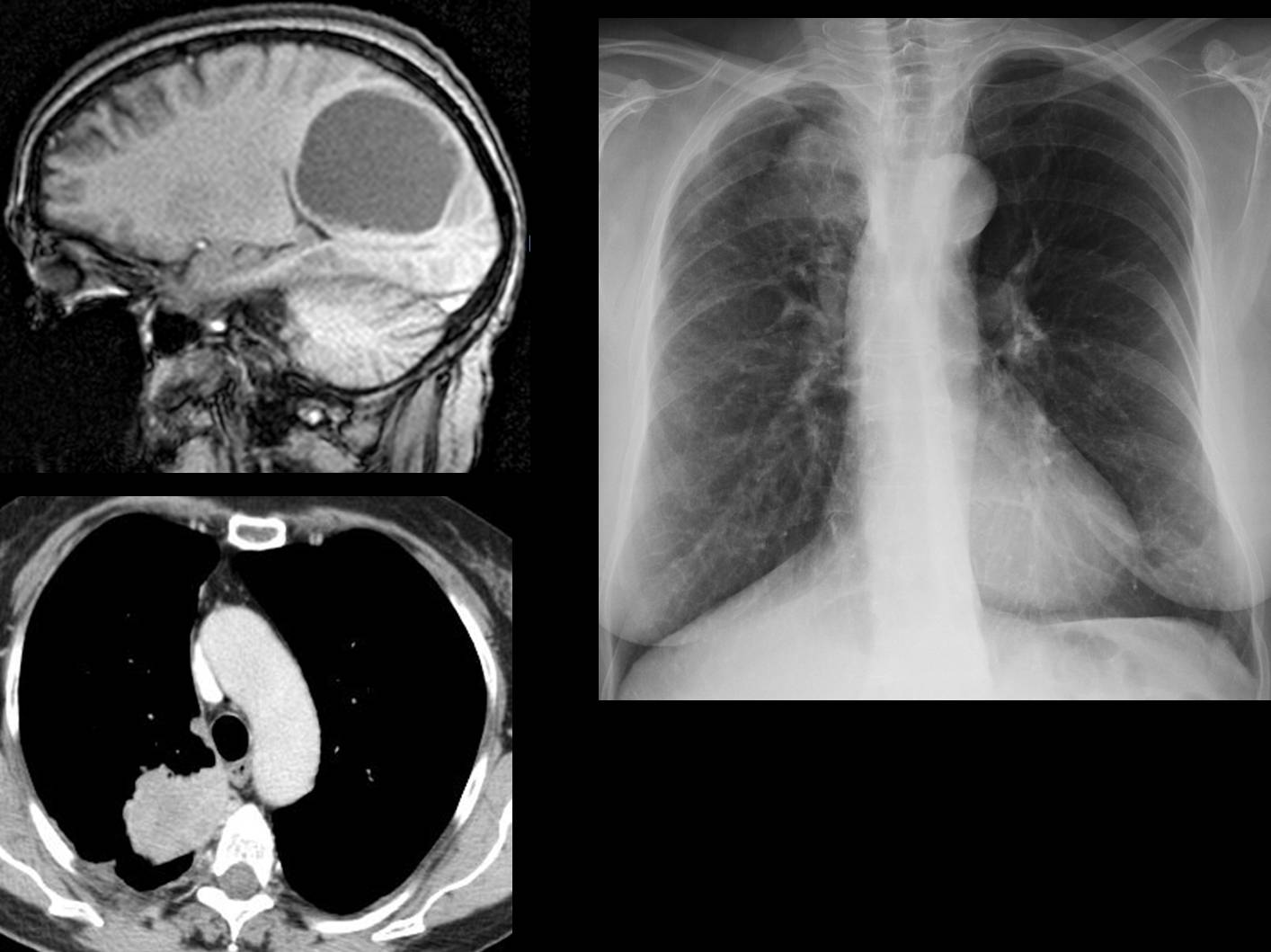
Below is a recent case (22 March), erroneously diagnosed as brain tumour, in which the primary lung tumour was discovered in the pre-op chest film.
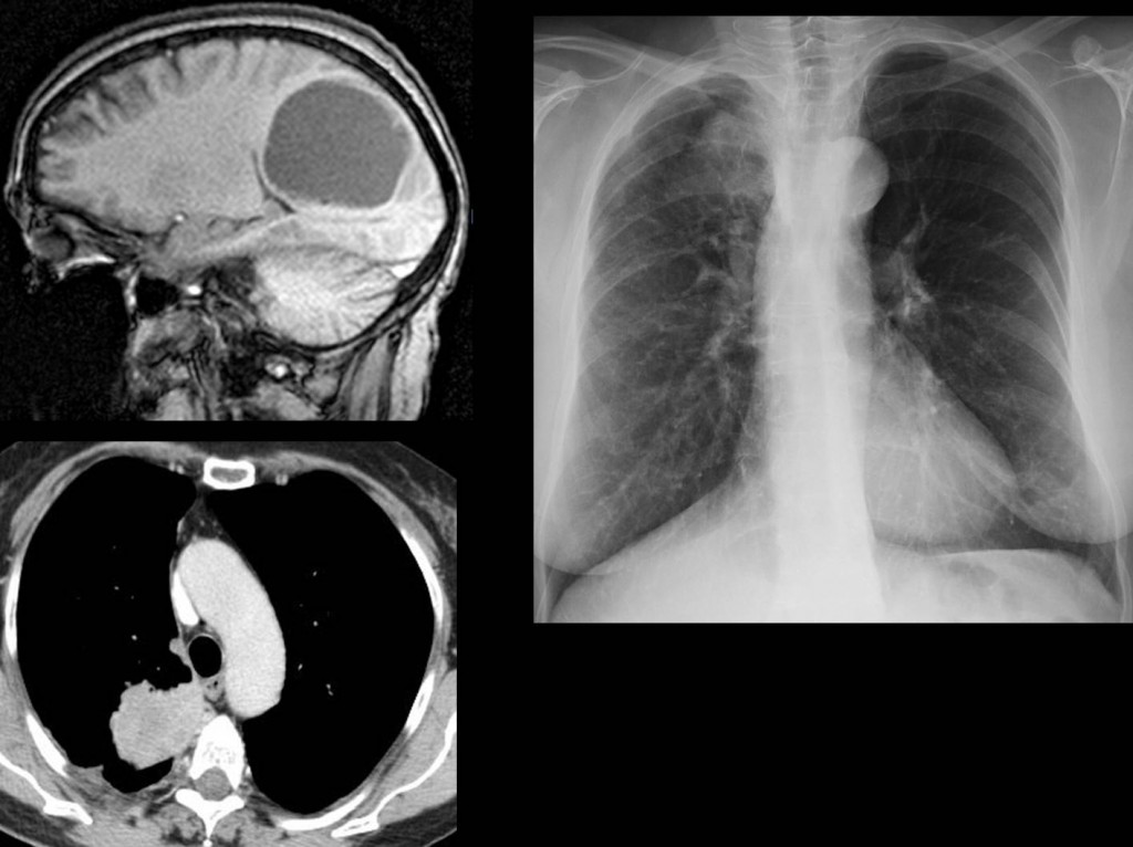
Fig. 4