Dr. Pepe’s Diploma Casebook: Case 110 – The Wisdom of Dr. Pepe (Chapter 3) – SOLVED!

Dear Friends,
To continue with the third chapter of The Wisdom of Dr. Pepe, I am showing radiographs of an asymptomatic 52-year-old man with previous history of asbestos exposure.
Check the images below, leave your thoughts in the comments section, and come back on Friday for the answer.
Diagnosis:
1. Fibrous tumour of pleura
2. Large pleural plaque
3. Pleural fat
4. Any of the above
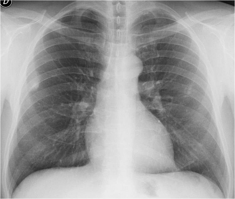
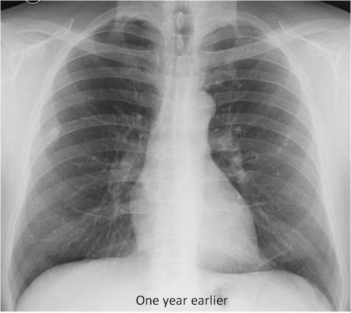
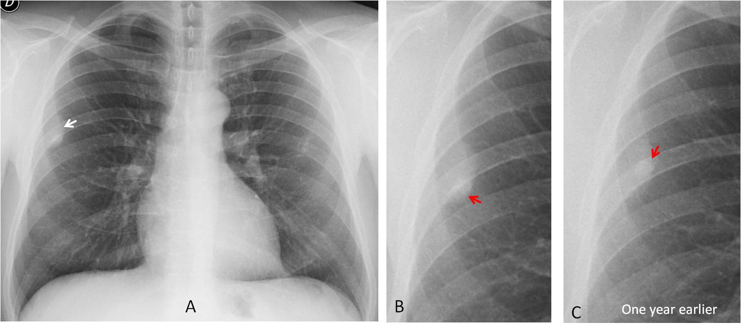
Findings: PA radiograph shows an extrapulmonary lesion in the right hemithorax (A, white arrow). There is a calcification in the lesion that appears to shift position in the comparison with a previous film (B and C, red arrows).
Enhanced axial CT shows that the extrapulmonary lesion corresponds to pleural fat (D, white arrow) The calcification is a superimposed pleural plaque (E, red arrow). Coronal reconstruction shows overlapping of fat and plaque (F, white and red arrows). In addition, there are other smaller plaques (D and E, yellow arrows).
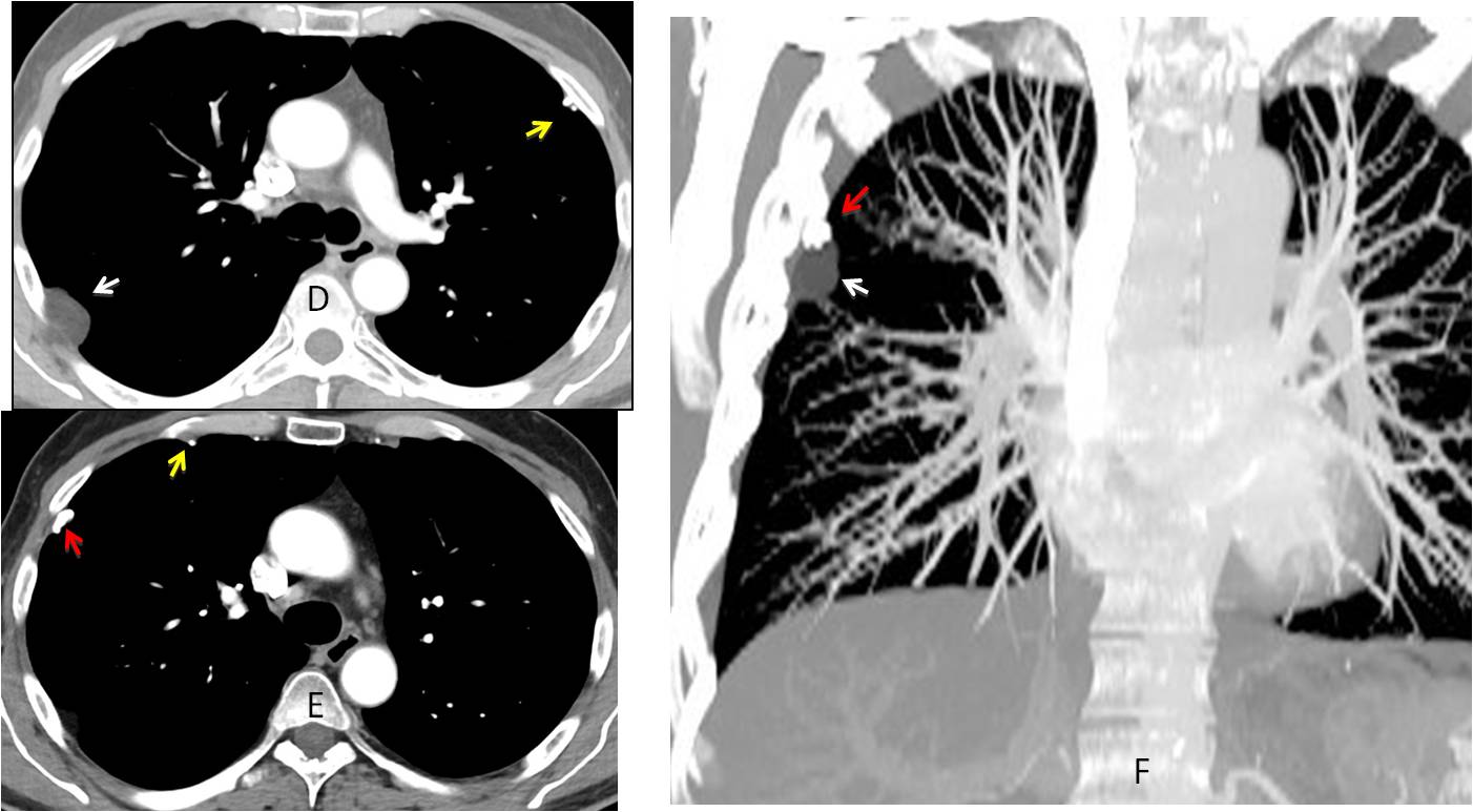
Diagnosis: pleural fat and superimposed pleural plaque
In the third chapter of The Wisdom of Dr. Pepe I intend to discuss previous studies (ps), the third component of the equation:
D = (bc + lv + ps + chl) e
Previous studies are crucial to determining whether or not a lesion was present and, if so, whether it has changed and the timing of the change. To my mind, this is one of the most important parts of the equation.
In the comparison with previous examinations there are four possible scenarios:
1. Abnormality was not present
2. Abnormality was present and is unchanged
3. Abnormality has regressed
4. Abnormality has progressed
If the abnormality was not present and previous films were not taken far in the past, we know that the condition is relatively recent (Fig. 1). Comparison with the previous normal appearance also provides the opportunity to discover subtle findings (Fig. 2).
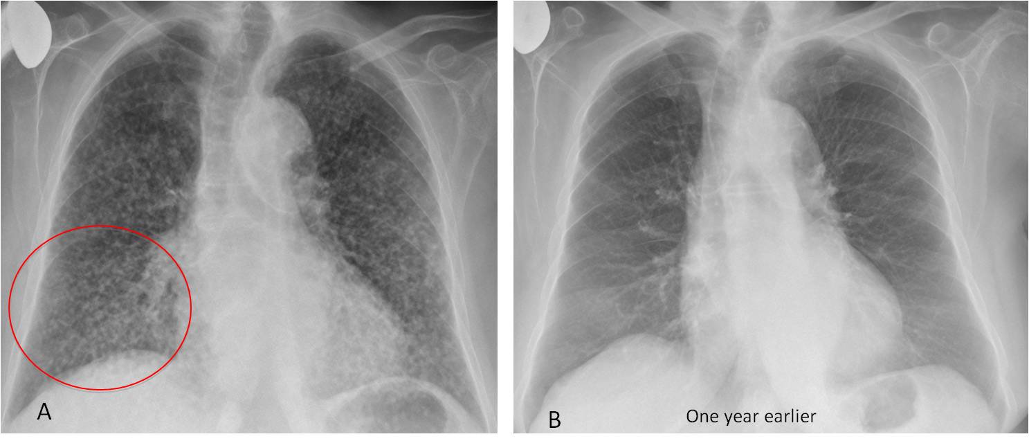
Fig. 1
Fig 1. 87-year-old woman with dyspnoea. Chest radiograph shows a diffuse micronodular pattern (A), not present one year earlier (B). This excludes chronic disease. The fact that the nodules are of different sizes (A, circle) suggests miliary metastasis or widespread adenocarcinoma.
Coronal and axial CT confirm the different sizes of the nodules (C and D, circles), and show a large nodule in the RLL (C, arrow) that was not visible in the chest radiograph. Diagnosis: widespread adenocarcinoma
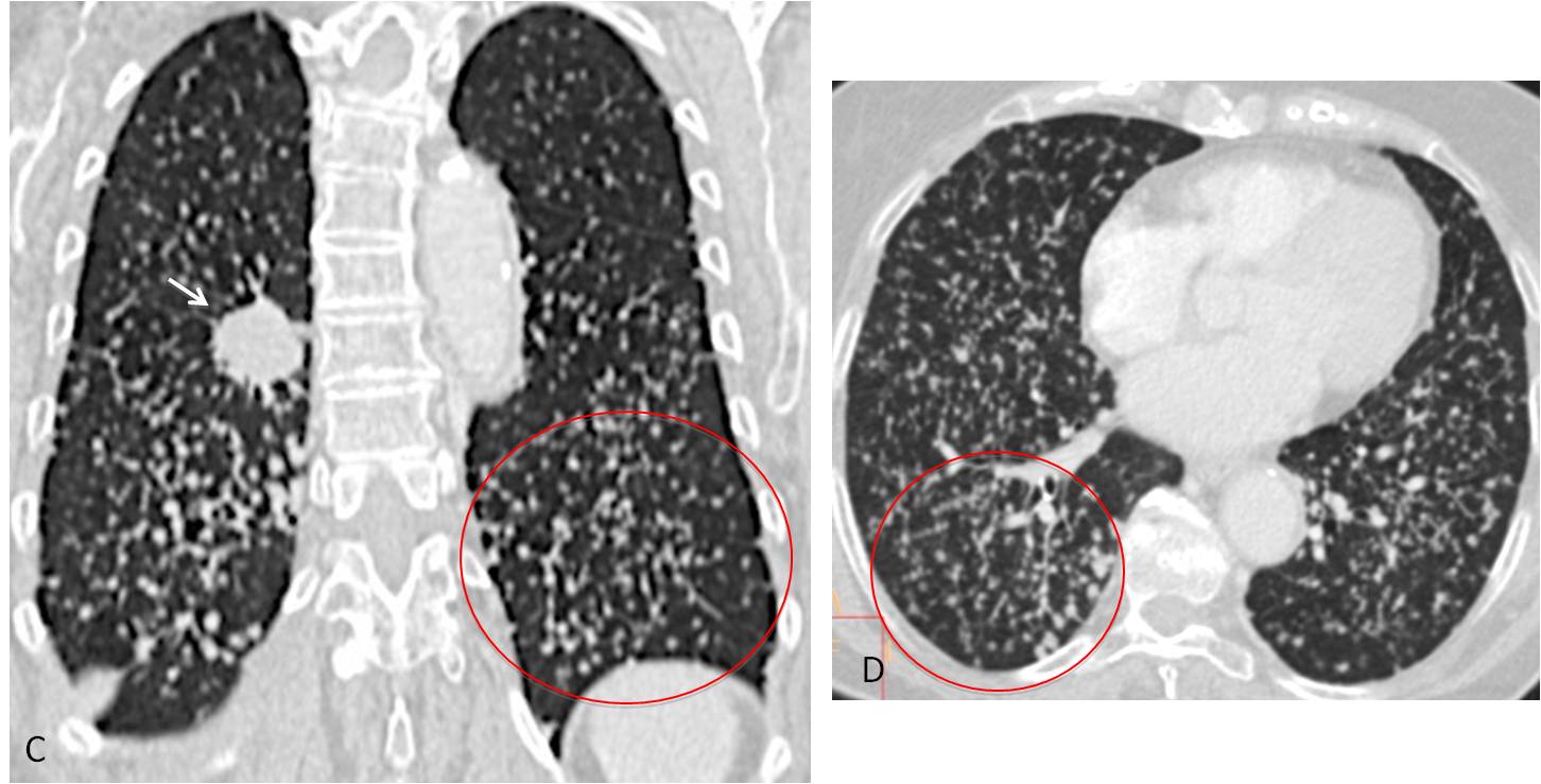
Fig. 1
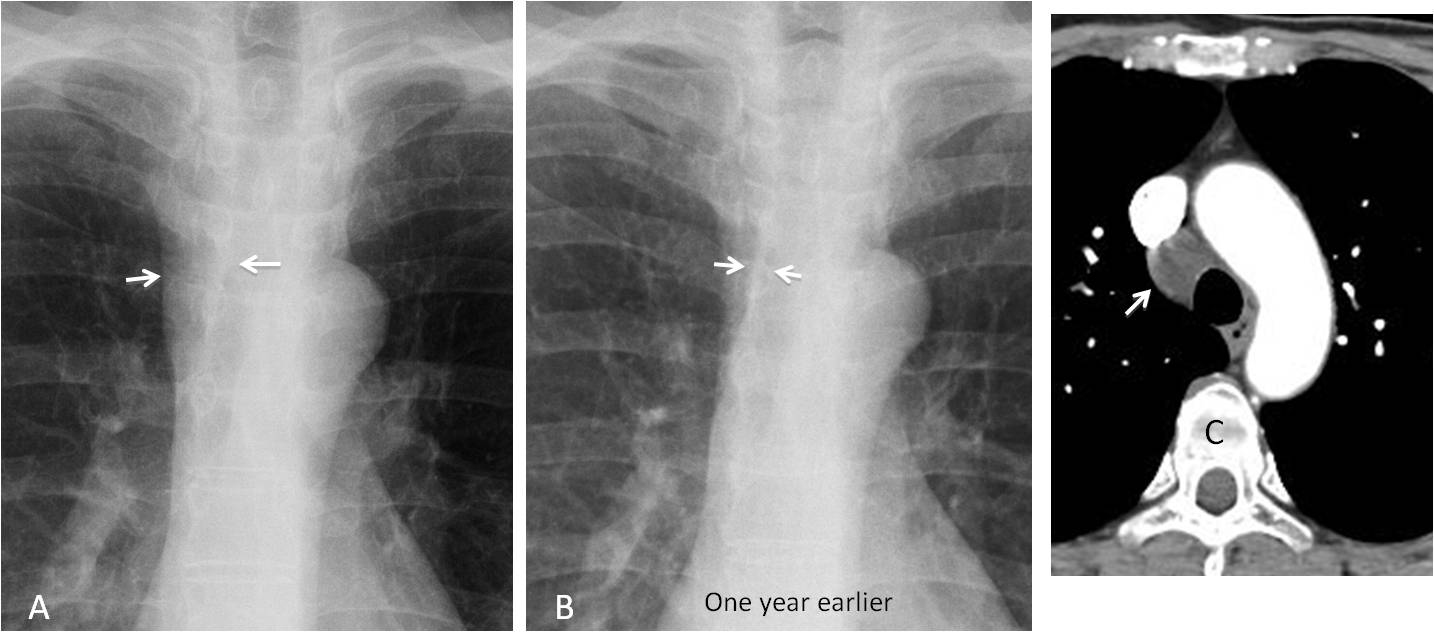
Fig. 2
Fig. 2. 53-year-old woman, surgically treated for breast carcinoma. PA radiograph shows obvious widening of the right paratracheal line (A, arrows), which is more evident than in the previous examination, one year earlier (B). Enhanced axial CT confirms the presence of a large right paratracheal lymph node (C, arrow). Diagnosis: sarcoidosis.
When the abnormality was present and is unchanged we are reassured, especially when the finding does not seem significant (Fig. 3). Of course, stability, although reassuring, does not mean that one should stop pursuing the correct diagnosis (Fig. 4).
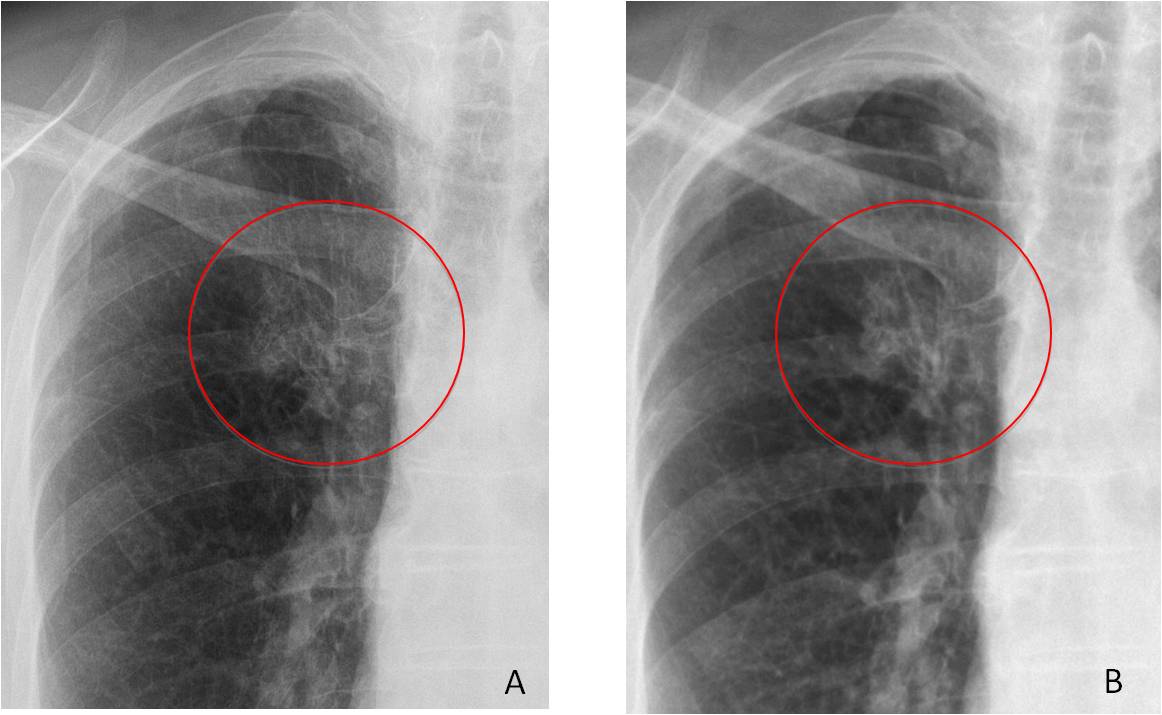
Fig. 3
Fig. 3. Preoperative film showing a poorly defined infraclavicular opacity (A, circle). PA chest film taken three years earlier shows that the opacity has not changed (B, circle). Diagnosis: exuberant calcification of the first costal cartilage
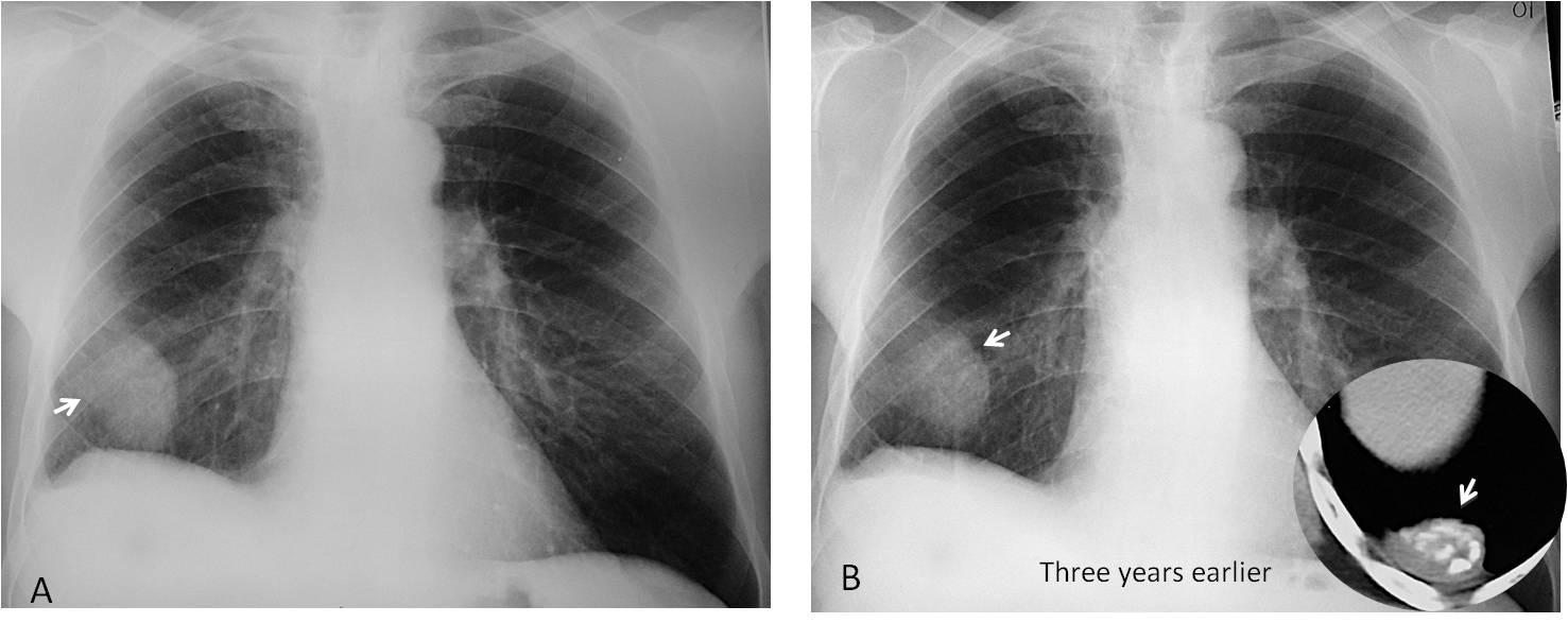
Fig. 4
Fig. 4. PA radiograph shows an extrapulmonary nodule with an incomplete border sign (A, arrow) and blunting of the costophrenic sinus, indicating a pleural lesion. The appearance is unchanged compared to that of a previous radiograph three years earlier (B, arrow). Axial CT shows a pleural mass with amorphous calcification (insert, arrow). Diagnosis: inflammatory pseudotumour of pleura.
Regression of the abnormality is synonymous with healing and virtually excludes malignancy. Regression usually indicates an inflammatory process (Figs. 5 and 6).
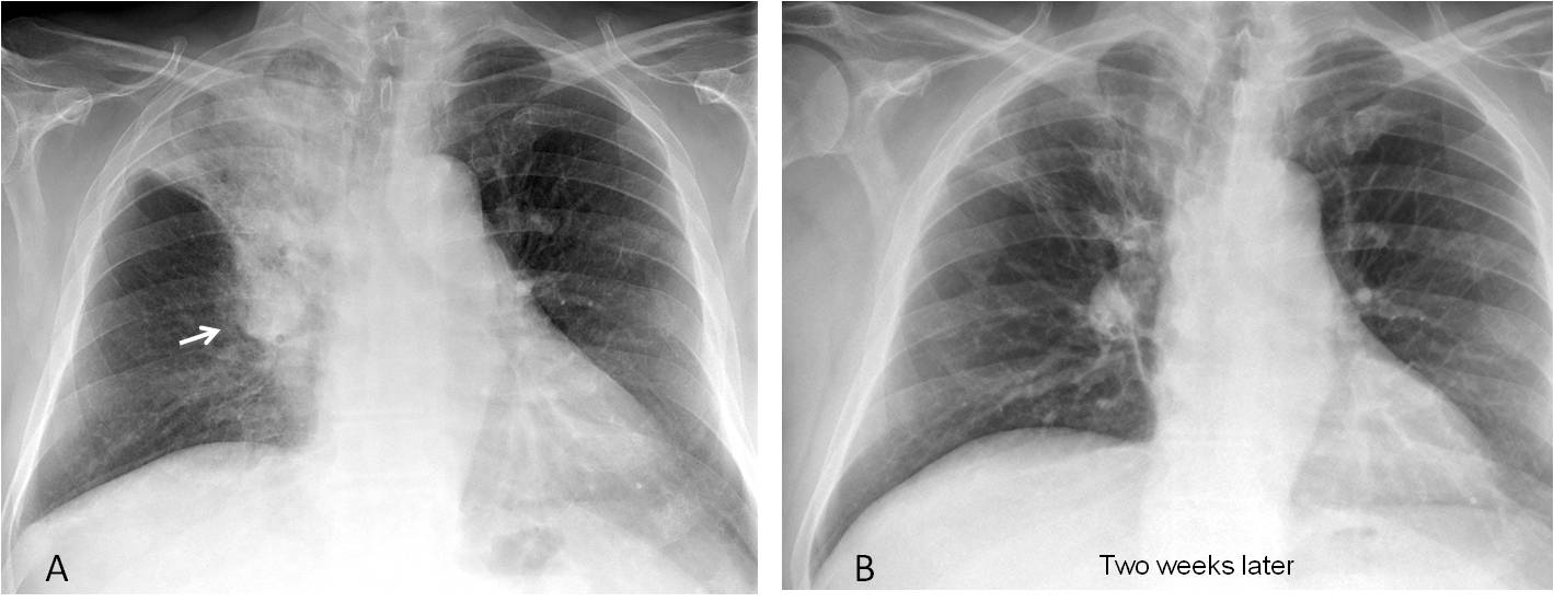
Fig. 5
Fig. 5. 63-year-old man with fever. PA chest film shows moderate RUL collapse with a Golden sign (A, arrow), highly suspicious of a central carcinoma. Two weeks later (B) the infiltrate has practically disappeared. Diagnosis: lobar pneumonia.
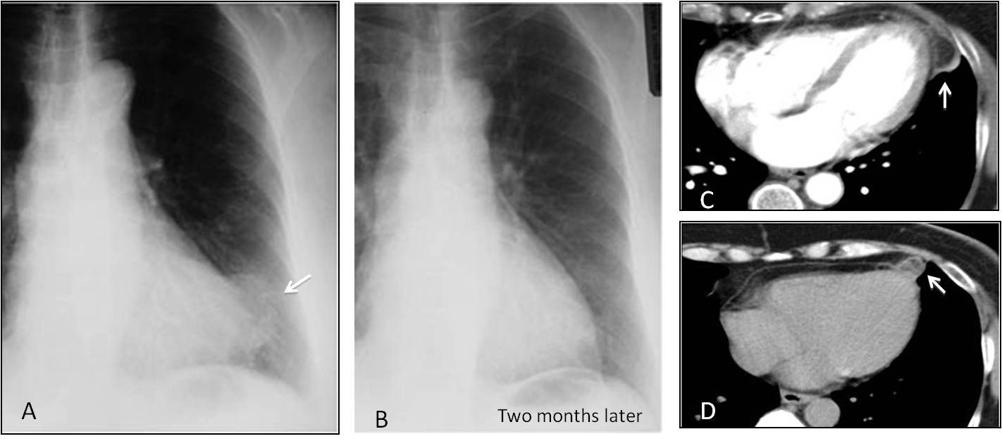
Fig. 6
Fig. 6. 45-year-old woman with acute chest pain. No fever. Chest radiograph shows a poorly defined paracardiac opacity (A, arrow) that has disappeared two months later (B). Initial CT shows encapsulated epipericardial fat (C, arrow), with a marked decrease in size at two months (D, arrow). Diagnosis: epipericardial fat necrosis.
Progression suggests an active process and indicates the need for further testing (Fig. 7). The speed of change is important because it helps to determine the aggressiveness of the abnormality (Figs. 8 and 9).
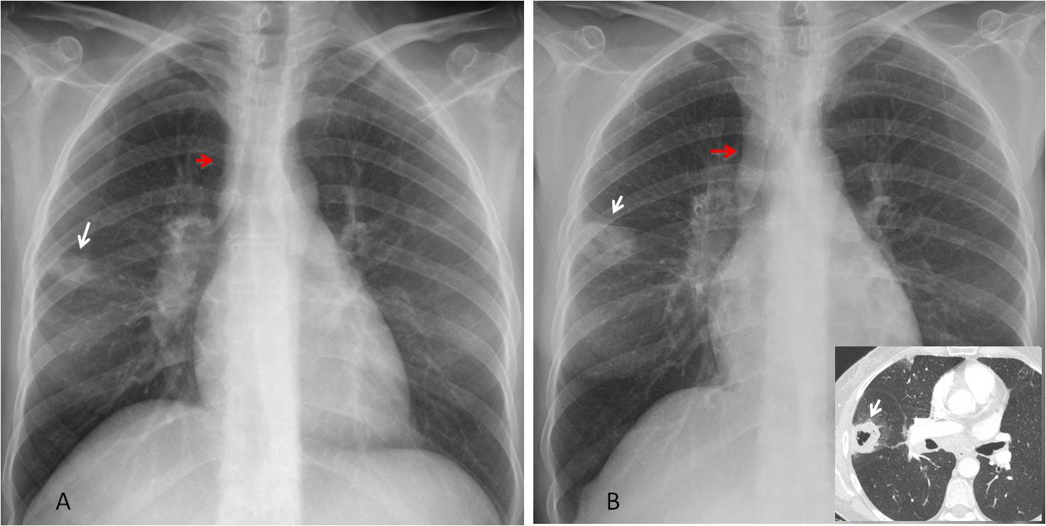
Fig. 7
Fig. 7. 45-year-old man with an initial diagnosis of pneumonia (A, white arrow). The widening of the paratracheal line was overlooked (A, red arrow). One month later the peripheral lesion has increased in size and cavitated (B, white arrow). The paratracheal line is now thicker (B, red arrow). CT shows a cavitated nodule (insert, arrow). Diagnosis: adenocarcinoma.
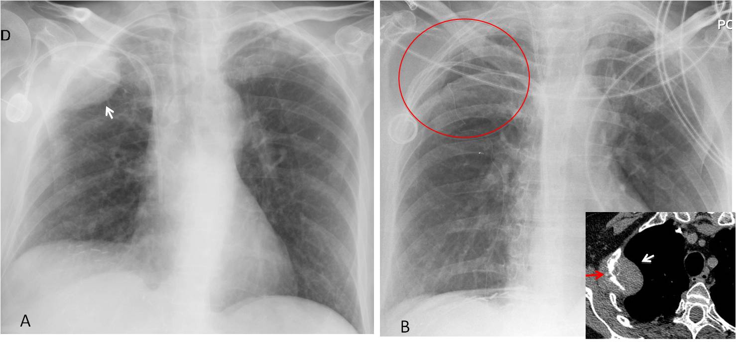
Fig. 8
Fig. 8. Extrapulmonary lesion in an oncologic patient (A, arrow), which was not present six months earlier (B, circle). The sudden appearance suggests metastasis. This was confirmed with CT (insert, white arrow), which also shows destruction of the underlying rib (insert, red arrow).
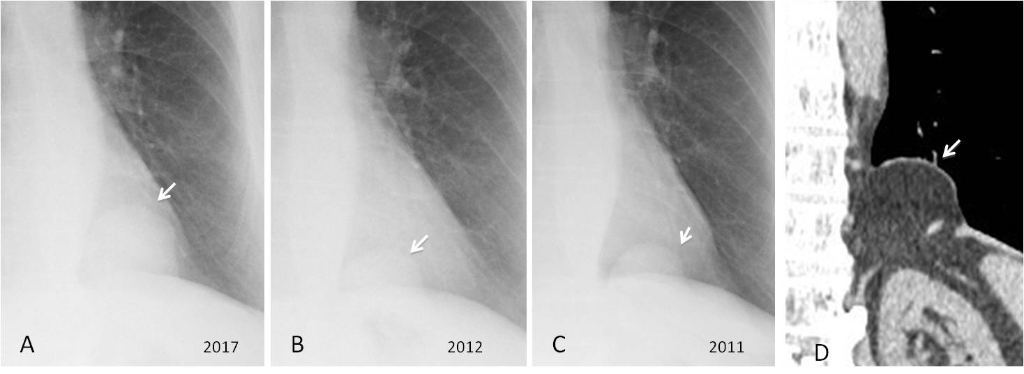
Fig. 9
Fig. 9. Extrapulmonary basal lesion (A, arrow), discovered in a preoperative film. Radiographs taken five and six years earlier show that the lesion was present, albeit smaller (B and C, arrows).
The location and progression of the lesion suggest a diaphragmatic hernia, confirmed with coronal CT (D, arrow).
The final vintage case is a practical example of how the speed of change helps in the diagnostic process (Fig. 10).
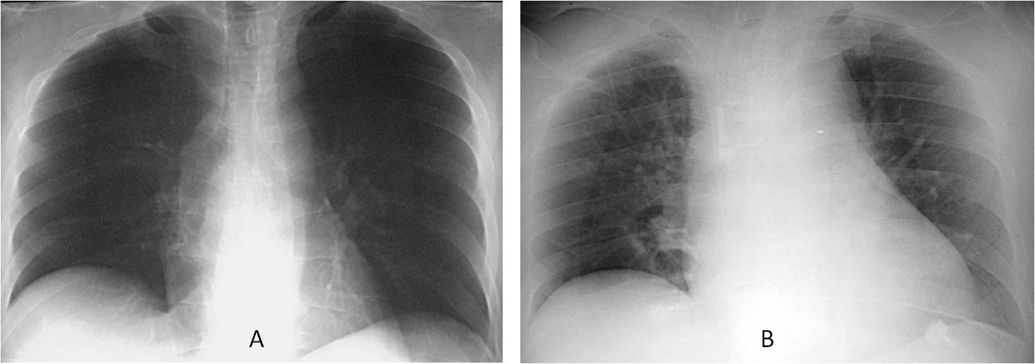
Fig. 10
Fig. 10. 52-year-old man with colon carcinoma. Preoperative film (A) shows a normal heart size. Two days post-op (B) there is marked enlargement of the cardiac silhouette, with no clinical signs of heart failure. The quick change in size of the heart raised the possibility of pericarditis. Pericardial tap returned clear fluid. Final diagnosis: iatrogenic pericarditis secondary to perforation of the pericardium by the central venous catheter.

Dr. Pepe’s third commandment:
Thou shalt always look at previous films:
- To determine whether there are any changes
- To evaluate the speed of change


