Dr. Pepe’s Diploma Casebook: Case 25 – SOLVED!

Dear Friends,
This week I’m presenting the case of a 9-year-old child with pain in the leg after trauma.
Diagnosis:
1. Aneurysmal bone cyst
2. Simple bone cyst
3. Giant-cell tumour
4. Osteosarcoma
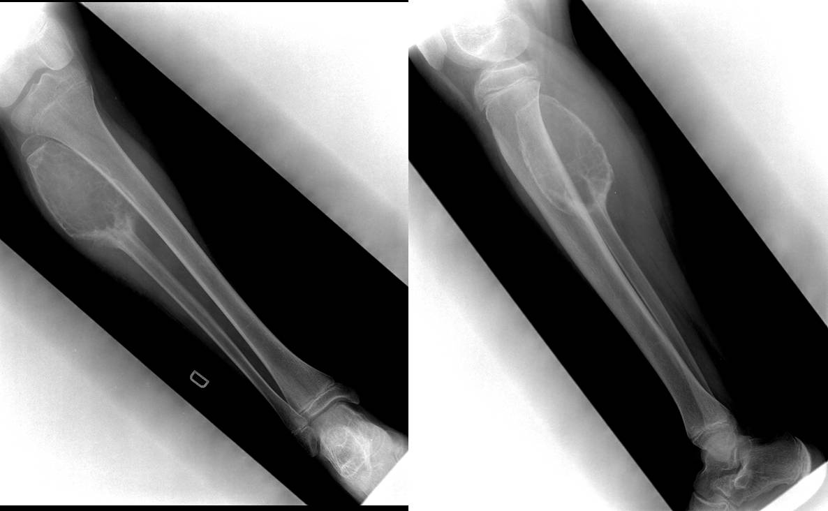
9-year-old child with pain in the leg after trauma
Findings: well-delimited, expansive intraosseous lytic lesion at the proximal peroneal diaphysis. Thinning of the periosteum and multiple internal septations (black arrows). Periosteal reaction at the distal end (white arrows).
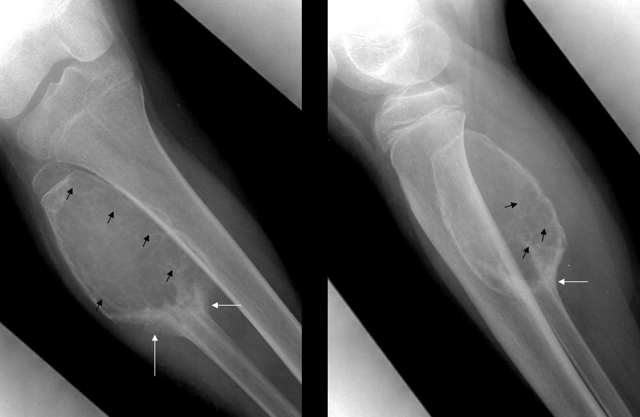
Fig. 1
Magnetic resonance shows the expansive intraosseous lesion with multiple small cavities (Fig. 2, black arrows). There is a larger cavity (Fig. 2, asterisk) with a fluid-fluid level (Fig. 3&4 red arrows), cortical thinning, and periosteal reaction (Fig. 4, yellow arrows). No soft tissue lesions are seen.
Diagnosis: Aneurysmal bone cyst.
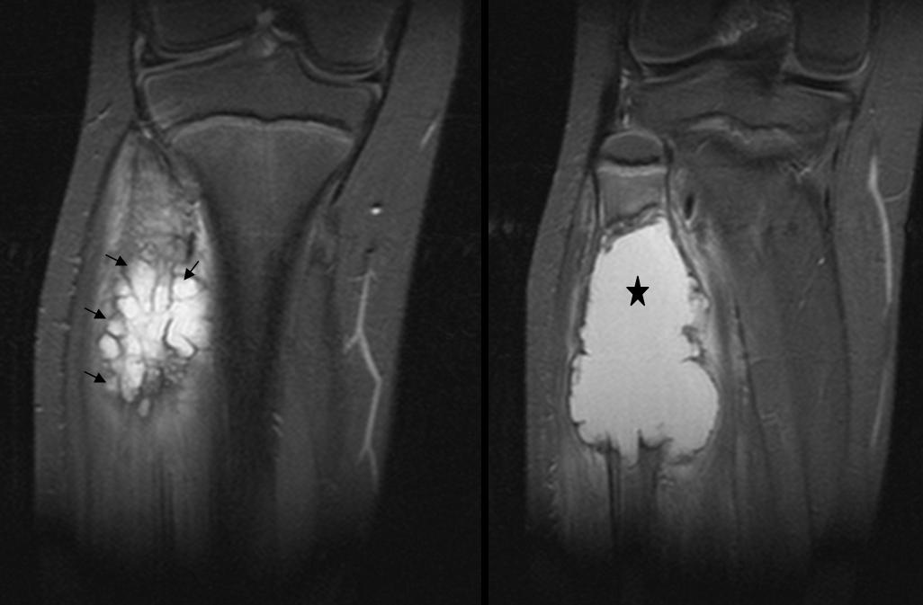
Fig. 2
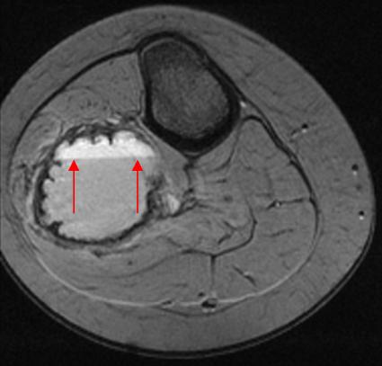
Fig. 3
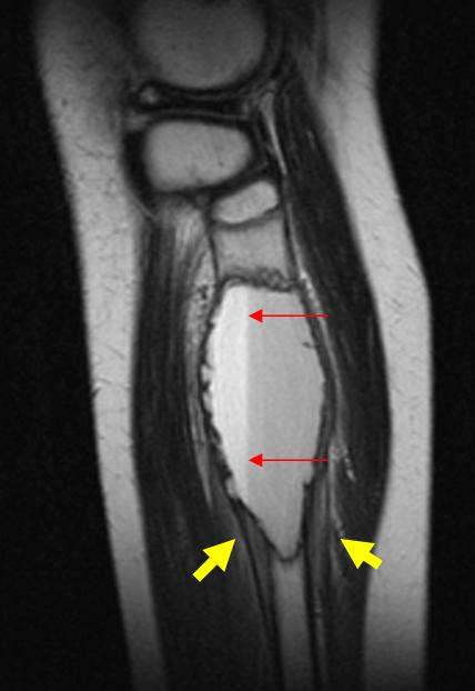
Fig. 4
Aneurysmal bone cyst, a common condition in children and adolescents, is characterised by the presence of large cavities filled with haematic material. It is usually identified incidentally following a trauma, or in studies investigating pain or increased bone volume. Aneurysmal bone cyst usually occurs in the long bones, although any bone of the organism can be affected. The most commonly affected flat bone is the pelvis. The size of the mass varies considerably from 2 to 20cm.
The radiologic appearance of long bone involvement is characteristic: well-delimited lytic area in the metaphysis with fine internal septations, cortical thinning, and bone expansion. There may be periosteal reaction and cortical rupture (pathologic fracture). The margin of the lesion is usually sharp and sclerotic. The radiologic features in flat bone involvement can simulate aggressive disease. On CT and MRI, fluid-fluid levels are often seen in the cavities. The soft tissues are seldom affected. In cases of pathologic fracture, there may be oedema of the soft tissues.
The etiopathogenesis of aneurysmal bone cyst is unknown. There is some debate regarding whether it is a radiologic and pathologic entity or the result of changes induced by a pre-existing lesion.
Simple bone cyst is a tumour-like lesion of unknown etiology, usually presenting as a well-defined, central lytic lesion of the metaphysis located adjacent to the epiphysis (Fig. 5, arrows). Any bone can be affected, but it most commonly occurs in the long bones, with 60% to 80% of cases involving the femur and humerus. The size of the cyst ranges from 2 to 15cm. The lesion may increase in length as the patient grows, but it never extends toward the adjacent epiphysis; hence, the growth cartilage remains intact. Pathologic fractures are common and when they occur, pain ensues. If there is no associated fracture, these lesions are usually asymptomatic.
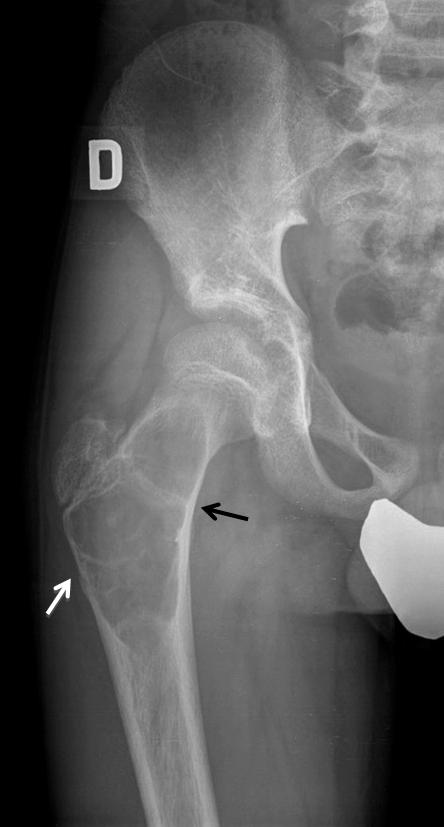
Fig. 5
Giant cell tumour is an epiphyseal bone tumour that affects patients when the growth cartilage has already fused. Most cases occur between the ages of 20 and 50 years; they are rare in children. The lesion mainly affects the long bones of the extremities (70-90% of cases), as well as the spine and pelvis. Giant cell tumour is usually a single lesion, 2 to 20cm in size. On radiographic study, it is seen as an expansive, lytic, epiphyseal lesion that extends to the subchondral bone, with well-defined borders, but no sclerosis (Fig. 6, arrows).

Fig. 6
The differential diagnosis with aneurysmal bone cyst is based on the age at presentation and the location of the tumour within the bone. Aneurysmal bone cyst rarely affects the epiphysis and occurs when the growth cartilage still persists. Giant cell tumour affects the epiphyses of mature skeletons.
Conventional osteosarcoma is usually an extensive lesion with an aggressive radiologic appearance, including bone destruction, poorly defined margins, cortical rupture, and soft tissue invasion. One variant of this tumour, telangiectatic osteosarcoma (less than 5% of osteosarcomas) presents cystic areas of necrotic or haemorrhagic nature. On MRI, the lesion may show blood-filled cavities with an internal fluid-fluid level that can mimic aneurysmal bone cyst. The presence of periosteal reaction and soft tissue invasion (Fig. 7, arrows) helps to differentiate between these conditions.

Fig. 7

Follow Dr. Pepe’s advice
Follow Dr. Pepe’s advice:
- Aneurysmal bone cyst appears as an expansive, lytic metaphyseal lesion in children.
- The characteristic MR findings are expansive lesion with cavities and internal fluid-fluid levels.
- Consider other conditions if the epiphyses have already fused or there is invasion of the soft tissues.
Suggested reading:
- Aneurysmal bone cyst: Value of MR imaging and conventional radiography. Eur Radiol 13:1118-24,2003.
- Imaging features of subperiosteal aneurysmal bone cyst. Acta Radiol 43:336-9, 2002.


