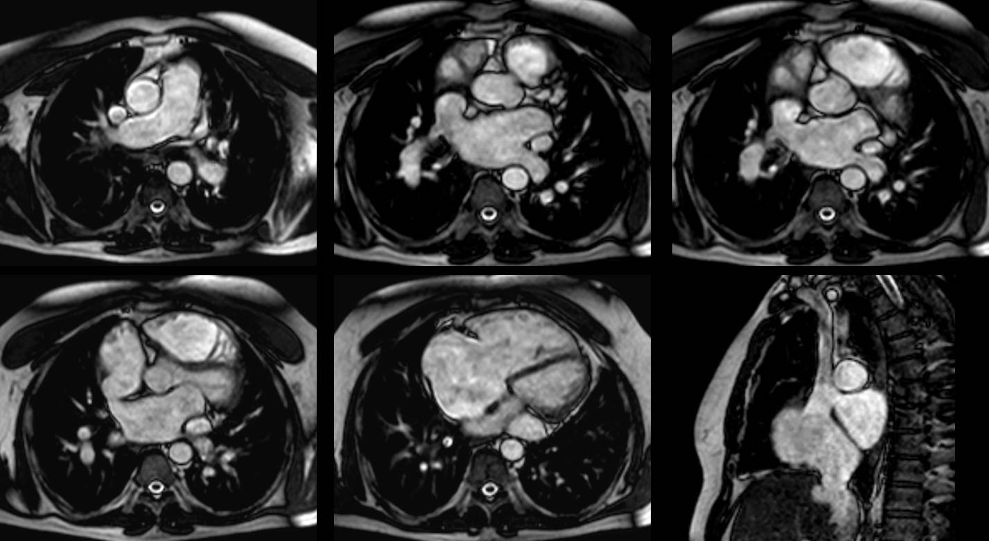
Moving forward on our journey through the systems, our next stop is the heart. I want to put your diagnostic skills to the test with the following cardiac case.
Our patient is a 60-year-old male with dyspnea and fatigability.
1. Pulmonary hypertension
2. Pulmonary valvular stenosis
3. Left-to-right shunt
4. None of the above
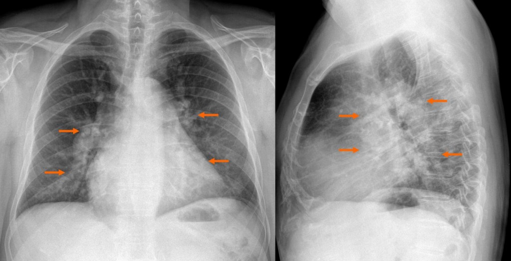
Fig. 3
Findings: Chest radiograph reveals enlargement of the pulmonary vasculature and the central pulmonary arteries (arrows)
- Pulmonary hypertension is a cause of central pulmonary arteries enlargement but with tapering of peripheral vessels
- Valvular pulmonary stenosis may cause enlargement of the central pulmonary arteries but not produce dilatation of peripheral vessels
- In a left- to-right shunt, the central and pulmonary vessels are homogeneously enlarged due to increased flow so the most likely diagnosis is left-to-right shunt
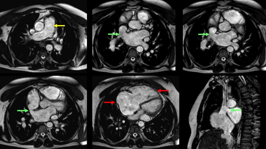
Fig. 4
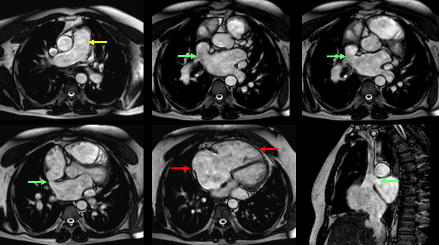
Diagnosis is confirmed by MR Axial ECG-gated cine-MR images which show:
- A defect communicating the right atrium-superior vena cava junction with the left atrium (green arrow)
- Enlargement of right-sided chambers (red arrows) and pulmonary arteries (yellow arrow) due to volume overload
- FINAL DIAGNOSIS: Superior sinus venosus atrial septal defect
Atrial septal defect is the most common left-to-right shunt detected in adulthood. It can be due to ostium primum, ostium secundum, sinus venosus and unroofed coronary. Ostium secundum is the most common type of ASD (Fig. 5), followed by ostium primum.
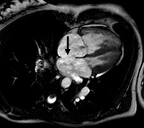
Fig. 5
Fig 5: 37-year-old woman with a two month history of angina of effort. MRI shows direct communication between the two atria located at the fossa ovalis region (arrow) due to ostium secundum atrial septal defect.
Sinus venosus defects account for 5–10% of all atrial septal defects. The superior sinus venous is more common than the inferior one (Fig. 6).
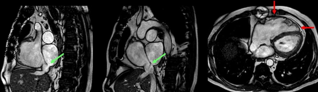
Fig. 6
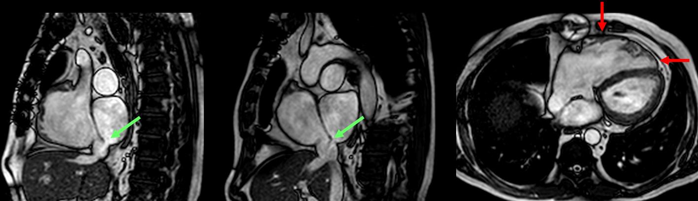
Fig. 6: 62-year-old woman with dyspnea. MRI shows a defect communicating the right atrium-inferior vena cava junction with the left atrium (green arrow) and enlargement of right ventricle (red arrows) due to inferior sinus venosus atrial septal defect.
Left-to-right shunts.
Velocity-encoded phase-contrast (VEC) MRI in ascending aorta and pulmonary artery is used to quantify the ratio pulmonary to systemic flow: Qp/Qs.
Qp/Qs is the hallmark for defining the indication for surgical correction of left-to-right shunts.
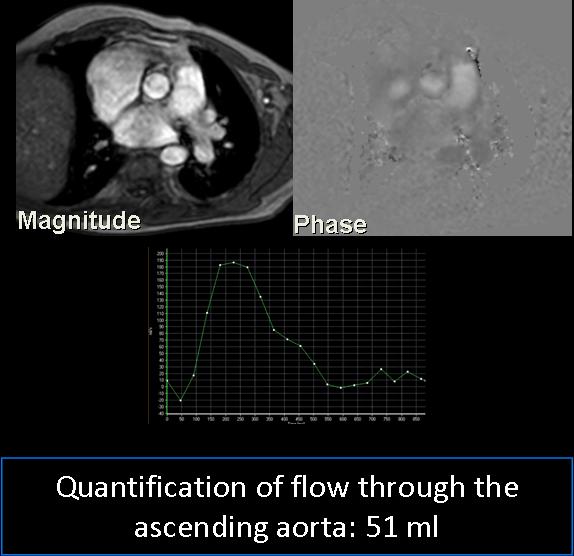
Fig. 7

Fig. 8

Follow Dr. Pepe’s advice!
Follow Dr. Pepe’s advice:
- Cardiovascular shunts can be suspected in chest radiography and confirmed by MRI.
- Ostium secundum is the most common type of atrial septal defect
- The severity of the shunt can be calculated by MR flow measurements within the pulmonary artery (Qp) and ascending aorta (Qs) and expressed as the Qp/Qs ratio
Suggested reading:
- Babar JL, Jones RG, Hudsmith L, Steeds R, Guest P. Application of MR imaging in assessment and follow-up of congenital heart disease in adults. Radiographics. 2010;30:1145. PMID: 20442335
- Rajiah P, Kanne JP. Cardiac MRI: Part 1, cardiovascular shunts. AJR Am J Roentgenol. 2011;197:W603-20. PMID: 21940532
Case prepared by Rafaela Soler MD