This week I want to show you a neuro/skeletal case, relating to a 25-year-old male with a 3-month history of sciatica.
1. Schwannoma
2. Perineural cyst
3. Sacrococcygeal teratoma
4. None of the above
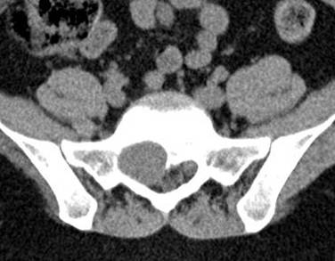
Findings: Non-enhanced CT shows a soft tissue mass with neural foramen enlargement and bone remodelling (arrows).
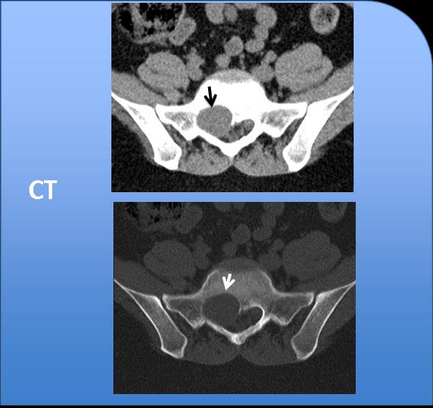
Fig. 4
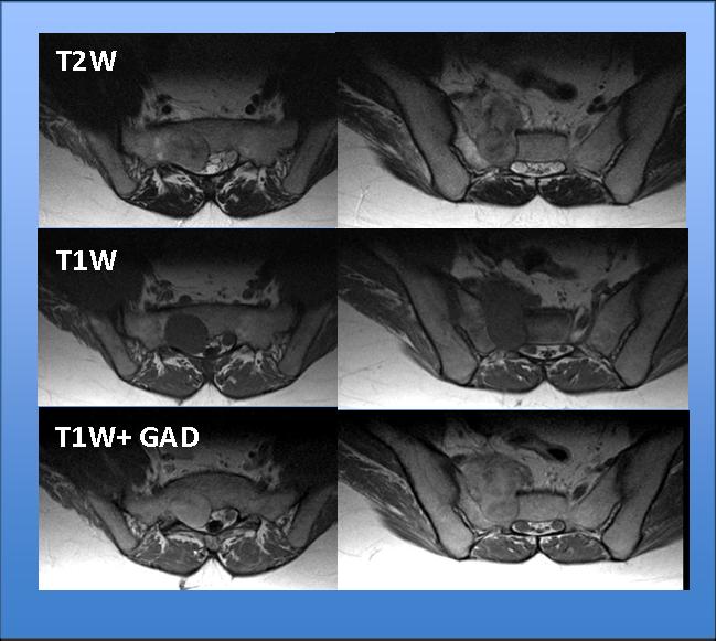
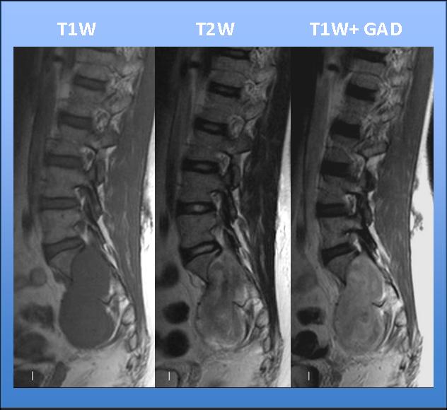
MRI depicts a well-defined bilobed soft tissue mass (arrows) with intraspinal and presacral components. The mass is hypointense on T1W, heterogeneous on T2W, and shows high and low signal intensities and uniform heterogeneous enhancement with gadolinium.
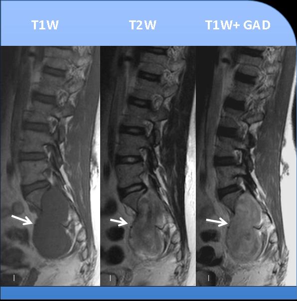
Fig. 5
These findings suggest schwannoma as the most likely diagnosis
Schwannomas are the most common intradural extramedullary masses. They grow slowly and cause bone remodelling, vertebral body scalloping, and neural foraminal enlargement (yellow arrow).
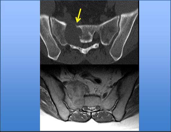
Fig. 6
On MRI imaging, schwannoma shows homogeneous or heterogeneous signal or density. Cystic areas are often seen within the tumour. The solid component enhances with contrast. Haemorrhage and calcification are rare.
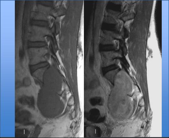
Fig. 7
Multiple schwannomas are usually found when the condition is associated with neurofibromatosis.
DIFFERENTIAL DIAGNOSIS
Perineural cyst: has homogeneous high signal intensity on T2W images following the CSF signal characteristics, and no enhancement with gadolinium.
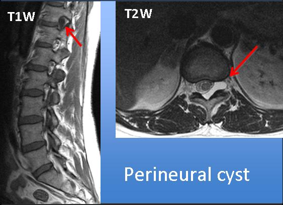
Fig. 8
Sacrococcygeal teratoma: important clues to look for are sacral malformations and heterogeneous content (eg. fat and calcium) within the mass.
Anterior sacral meningocele: important to look for associated sacral anomalies. Fluid content, highly hyperintense on T2W images, no enhancement with gadolinium.
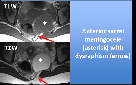
Fig. 9

Follow Dr. Pepe’s advice:
Follow Dr. Pepe’s advice:
- Spinal schwannomas present as dumb-bell tumours and cause neural foramina enlargement and bone remodelling
- High signal intensity on T2W imaging and gadolinium enhancement differentiate neural tumours from cystic masses (perineural cyst, or meningocele).
- Associated sacral or coccygeal anomalies are important clues to differentiate neural tumours from teratoma or anterior sacral meningocele.
Suggested reading: Wald JT. Imaging of spinal neoplasm. Radiol Clin North Am. 2012; 50:749-76.
Case prepared by Laura Oleaga, MD