This year I intend to discuss the basic principles of interpreting chest radiographs, under the heading, “The Beauty of Basic Knowledge”.
I plan to structure the discussion into three main parts, which will take us through the whole academic year:
This week’s case is the first chapter of my ‘painless approach to interpretation’. Interpreting chest radiographs is not difficult if we follow Confucius’ saying: “ A journey of a thousand miles begins with a single step”. As a clinical radiologist, my first step is to ask myself three questions:
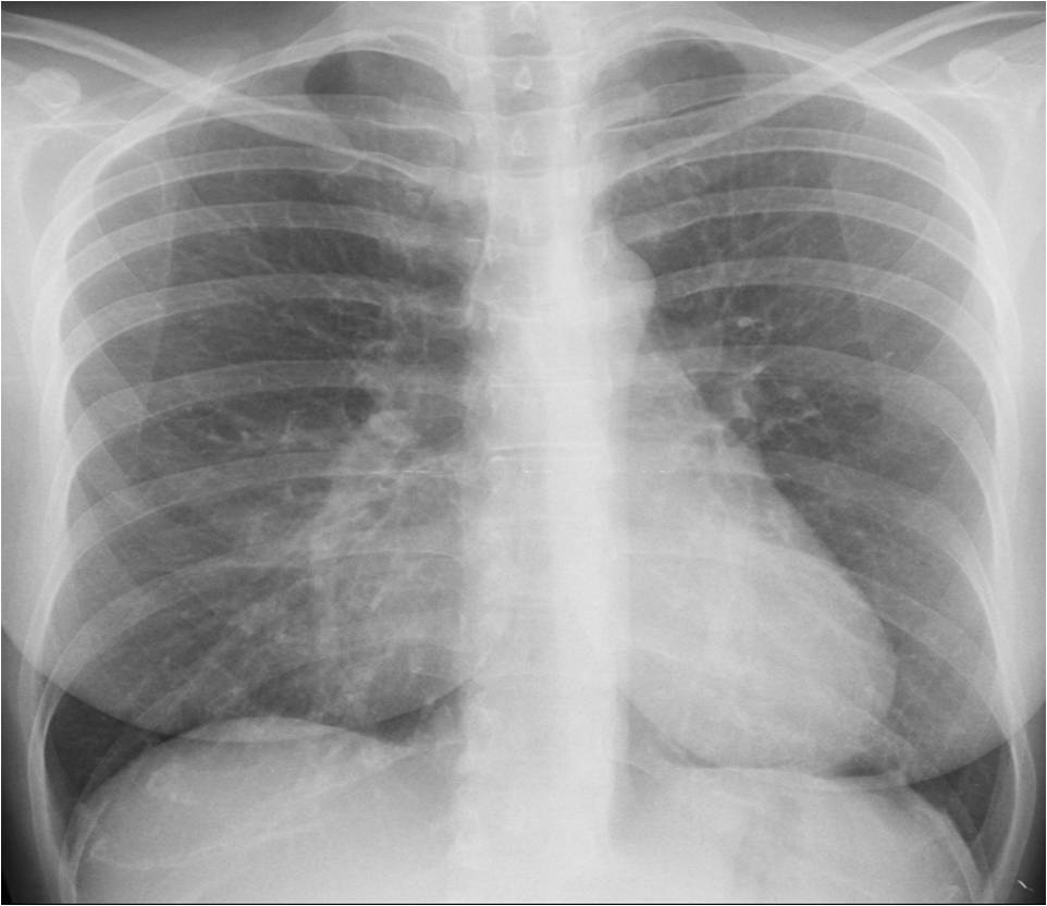
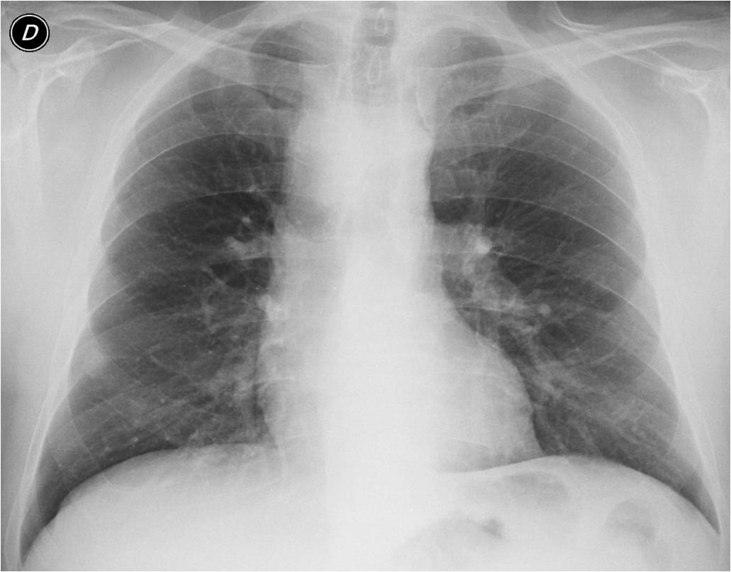
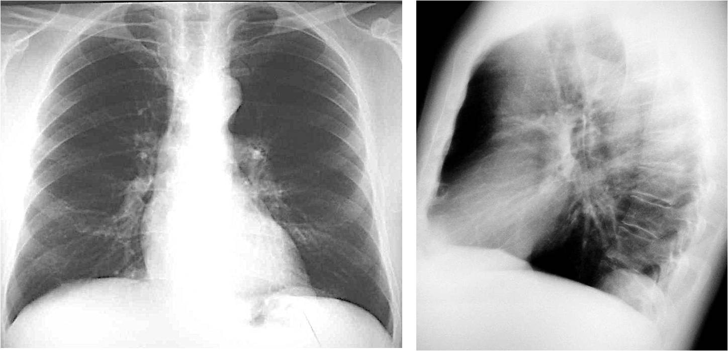
Today I’ll discuss the first question. Below you can see the chest radiographs of three different patients. Do you see any visible abnormality in any of them? Let me know in the comments section and come back on Friday for my answer.
First question: Is there any visible abnormality?
If you answered yes in all three cases, you’re right: Case 1 shows a pulmonary para-cardiac opacity (A, arrow), Case 2 shows a right mediastinal abnormality (B, arrow) and Case 3 shows a posterior extrapulmonary opacity (C, arrow).
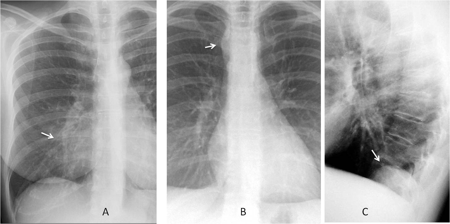
Once an abnormality is found, the next step is to determine whether it is significant or not because this decision will influence our diagnostic approach.
We should mentally review the three main causes of non-significant findings:
1. Normal variants
2. Congenital malformations
3. Changes secondary to ageing
1. NORMAL VARIANTS
A number of normal anatomical variants may cause a confusing appearance on chest radiographs. It is important to know the most common ones to avoid mistaking them for pathology. Normal variants are well described in most textbooks and I cannot show them all here. As examples, I will cite two of the most common ones.
Case 1 is an example of a common normal variant: pectus excavatum (Fig. 1). It occurs in about 0.3% of the population and can cause findings of apparent cardiomegaly or blurring of the right heart border, simulating RML disease. In the PA view pectus excavatum can be suspected by the vertical path of the anterior ribs. The diagnosis is easily made in the lateral view.
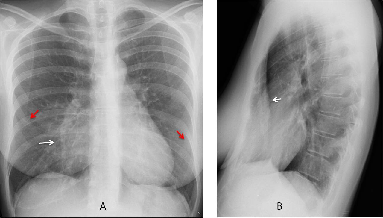
Fig. 1
Fig. 1: PA chest film shows an apparent opacity in the RML (A, white arrow). Note the vertical path of the anterior ribs (A, red arrows). The lateral view confirms that pectus excavatum (B, arrow) is the cause of the opacity.
Rhomboid fossa of the clavicle is another common variant that should be mentioned because it can simulate a lytic lesion, especially when it appears on only one side (Fig. 2). Rhomboid fossa results from the insertion of the costo-clavicular ligaments. It occurs in 30% of males and 5% of females and is more commonly seen in younger individuals, as it becomes less visible with age.
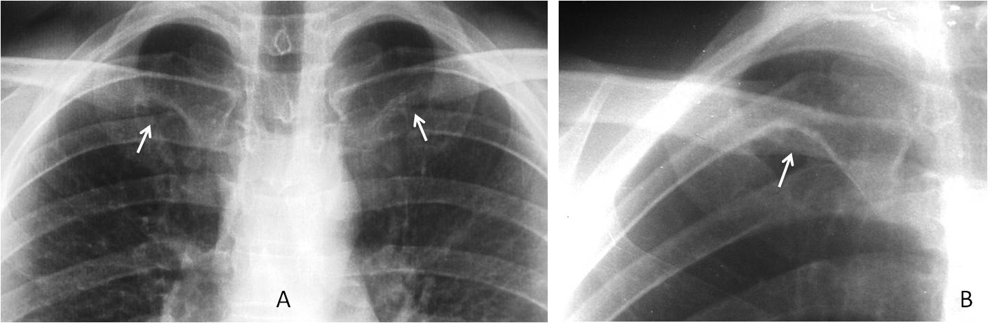
Fig. 2
Fig. 2: Examples of rhomboid fossa (RF) in two different patients. The first has bilateral RF (A, arrows), whereas the second has a single RF (B, arrow), which could easily be confused with a lytic lesion.
2. CONGENITAL MALFORMATIONS
Not uncommon in adults. It is important to be aware of these malformations because they may not be suspected in adult patients. Most are asymptomatic and easy to detect on chest radiographs if we aware of their existence.
Case 2 shows one of the most common congenital vascular malformations in adults: right aortic arch with an anomalous left subclavian artery (Fig. 3). It is asymptomatic and occurs in less than 1% of the population. Do not mistake it for a mediastinal mass or paratracheal lymph nodes.
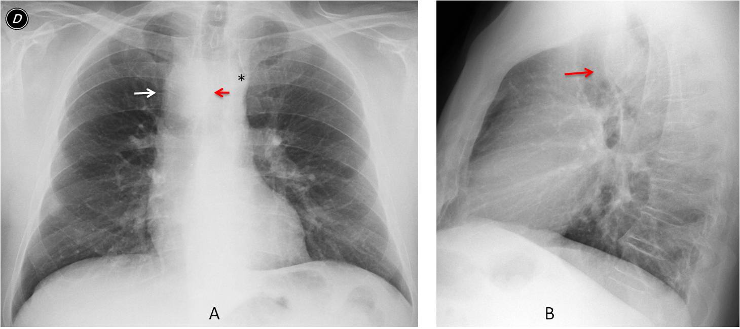
Fig. 3
Fig. 3: An apparent right mediastinal mass was found (A, white arrow) in this pre-op patient. The appearance is typical of a right aortic arch, with deviation of the trachea to the left (A, red arrow) and absence of the aortic knob on the left side (A, asterisk). The lateral view shows anterior displacement of the trachea by the posterior aortic arch (B arrow).
The diagnosis can be made with enhanced CT, but it’s cheaper to compare the image with previous films, which will show identical findings (Fig. 4).
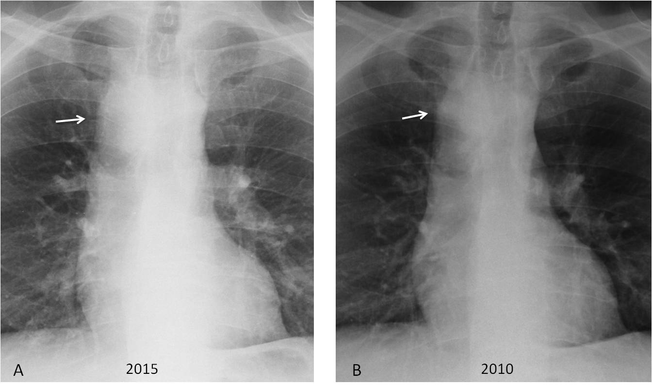
Fig. 4
Fig. 4: (same patient as Fig. 3). PA chest film taken five years earlier shows identical findings. The right aortic arch remains unchanged (A and B, arrows).
Many congenital malformations involve the lung. Most are asymptomatic and are first discovered in the adult age. Some of them were reviewed in Dr. Pepe’s Diploma Casebook Case #73. One of the most common is hypogenetic right lung (scimitar syndrome), which gives the chest radiograph a bizarre appearance (Fig. 5).
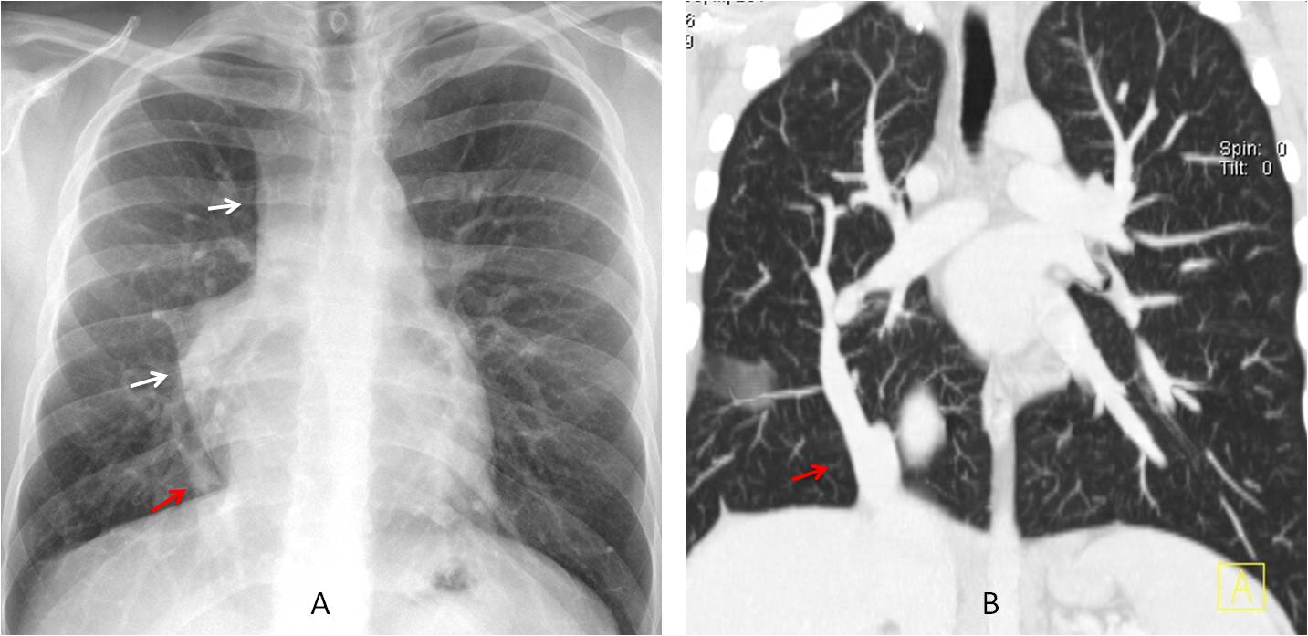
Fig. 5
Fig. 5: Hypogenetic right lung showing an abnormal mediastinal contour in the PA chest film (A, white arrows). Coronal CT shows a smaller right lung and the scimitar vein (A and B, red arrows), which is typical of this malformation.
3. CHANGES WITH AGEING
As we get older, morphologic and physiologic changes occur (aortic elongation, kyphosis, etc.) that alter the appearance of the chest. We should be familiar with the various changes that occur in the elderly chest to enable their differentiation from pathological conditions.
CASE 3 is an example of a normal occurrence in older persons: Bochladek hernia (Fig. 6), secondary to weakening of the muscle fibres of the diaphragm as a consequence of ageing. It creates bumps in the diaphragm that should not be confused with disease.
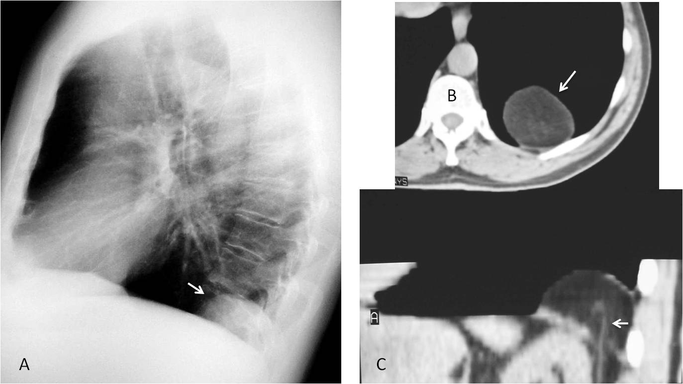
Fig. 6
Fig. 6: Bochladek hernia in a 65-year-old man, simulating a posterior mass (A, arrow). Axial CT demonstrates the fatty component (B, arrow). Sagittal CT demonstrates the rent in the diaphragm and a crossing abdominal vessel (C, arrow).
Calcification of the mitral annulus occurs in about 10 % of old persons, and is usually very obvious in the chest radiograph (Fig. 7). It should not be confused with calcium in the valve. Usually asymptomatic, it can occasionally cause mild valve dysfunction.
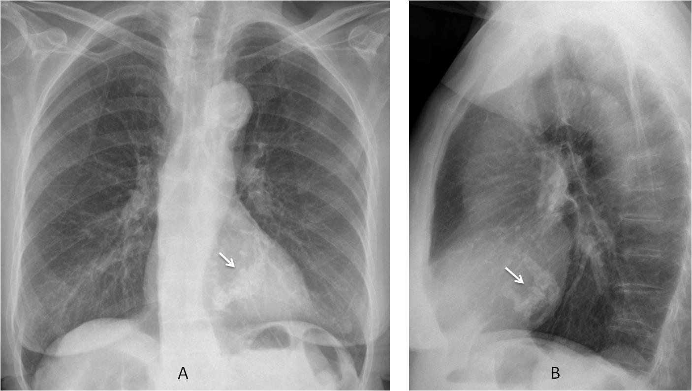
Fig. 7
Fig. 7: Calcification of the mitral annulus in a 78-year-old woman. Note the typical reversed C-shape of the calcium (A and B, arrows).
In conclusion, the first step when looking at a chest radiograph is to determine if any visible anormality is present.
If an abnormality is detected, our next concern should be to exclude a non-significant finding to avoid an unnecessary (and costly) work up. To do that, we should be familiar with the most common normal variants and congenital malformations, as well as the appearance of the chest in persons of advanced age.

Dr. Pepe’s Teaching Points
Your first step when looking at a chest radiograph is to detect visible abnormalities
When abnormal features are present, exclude the following:
– Normal variants
– Congenital malformations
– Changes secondary to ageing