Dr. Pepe’s Diploma Casebook: Case 81

Dear Friends,
This week’s case follows the pattern of a “Meet the examiner” presentation, with questions and answers similar to a real examination. Take your time before scrolling down for the answer. No peeking! My good friend Dr. Jordi Andreu has prepared the case.
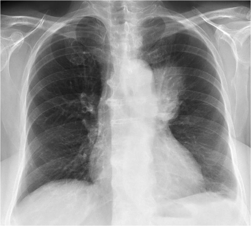
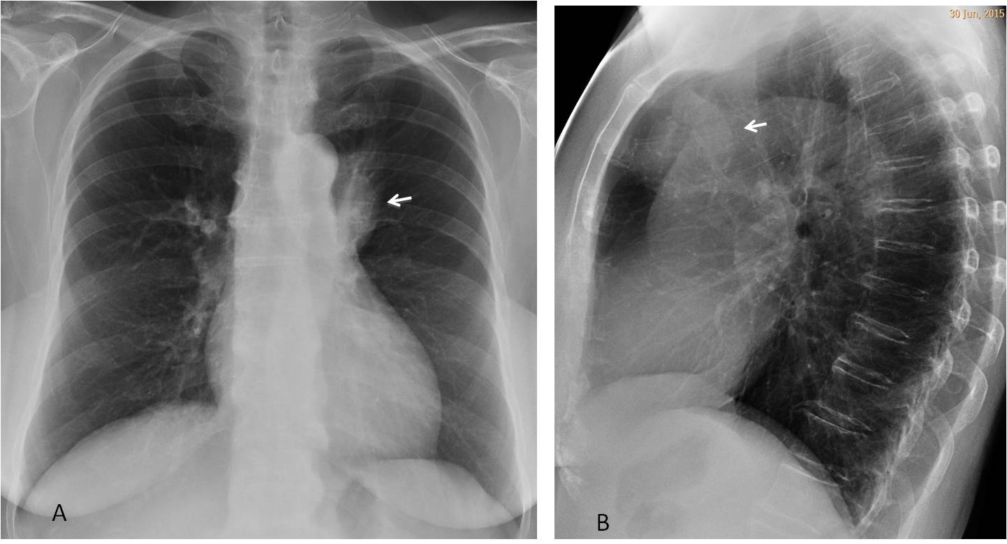
Chest radiographs belong to a 69-year-old woman with cough, fever and purulent sputum for the last ten days. History of TB thirty years ago.
Diagnosis:
1. Reactivation TB
2. Mediastinal cyst
3. Mediastinal tumour
4. None of the above

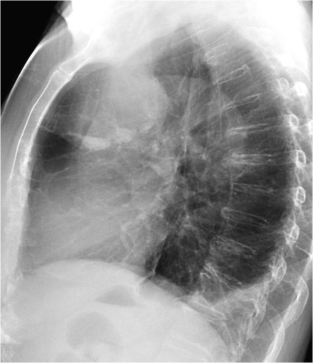
Chest radiographs show a left pleural effusion and poorly-defined opacity in the PA view (A, white arrow) that is rounded and better defined in the lateral view (B, white arrow). The clue to the diagnosis lies in identifying the high density area(s) with a straight upper border in the lower aspect of the mass (A and B, red arrows). This appearance is very suggestive of milk of calcium, which only occurs in long-standing cystic structures. Therefore, the correct answer should be: 2. Mediastinal cyst. If you feel adventurous, you may even suggest a multicystic lesion, since two levels are seen in the lateral view.

What would be your diagnosis?
1. Infected cyst
2. Teratoma
3. Residual changes from previous TB
4. None of the above
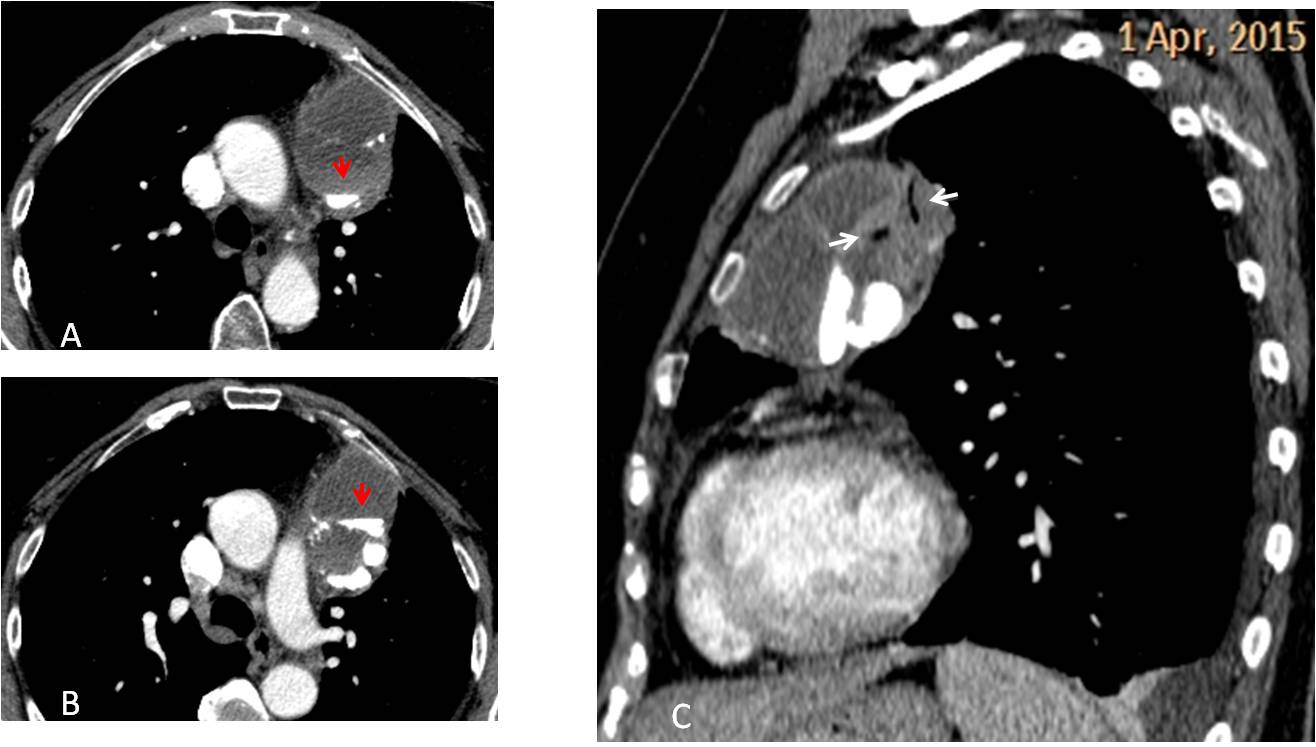
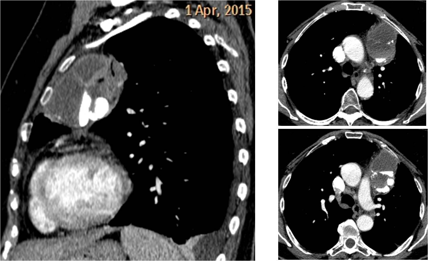
Enhanced axial and sagittal CT confirm a multicystic lesion with milk of calcium layered at the bottom of the cysts (A and B, red arrows). The sagittal reconstruction shows air within the mass (C, arrows), confirming a connection with the lung parenchyma as the probable cause of infection. The correct answer is: 1. Infected cyst
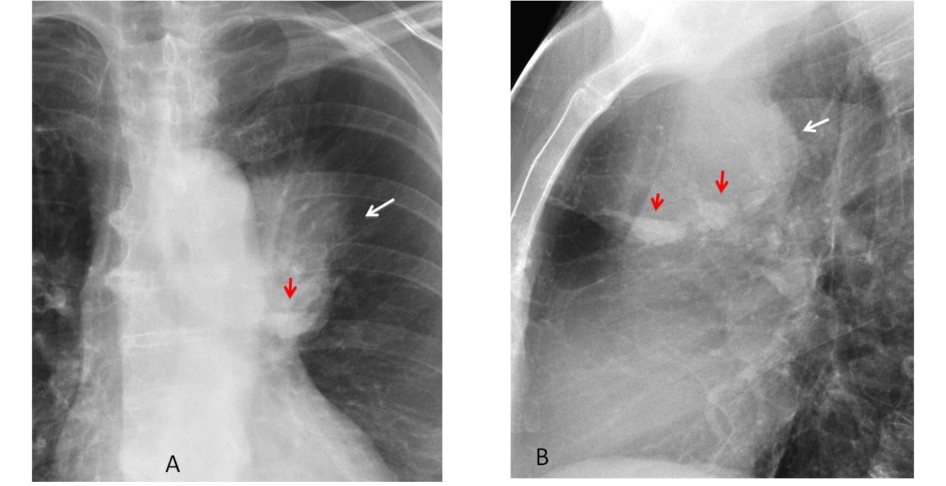
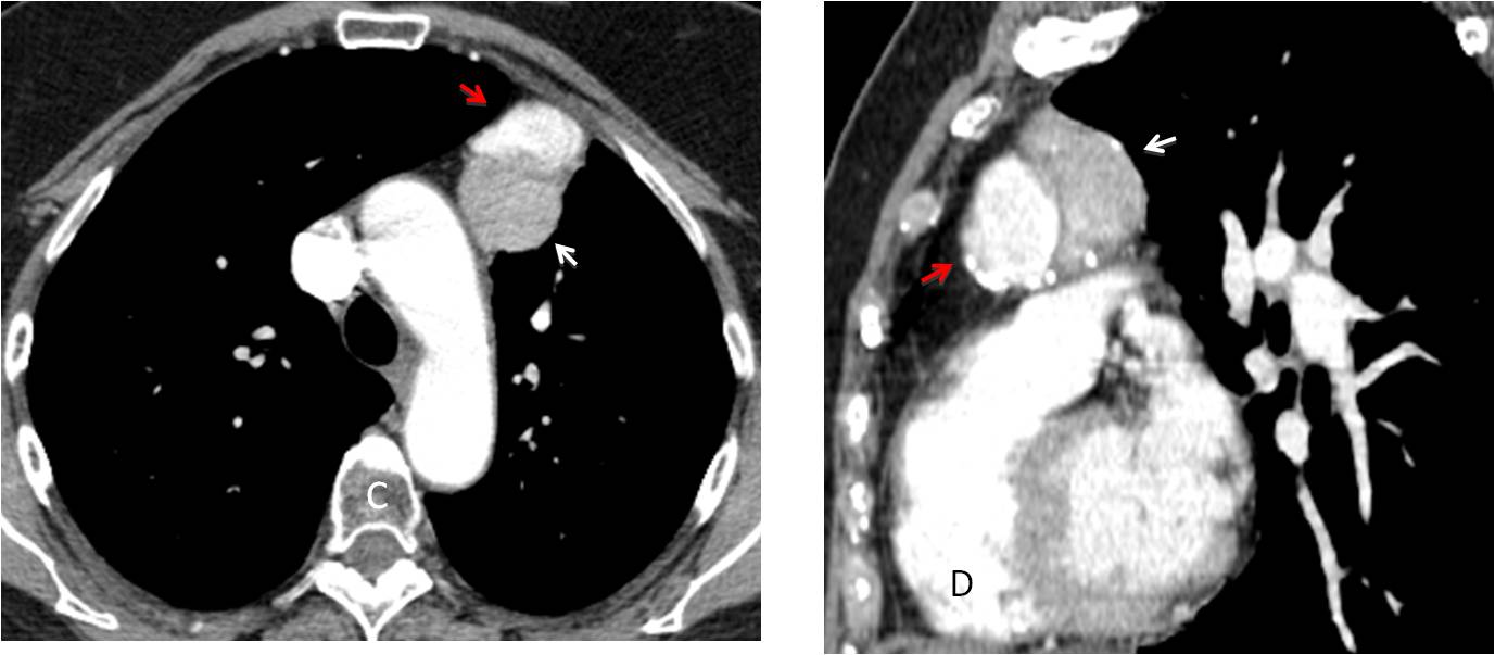
The patient improved markedly with antibiotic treatment. Chest radiographs three months later showed a marked decrease in the size of the lesion (A and B, arrows). The left pleural effusion has disappeared.
Enhanced axial and sagittal CT confirm the decreased size of the cysts, probably due to the patient coughing up part of their contents. It is interesting to note that milk of calcium is now filling the anterior cyst (C and D, red arrows) whereas none remains in the posterior one (C and D, white arrows).
The patient underwent surgery and the final diagnosis was bronchogenic cyst.

Follow Dr.Pepe’s Advice:
1. Milk of calcium is seen as a calcium level in the chest radiograph.
2. When it is found, it indicates a long-standing benign cystic lesion.
3. CT easily confirms the diagnosis.


Great case as always. Thank you very much.
Thank you. I strive to give satisfaction(( Jeeves )
Hello,
I am not a radiologist (pulmonologist) but I find your cases really interesting. I think we don`t usually pay much intention to small details, which would be sometimes extremely helpful. Thank you so much and more of these please.