Dr. Pepe’s Diploma Casebook: Case 99 – A painless approach to interpretation (Chapter 6) – SOLVED!

Dear Friends,
We’re moving on to a new chapter of the Painless Approach to Interpretation, and this week I’m showing the routine control radiographs of a 48-year-old woman, surgically treated for carcinoma of the breast ten years ago.
What do you see?
Check the image below, leave your thoughts in the comments section, and come back for the answer on Friday.
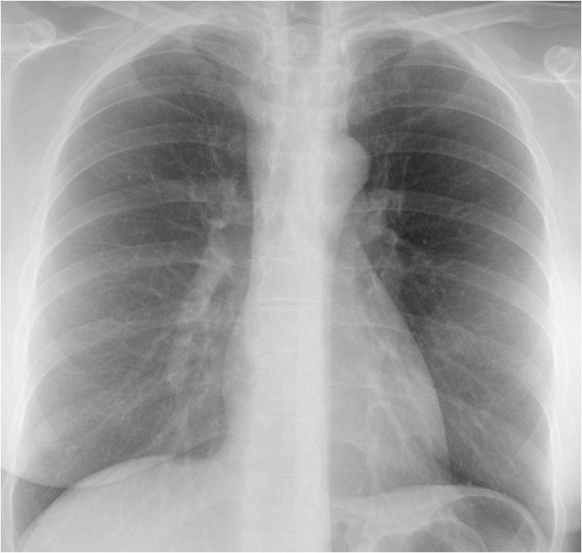
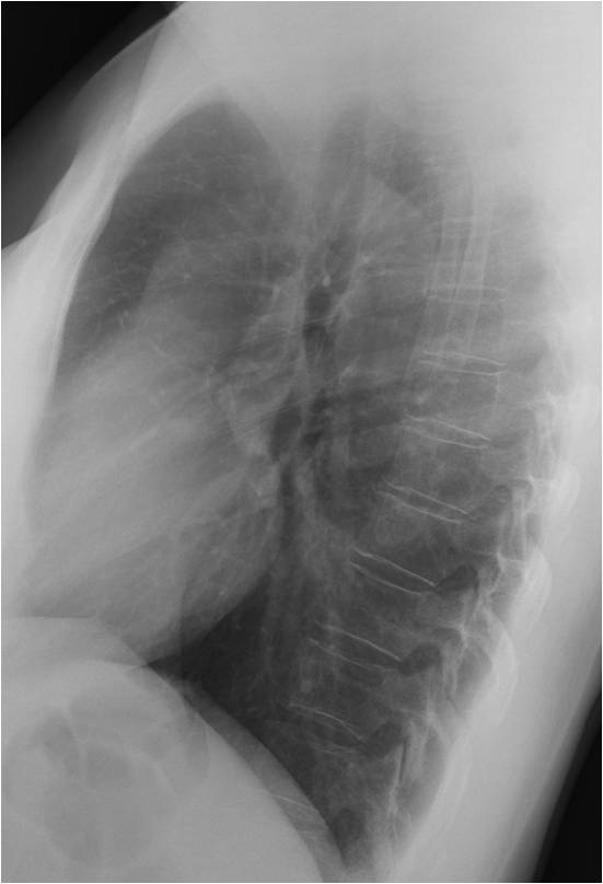
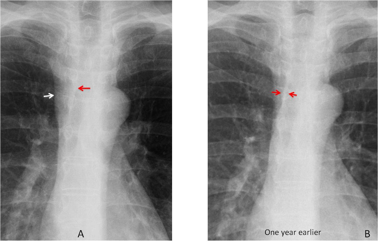
Findings: the PA radiograph shows obvious widening of the right paratracheal line (A, white arrow), which is more evident when compared with a previous examination made a year earlier (B). Note that both borders of the line were seen in the previous radiograph (B, red arrows), whereas only the inner border is visible now (A, red arrow).
Enhanced axial CT confirms the presence of a large right paratracheal lymph node (C, arrow) and bilateral hilar nodes (D, arrows). PET was negative. The nodal distribution in an asymptomatic patient raises the possibility of sarcoidosis, which is known to occur after treatment for solid tumours.
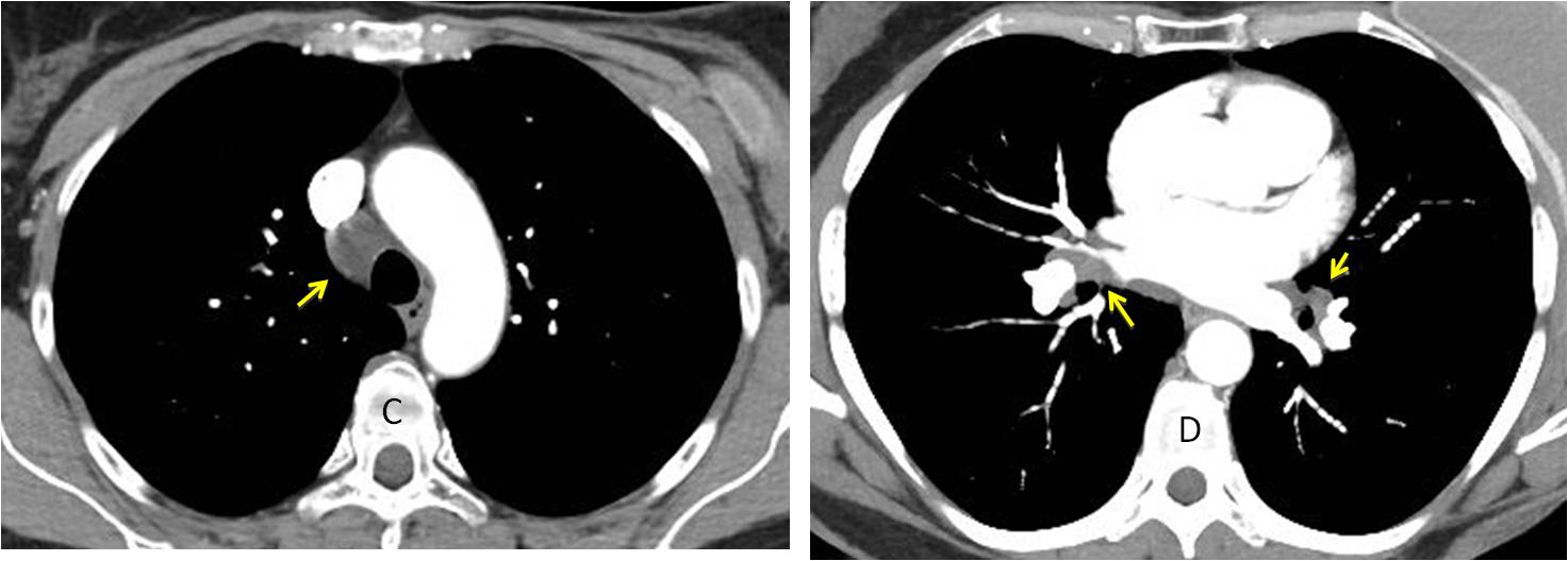
Final diagnosis: sarcoidosis following breast carcinoma treatment.
As we all know, an obvious mediastinal mass is easily seen, but it is equally important to detect subtle mediastinal abnormalities that may uncover serious disease. In this presentation I’d like to discuss a checklist of structures to be examined in the mediastinum to avoid overlooking abnormalities.
The basic checklist for the mediastinum includes the following structures:
1. Mediastinal lines:
– Right paratracheal line
– Paraesophageal line
2. Mediastinal contour:
– Right side: azygos vein and ascending aorta
– Left side: aortic knob and pulmonary arch
Of all the mediastinal lines, the right paratracheal line is the most useful for detecting disease. It is less than 3mm thick and visible in about 95% of PA chest films. Any increase in width over 3mm should be investigated, the most common cause being enlarged lymph nodes (Fig. 1).
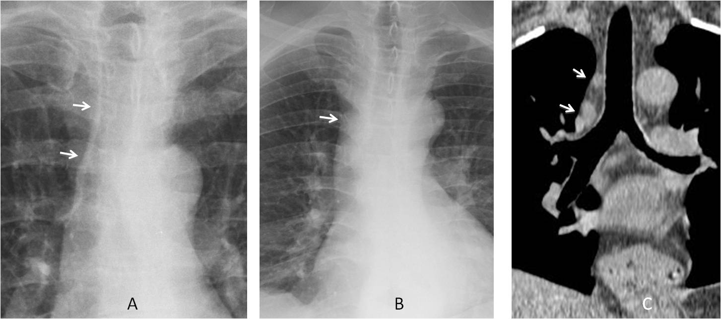
Fig. 1
Fig. 1. A. Normal paratracheal line (A, arrows). B. Widened paratracheal line (B, arrow) in a patient with fever and malaise. Coronal CT confirms mediastinal lymph nodes (C, arrows). Diagnosis: lymphoma.
The paraesophageal line is created by contact of the right lung with the oesophagus and it is seen as an oblique line projected over the spine. Displacement of the interface suggests oesophageal dilatation or hiatal hernia (Fig. 2). Focal bulging may be due to enlarged lymph nodes, oesophageal tumour, or mediastinal cyst.
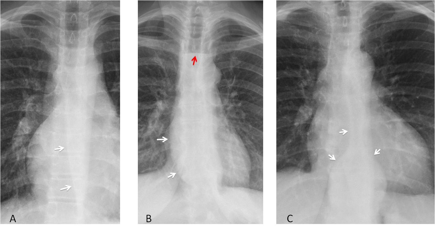
Fig. 2
Fig. 2. A. Normal paraesophageal line (A, arrows).B. Bulging of the oesophageal interface due to achalasia (B, white arrows). Air-fluid level in oesophagus (B, red arrow). C. Hiatal hernia opening the distal paraesophageal line (C, arrows).
The right mediastinal contour has four components: superior vena cava, azygos arch, ascending aorta and right atrium. In my opinion, the two that should be included in the checklist are the azygos arch and the ascending aorta (Fig. 3).
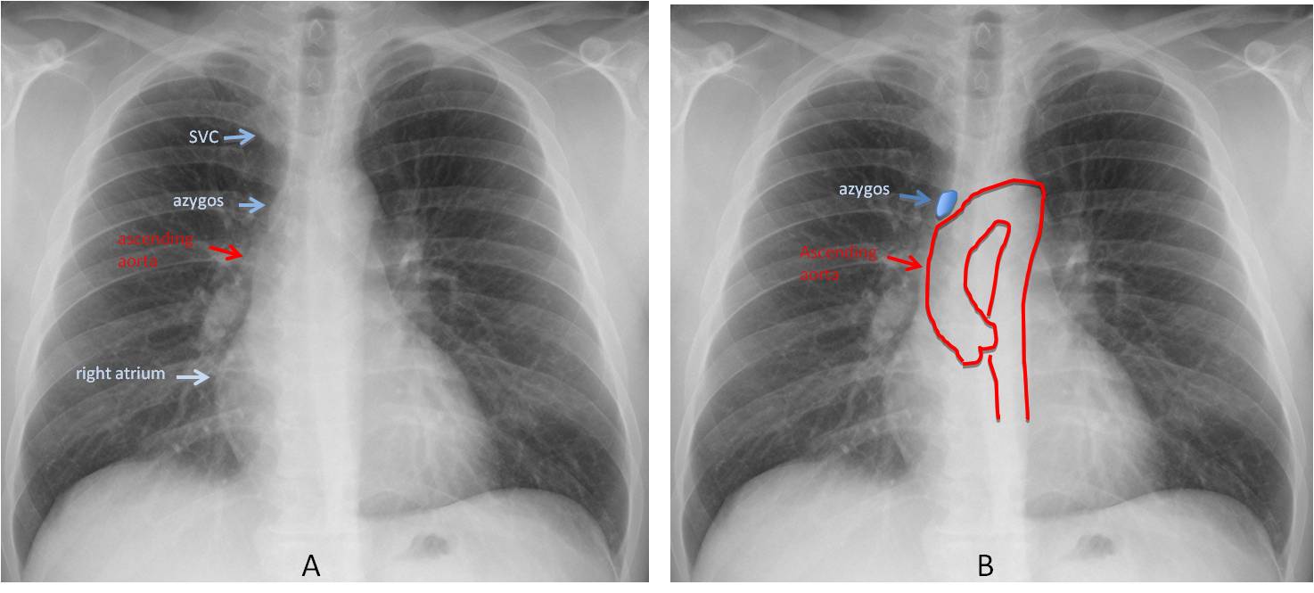
Fig. 3
Fig. 3. Normal chest film showing the four components of the right mediastinal border (A,B).
The azygos arch is the only part of the azygos vein visible in the PA radiograph. It is seen in about 10% of upright PA views. An azygos arch larger than 1cm usually indicates increased blood flow in the azygos system. It may be non-obstructive or secondary to obstruction of the superior or inferior vena cava, in which case the azygos system acts as collateral drainage (Fig. 4).
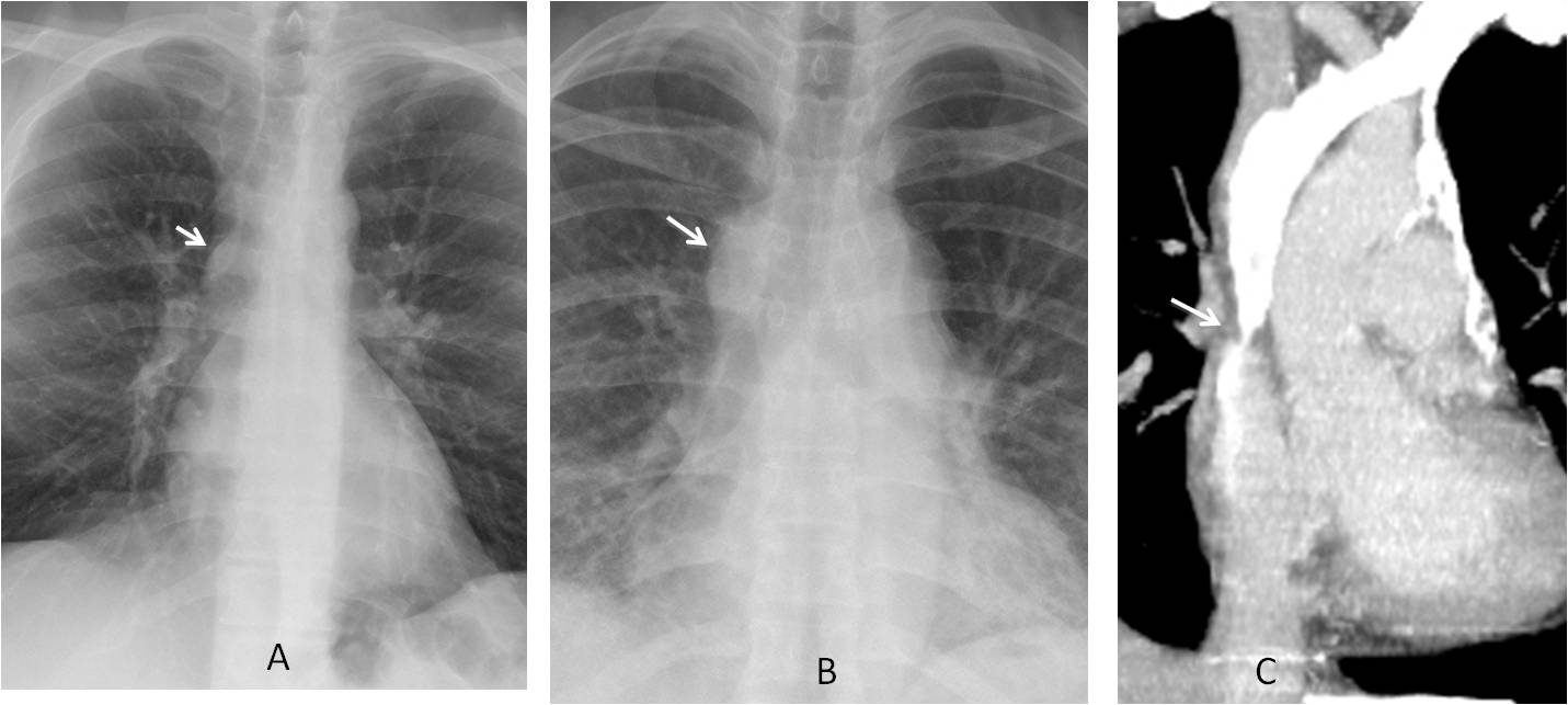
Fig. 4
Fig. 4. A. Normal azygos arch, with typical lenticular shape (A, arrow). In B, the azygos arch is obviously enlarged (B, arrow), as it is acting as a collateral channel for SVC obstruction (C, arrow).
Prominence of the right mediastinum middle segment is usually due to dilatation of the ascending aorta. Four conditions can lead to isolated ascending aorta dilatation: bicuspid aortic valve, ascending aorta aneurysm, type A aortic dissection, and aortic coarctation (Fig. 5).
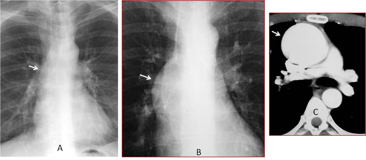
Fig. 5
Fig. 5. A: Normal appearance of ascending aorta (A, arrow). B: Marked prominence of the ascending aorta in an 18-year-old male with Marfan syndrome (B, arrow). Enhanced CT shows an ascending aorta aneurysm (C, arrow).
The left mediastinal contour has three components: the uppermost is the aortic knob, the middle one corresponds to the main pulmonary artery, and the lower one represents the left ventricle (Fig. 6). In my opinion, the aortic knob and pulmonary artery arch should be included in the checklist.
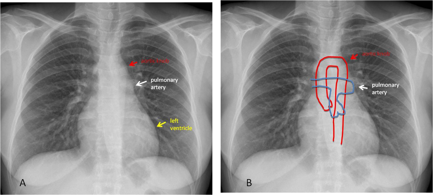
Fig. 6
Fig. 6. Normal chest films (A and B) showing the three components of the left mediastinal border.
The aortic knob should be examined in every chest radiograph. The most obvious finding is a change in size. The knob increases in size with aging, but a large aortic knob in a relatively young person should bring to mind an aneurysm or a type B dissection (Fig. 7).
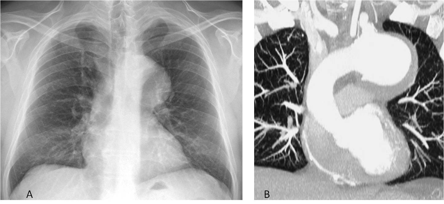
Fig. 7
Fig. 7. 54-year-old patient with a large aortic knob (A, arrow). Enhanced coronal CT confirms an aneurysm of the aortic arch (B, arrow).
In my experience, one of the most common causes of a small left aortic knob in adults is a right aortic arch. This congenital variation is easily detected by recognising the knob at the right mediastinal border and detecting the imprint in the right tracheal wall (Fig. 8).
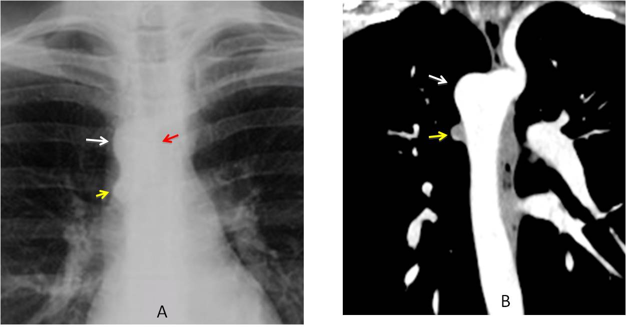
Fig. 8
Fig. 8. 27-year-old woman has no obvious left aortic knob; instead, the knob is located on the right side (A and B, white arrows). Note the imprint in the right tracheal wall (A, red arrow). The small bump under the right arch represents the azygos vein (A and B, yellow arrows).
Occasionally, a small mediastinal mass may hide behind the aortic knob, and careful inspection is needed detect it (Fig. 9).
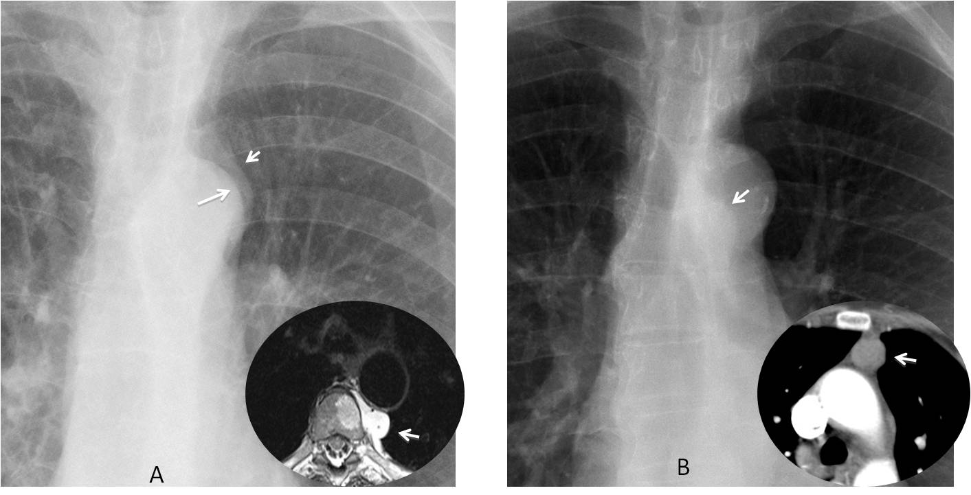
Fig. 9
Fig. 9. A: Control radiograph in a patient with leg sarcoma. The aortic knob shows a double contour (A, arrows). Axial MRI confirms a metastatic mass invading the spine (insert, arrow). In another patient, a small mass is seen through the aortic knob (B, arrow), confirmed with CT (insert, arrow). Diagnosis: thymoma.
Bulging of the pulmonary artery arch usually indicates dilatation of the main pulmonary artery, which can be caused by 3 conditions. The most common is pulmonary arterial hypertension, followed by left-to-right shunt and pulmonary valve stenosis (Fig. 10).
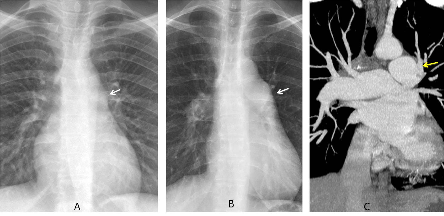
Fig. 10
Fig. 10. A: Normal pulmonary artery arch (A, arrow). B: Bulging of this segment in another patient (B, arrow). Coronal enhanced CT shows enlargement of the main pulmonary artery (C, arrow), which is wider than the aorta. Diagnosis: pulmonary arterial hypertension
In conclusion, the following mediastinal areas should be investigated in the PA view:
The paratracheal line, to evaluate changes in width usually due to enlarged lymph nodes, and the paraesophageal line, which may be displaced by esophageal dilatation, enlarged lymph nodes, or congenital cysts.
The azygos vein. Enlargement is usually due to thrombosis of the superior or inferior vena cava.
The ascending aorta. Enlargement can be due to bicuspid aortic valve, ascending aorta aneurysm, type A aortic dissection, or aortic coarctation
The aortic knob, to detect changes in size and, occasionally, hidden mediastinal masses.
The pulmonary artery arch. The most common cause of prominence is pulmonary arterial hypertension.
Next week I will continue with further checklists.

Follow Dr. Pepe’s advice:
1. The mediastinum checklist includes mediastinal lines and bulges of the outline.
2. The main lines to examine are the paratracheal line and the paraesophageal line.
3. On the right mediastinal contour, look at the azygos vein and ascending aorta.
4. On the left mediastinal contour, look at the aortic knob and pulmonary arch.


