
Hola amigos, el caso de hoy corresponde a una mujer de 54 años con cefalea vespertina, sensación de inestabilidad y episodios de minutos de evolución de afasia motora, a lo largo de los últimos seis meses. Exploración física y neurológica sin alteraciones.
¿Cuál sería el diagnóstico?
1. Metástasis dural
2. Tumor fibroso solitario
3. Meningioma
4. Hemangiopericitoma
Podéis dar vuestra opinión hasta el viernes, día en el que se publicará la solución.
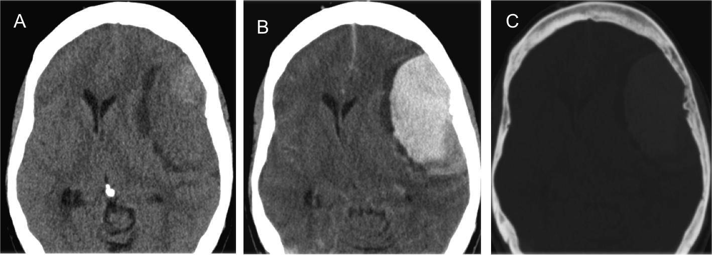
Imágenes de TC craneal sin contraste (A) y con contraste endovenoso (B), y con ventana de hueso (C)
Haz click aquí para ver la respuesta
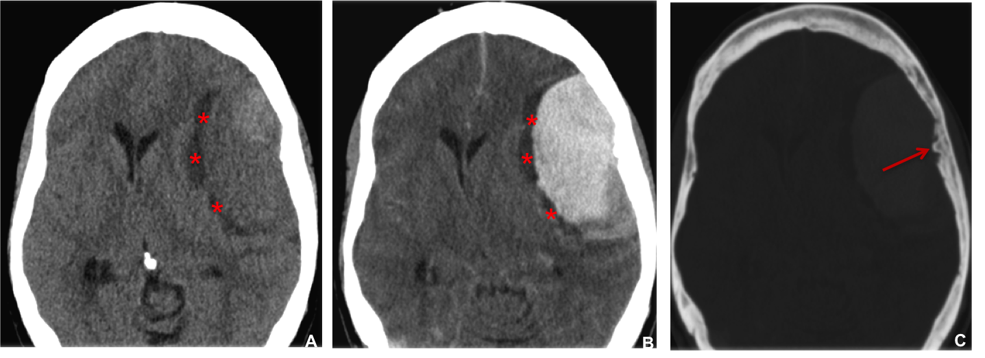
Hallazgos: masa extraparenquimatosa situada en la convexidad hemisférica cerebral fronto-temporal izquierda, isodensa en relación al parénquima cerebral (A) con un intenso y homogéneo realce tras la administración de contraste e.v (B), y un ribete medial a la masa hipodenso (asteriscos A-B). Hiperostosis focal en la calota craneal adyacente (C) en relación a la presencia de un spur enostótico (flecha).
Diagnóstico definitivo: meningioma cordoide.
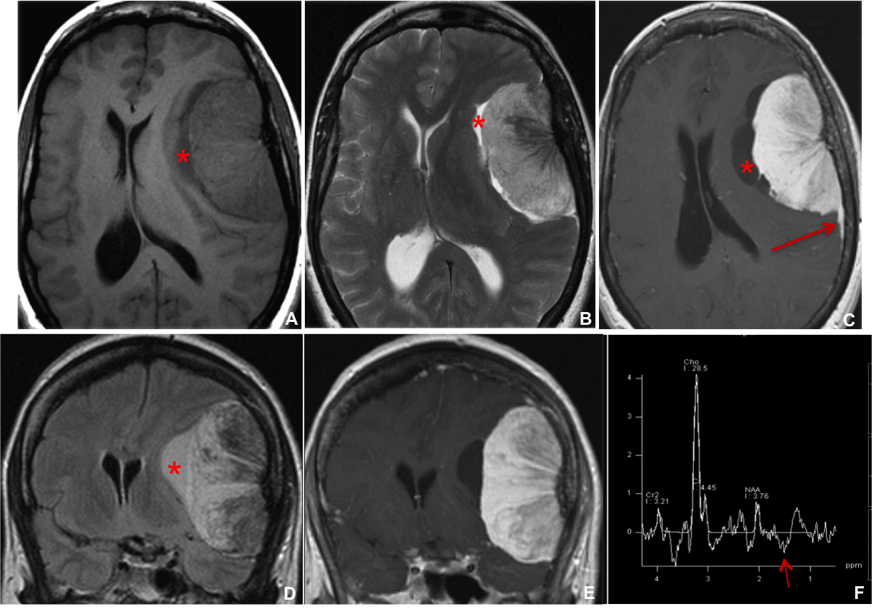
Imágenes de RM craneal en el plano axial y secuencias T1, T2 Y T1 con contraste (A, B, C) y en el plano coronal y secuencias FLAIR (D) y T1 con contraste endovenoso (E). Se incluye estudio de espectroscopia univoxel eco largo (F).
Masa extraparenquimatosa situada en la convexidad hemisférica cerebral fronto-temporal izquierda, isointensa en T1 en relación al parénquima cerebral (A), hiperintensa en T2, con imágenes lineales de baja señal en su interior por la presencia de vasos intratumorales (B). Intenso realce tras la administración de contraste e.v, con realce dural lineal en el margen posterior de la lesión (flecha en C). Ribete periférico y medial a la masa hipointenso en T1, sin y con contraste, e hiperintenso en T2/FLAIR, en relación a loculaciones de LCR atrapado en el espacio subaracnoideo (asteriscos). El estudio espectoscrópico muestra el patrón de metabolitos característico del meningioma: pico de colina, mínima presencia de N-acetilaspartato y doblete invertido de alanina en la posición 1.5 (flecha, F).
HISTOPATOLOGÍA
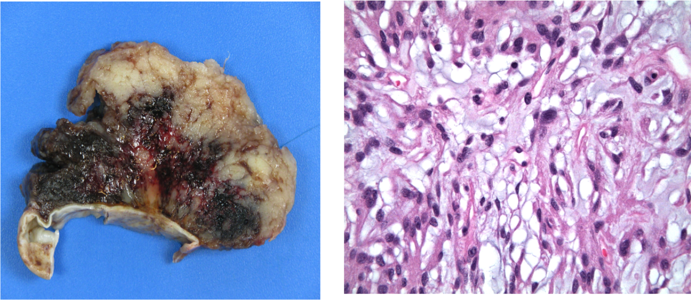
Fragmento hístico de siete centímetros de diámetro máximo, consistencia elástica y aspecto granular, y de color terroso con focos hemorrágicos.
MENINGIOMA: SEMIOLOGÍA RADIOLÓGICA
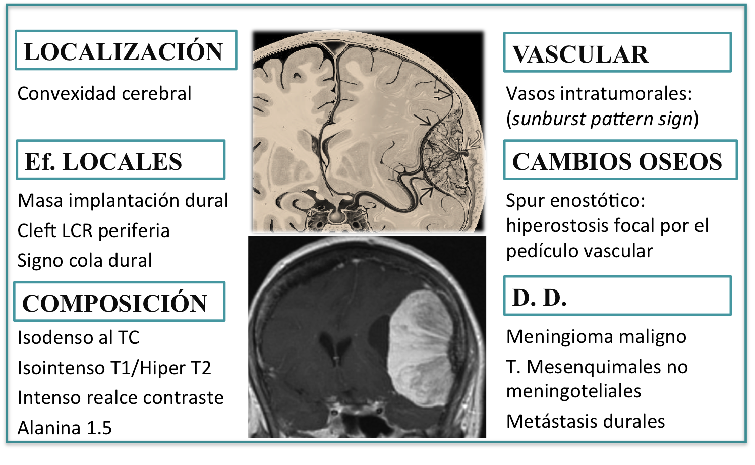


Meningioma convexidad
Meningioma
….la presencia de un área hipo densa y ángulos agudos de contacto me hacen descartar meningioma, el realce con el contraste podría corresponder a hemangiopericitoma….creo, veremos que dirá el Dr. Pepe. Saludos
Hemangiopericitoma
tumor fribroso solitario meningeo
hemangiopericitoma
Meningioma
The presence of a hypodense area and sharp contact angles lead me to believe that hemangiopericytoma is more likely than meningioma. On the other hand, the contrast enhancement may be indicative of hemangiopericytoma. I believe that we should wait to hear what Dr. Pepe has to say. Greetings
Great article! It’s well-written, informative, and provides valuable insights on the topic.
Thank you for your post; I’ve been looking for such an article for a long time, and today I’ve found it. This post provides me with a wealth of advice; it is very helpful to me
I like how lucid your writing is. A broad spectrum of individuals should find it simple to read since the explanations are clear and succinct.
Absolutely thrilled with our solar panel installation in Richmond, Virginia! We found this top-notch installer through Solar Power Systems website. They wowed us with their efficiency and smooth process – a shining example of professionalism. It’s been a brilliantly sunny decision for our home!
Given the symptoms described, especially the evening headaches and episodes of motor aphasia, my guess would lean towards meningioma.
I’m also a doctor so I can confirm that the diagnoses you gave are correct.
Additionally, as a medical professional, I can verify the accuracy of the diagnoses provided.
Based on the presenting symptoms and the absence of abnormal findings on physical and neurological examination, along with the progressive nature of the symptoms over six months, the most likely diagnosis would be meningioma.
What a wonderful piece! The article is not only well-written but also offers light on the subject at hand.
Furthermore, speaking from my expertise in the medical field, I can confidently confirm the precision and reliability of the diagnoses offered. And for a quick break from the demanding world of medicine, why not indulge in some thrilling gameplay with Red Ball 4? With its engaging mechanics and vibrant visuals, it’s the perfect way to unwind and recharge between hectic shifts.
Moreover, drawing upon my extensive experience in the medical domain, I can assert with certainty the accuracy and dependability of the diagnoses provided. And when it’s time to take a breather from the rigors of the medical profession, why not immerse yourself in the exhilarating gameplay of Red Ball 4? Its captivating mechanics and dynamic visuals offer the ideal escape to rejuvenate and revitalize amidst busy schedules.
In Bad Time Simulator, players face challenging combat scenarios where survival is the main goal. Dodge attacks, strategize your moves, and see how long you can last in this intense game.
I would want to express my genuine gratitude for your considerate focus on my request for this information. I am grateful for the wisdom you have shared with me.
Slither.io challenges you to think ahead and anticipate other players’ moves to avoid being trapped.
Los meningiomas son tumores generalmente benignos que se originan en las capas de tejido que cubren el cerebro y la médula espinal (las meninges). Pueden causar síntomas como cefalea, cambios en la función cerebral dependiendo de su ubicación, y pueden crecer lentamente a lo largo del tiempo.
¿Cuál es la conclusión final de este diagnóstico?
Me acuerdo perfectamente de esté caso , todos los días era el tema de todos los noticieros de la tele, nunca más apareció la Dra
Es un verdadero misterio.
Marvelous, what a blog it is! This website provides valuable facts to us, keep it up.
If you’re looking for a game that combines fun and skill, Eggy Car is perfect for you! The challenge of keeping the egg safe while navigating through bumpy roads is thrilling. I highly recommend it!
Each mod in FNF has its own storyline. It offers all-round enjoyment with engaging gameplay, distinctive visuals, and an engaging storyline, cementing it as a standout entry in the rhythm game genre.
En este caso, la combinación de cefalea vespertina, sensación de inestabilidad y episodios de afasia motora en una mujer de 54 años es muy preocupante y sugiere la posibilidad de un proceso intracraneal significativo. Personalmente, creo que un meningioma podría ser una de las explicaciones más probables, ya que estos tumores pueden presentarse con síntomas neurológicos sutiles y progresivos. Estoy ansioso por conocer la respuesta final, ya que estos casos siempre brindan una gran oportunidad para aprender y reflexionar sobre el diagnóstico diferencial.
I am impressed. You are truly well informed and very intelligent. You wrote something that people could understand and made the subject intriguing for everyone.
“The Cases of Dr. Pepe – Diploma Case 9” offers readers an exciting challenge in diagnosing pathology from complex symptoms, while also arousing an interest in medicine and the reasoning process needed to find the cause of disease.
Whether you’re a casual gamer or a die-hard football fan, Retro Bowl College provides an addictive blend of strategic gameplay and classic visuals that keeps you coming back for more.
Your article is excellent! The content is very deep and easy to understand.
Friday Night Funkin’ combines engaging gameplay with a rich musical experience, making it a standout title in the rhythm genre. Its unique characters, catchy soundtrack, and strong community support contribute to its ongoing popularity.
Using this chance, I would want to express my thanks to you especially for the great help you have given in developing a good attitude and increasing my professional level.
I would like to present to you a recently discovered gaming category. A game with vintage gameplay that you can enjoy in your spare time is called a Retro bowl 2 game.
Dr. Pepe’s case was really interesting and insightful, especially how the details were resolved in Diploma 9. It reminded me of strategy and quick thinking, much like the challenges we face in crazy games. These kinds of problem-solving experiences are both fun and educational.
No matter if you’re a casual gamer or a dedicated football fan, Retro Bowl College offers a captivating mix of strategic play and nostalgic visuals that will have you hooked every time.
Whether you are a casual gamer or a hardcore football fan, Retro Bowl College blends strategy and nostalgia in a way that keeps you coming back for more.
Su artículo es una combinación perfecta de contenido reflexivo y fácil lectura. Logras cubrir temas complejos sin hacerlos sentir abrumadores.
In the last ten years, Five Nights At Freddy’s has been the best scary game. As time goes on, this line of battle royale games keeps people interested by making things more dangerous. Let’s look at the well-known game name. There are no limits on how you can play FNAF Unblocked, a scary survival game.
I recommend you join Retro Bowl 25, where you can not only play football but also manage your team, create unique strategies, and conquer your opponents.
Whether you are a casual gamer or a football fan, Retro Bowl College blends strategy and nostalgia, keeping you hooked every time. It’s an exciting experience for all!
Get ready for exciting challenges in Golf Orbit, where you will control the golf ball through creative levels full of obstacles. Do you want to experience the thrill of nailing the perfect shot?
Sunwin mang đến một hệ sinh thái giải trí toàn diện, từ game bài, slots, minigame, đến bắn cá và cá cược thể thao. Với giao diện hiện đại, công nghệ bảo mật tiên tiến cùng những chương trình ưu đãi hấp dẫn
69VN – Nhà cái cao cấp cung cấp hàng nghìn chương trình ưu đãi đến từ nhiều sảnh cược khác nhau. Những trò chơi tại 69VN mang đến vô vàn khuyến mãi mà bạn không nên bỏ lỡ. 69vn69.pro
How many times will I read your article? It opened my eyes to the world. There is always new knowledge inserted.
EscapeRoad is a fun and simple puzzle game. Just draw a road, avoid obstacles, and guide your character to the exit
Gracias por compartir estos detalles aquí. Son útiles para los usuarios que necesitan saber más sobre el tema. Me gusta que ayudes a las personas a encontrar estas ideas útiles.
With sharp graphics and lively sound effects, Traffic Rally creates a realistic and captivating racing environment.
Sunwin – cổng game đổi thưởng được yêu thích hàng đầu năm 2025. Cổng game SunWin cung cấp nhiều thể loại game mới như tài xỉu, xóc đĩa, quay hũ, game bài.
Crossy Road was developed by Hipster Whale, an Australian video game developer, and was first released on iOS on November 20, 2014, before being released on many other platforms.
In Tunnel Rush, you control a moving ball inside a bright tunnel. You must dodge obstacles and stay on track while the speed gets faster. Try to last as long as you can.
Xoilac TV – Xôi Lạc TV là website xem bóng đá trực tiếp chất lượng HD. XoilacTV phát sóng Ngoại Hạng Anh, Cup C1, V-League, AFF Cup miễn phí cho người Việt Nam. 489/12 Mã Lò, Bình Hưng Hoà A, Bình Tân, Thành phố Hồ Chí Minh
I am happy every time I come to read your articles. I keep reading.
asd
sd
dfgbn
sdfg
asdv
jhgfd
rfv
iuytr
yhntgb
edcrfvtgb
wallet
wallet exodus
edrfghbnm
fhgjhcgfn
sdgaer
nbvcx
asdfghbgf
sdfgbgfd
sdfghnhfgdfs
ertghgjghgf
aegwse4gsef
Bring back the beauty of your old memories by sharpening and upgrading them with Remini.