
Dear Friends,

This time I have the privilege to count with the help of Dr. Lucía Flors, and Dr. Jeff Kuni from the Department of Radiology, University of Missouri, Columbia.
Dr. Flors was a brilliant radiology resident not long ago at Hospital Universitario Dr. Peset.
Lucía and Jeff have lots of interesting cases and, to prove it, here is one of them.
These radiographs belong to a 65-year-old female with chest pain.
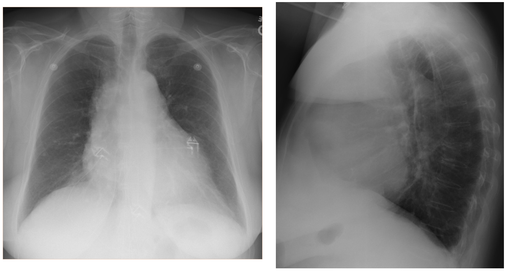
Actual radiographs.
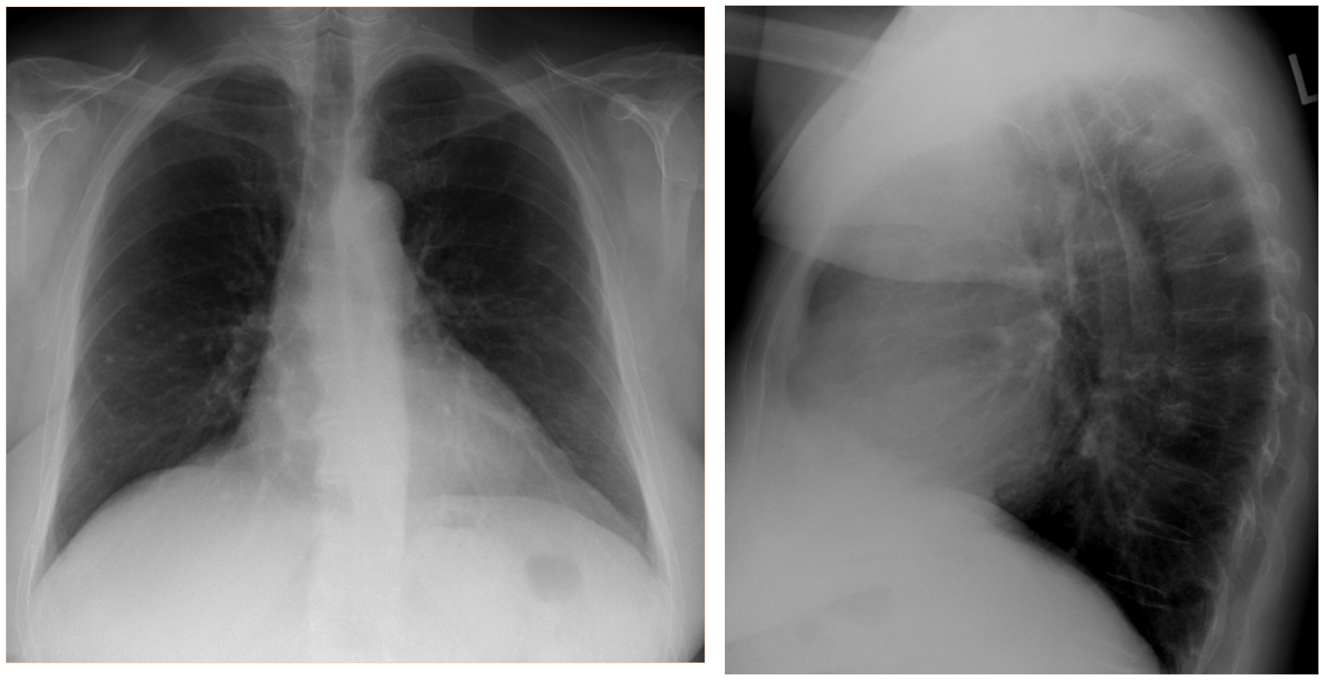
Previous examination one year before.
Let us hear your thoughts and remember that the main objective of this blog is not to get the exact diagnosis (which is not bad of course) but to find the correct approach to the images and the clinical scenario.
Click here for the answer
Here are the main findings as described by Drs Flors and Kuni.
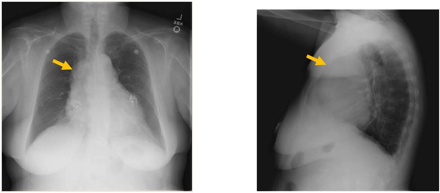
- Main findings:
- CXR: New mediastinal widening, specifically with enlarged cardiac silhouette, widening of the right paratracheal stripe and retrosternal mass like opacity. (Arrows)
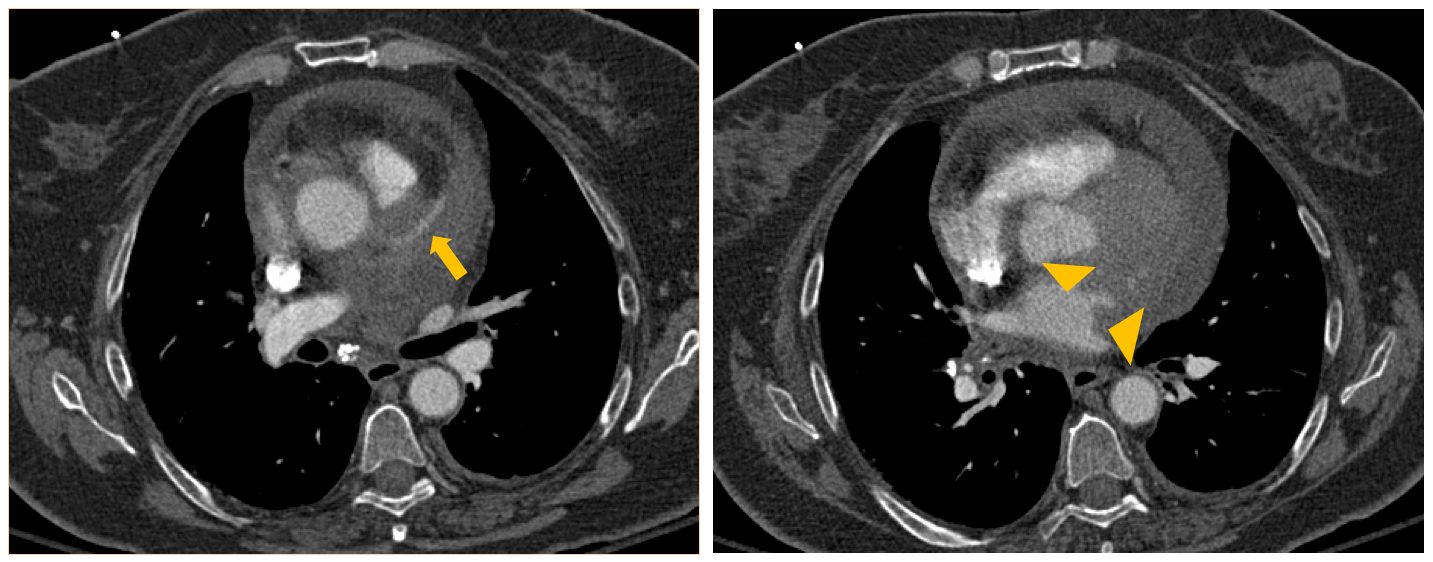
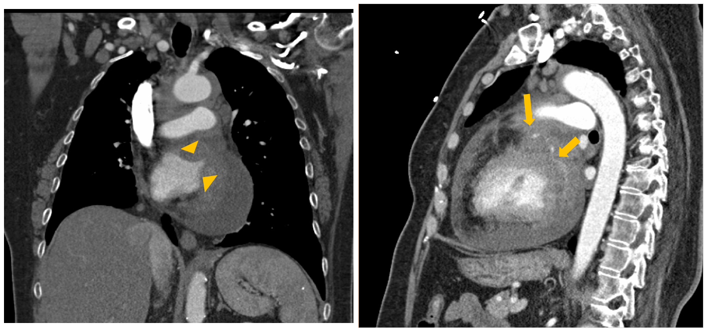
- CT:
- Diffuse pericardial thickening with soft-tissue masses seen encasing the coronary arteries (arrows) and infiltrating the left atrium (arrow heads) and anterior left ventricular wall
- Mediastinal lymphadenopathy
- Moderate pericardial effusion
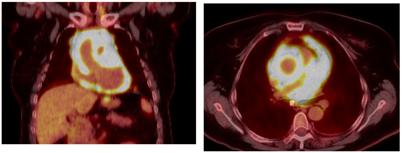
- PET-CT: Extensive circumferential pericardial FDG uptake
Diagnosis: Pericardiocenthesis: Large B-cell Lymphoma
Large B-cell Lymphoma
- Primary cardiac lymphoma is rare, representing only 1.3% of primary cardiac Secondary involvement of the heart in patients with systemic lymphoma is more common, but is still rare.
- Cardiac lymphomas are most commonly diffuse large cell lymphomas and frequently manifest as an ill-defined, infiltrative mass.
- The right atrium is the most common location
- Pericardial thickening or effusion is often a common early feature of the disease. At CT, lymphomas commonly appear as an infiltrating epicardial or myocardial mass that is often isoattenuating to hypoattenuating relative to myocardium. Heterogeneous enhancement after administration of intravenous contrast material is routinely demonstrated.
- A curious feature of cardiac lymphoma is the tendency of the tumor to extend along the epicardial surfaces of the heart, primarily encasing adjacent structures including coronary arteries and the aortic root and with frequent infiltration of the atrial or ventricular walls as seen in this case
- Treatment most commonly includes anthracycline-based chemotherapy and anti-CD20 treatment. Chemotherapy has been used alone or combined with radiation therapy. Palliative surgery has been performed, mainly for tumor debulking.
- The prognosis for patients with either primary or secondary lymphomatous heart involvement is usually poor; late diagnosis is one of the major factors affecting the outcome.
Radiographics. 2012 Sep-Oct;32(5):1369-80. doi: 10.1148/rg.325115126.
From the radiologic pathology archives: cardiac lymphoma: radiologic-pathologic correlation.
Jeudy J1, Kirsch J, Tavora F, Burke AP, Franks TJ, Mohammed TL, Frazier AA, Galvin JR.
- Some points to remember:
- Always compare with previous studies.
- False cardiomegaly may be caused by mediastinal tumours, especially Thymoma and Lymphoma
- Lymphomas often tend to “envelop” structures (heart, kidney, others), especially the most aggressive types.


