This week I am hiking in the mountains, but I have managed to send you this case.
Below you’ll find two radiographs belonging to a 38-year-old woman. Incidental finding.
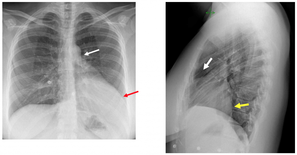
The PA chest radiograph shows apparent cardiomegaly with the prominence of the cardiac apex (Red arrow) and displacement of the heart to the left without pulmonary volume loss.. A lucency or cleft is seen between the aortic knob and the pulmonary artery and also in the retrosternal region in the lateral radiograph (White arrows). In the lateral radiograph, we can see that there is no pathology in the lower lobes but that left ventricle seems to grow posteriorly (Yellow arrow). The right heart border is not seen due to the cardiac displacement to the left.
CT showed very the penetration of lung tissue between the aorta and the pulmonary artery. (Red arrow) In this case, there was some pericardium present (partial absence) seen anteriorly in the contrast CT study. (Green arrows)
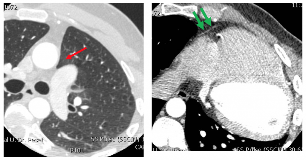
These findings are characteristic of congenital pericardial agenesis. The absence of the pericardium may be partial or total and if asymptomatic does not need treatment. Few of these patients may have cardiac pathology.
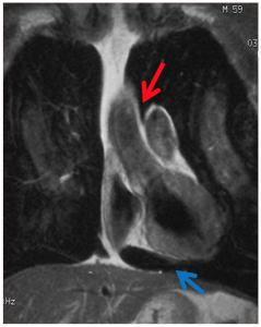
MRI in pericardial agenesis can demonstrate all the mentioned findings, especially the interposition of lung tissue in the aorto pulmonary window (red arrow) and under the left ventricle ( Blue arrow). Cine MR may show abnormal motility of the heart, and in some cases other associated cardiac pathology.
Point to remember: Congenital pericardial agenesis may mimic cardiac, pulmonary or mediastinal pathology. Try to remember the typical disposition of the cardiac silhouette along with a lucency in the aorto pulmonary region and think about this diagnosis.
Here is a good paper related to our case:


PD: By the way, the tree in my picture is a Cork Oak. You can see that the lower part of the trunk has been peeled off to obtain the cork. In a way, the cork layer is like the “pericardium” of the tree…

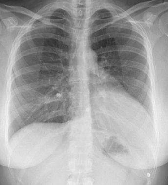
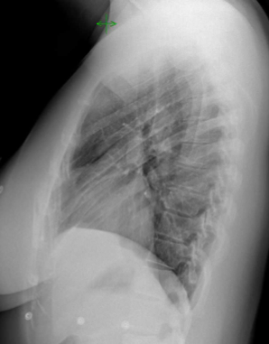


Displacement of cardiac silhouette to the left without any other signs of lung volume loss. Loss of right cardiac border and “Snoopy sign”.
These findings are consistents with partial pericardium agenesis.
The Heart
Absence of pericardium
OT