You did a great job with case 12. Here is our next case, to confirm or prove your diagnostic abilities.
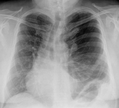
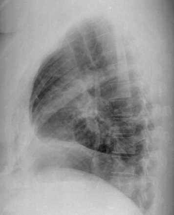
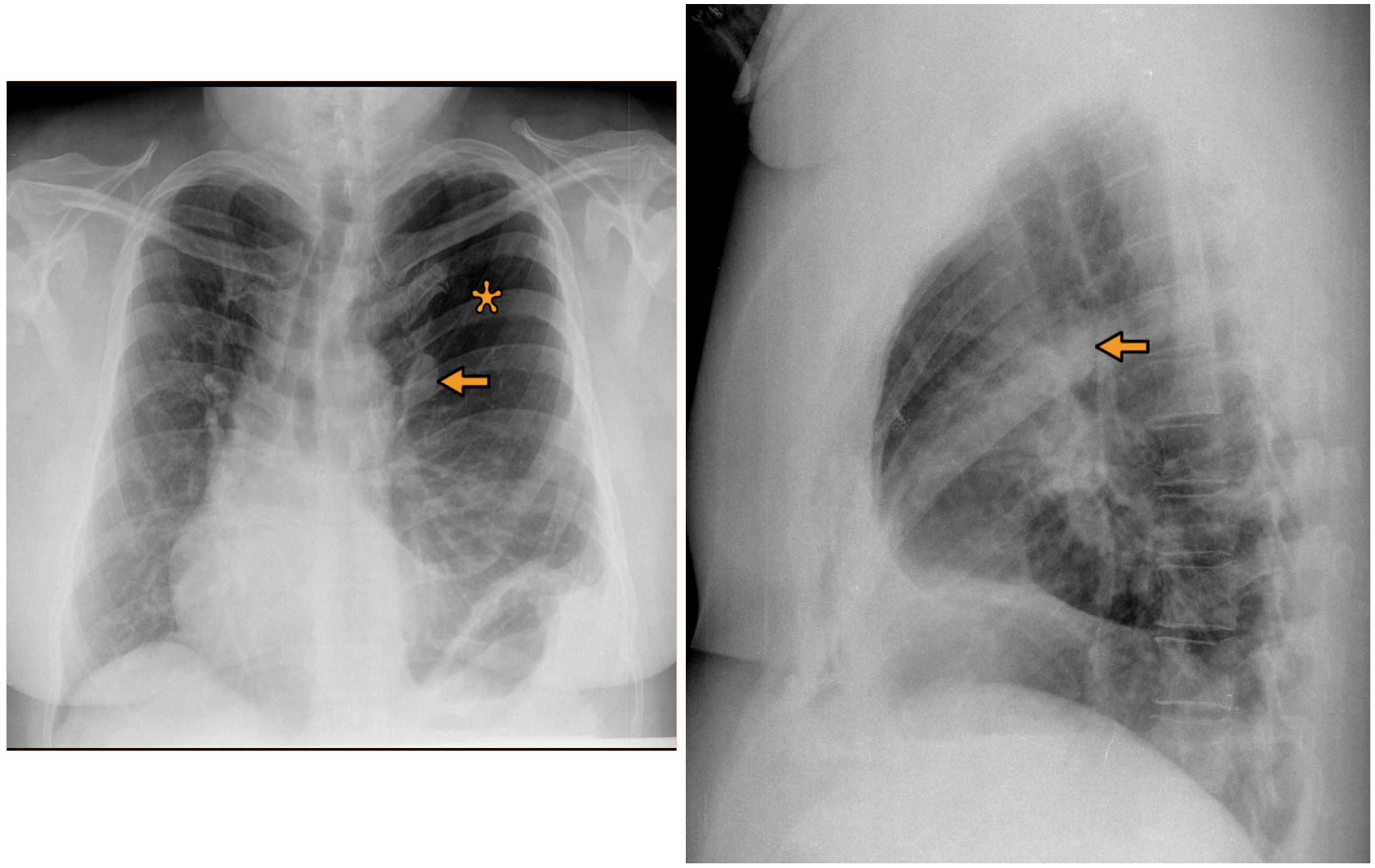
This is a 53 year old woman that had a chest radiographic study for suspicion of respiratory infection.
Let us see what diagnostic possibilities you suggest.
No previous surgery.
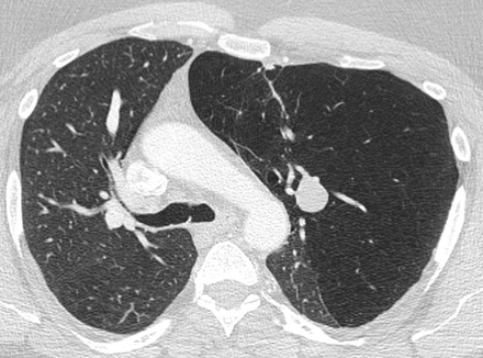
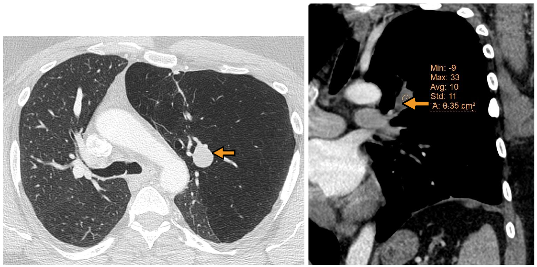
Following your suggestions here you have a couple of CT images.
Final solution:
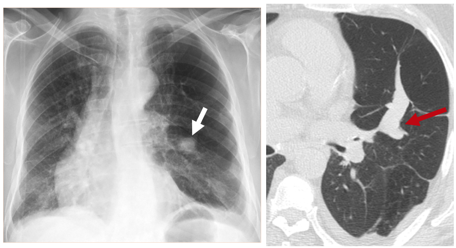
As very well indicated by some of you there is overinflation in the left upper lobe (Asterix) which explains a rightward deviation of the mediastinum. There is also a nodular ovoid lesion in the left hilar region (arrow).
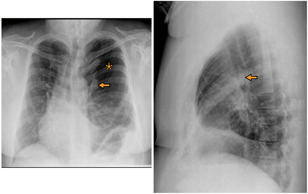
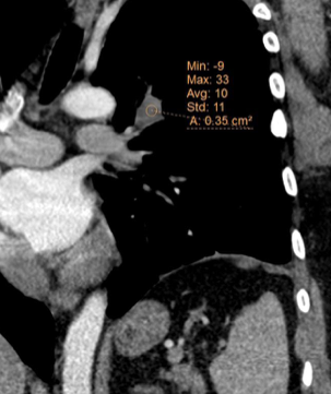
In the CT we can see that the “nodule” has a low density (-9) as it occurs in mucus, and it points to the hilum as it happens with bronchi or vessels.
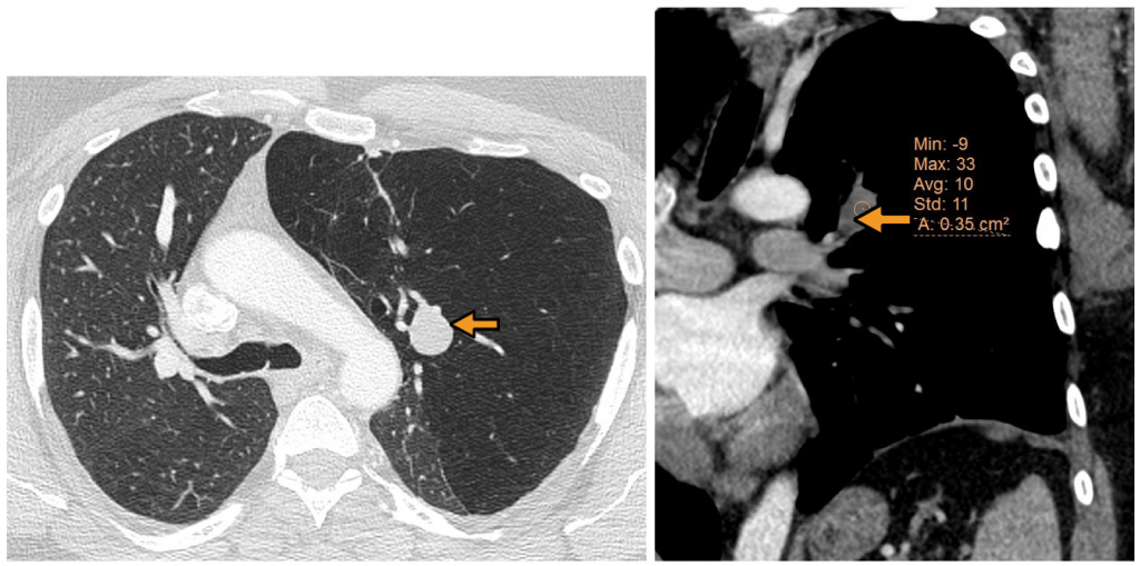
These findings are characteristic of bronchial obstruction with accumulation of mucus in the dilated obstructed brochocele. Air trapping is present with a reduction of vascularity in the left upper lobe.
Final diagnosis: Bronchial atresia
The two main findings of bronchial atresia are:
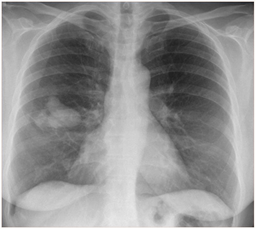
- Bronchocele due to mucus accumulation in the obstructed bronchus. Bronchoceles may look like a well defined nodule, and often giving the image of a “finger in glove” (fig 5).
- Air trapping proximal to the obstructed bronchus.
It is important to recognize this entity, because it may be confused with lung cancer and other significant pathologies.
CT is very useful, especially because it may reveal better that the radiograph the typical tubular morphology of the bronchocele.
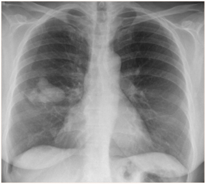
Right upper lobe bronchial atresia: “finger in glove” sign.
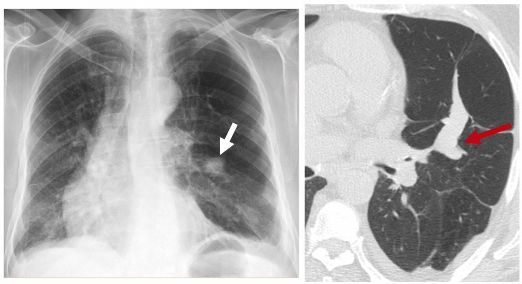 Left bronchial atresia. The chest radiograph shows a nodule ( white arrow), but in CT we may see very well the tubular feature /finger in glove ( red arrow). Air trapping is also present.
Congratulations to all the participants that mentioned the correct diagnosis.
I will be back in September, meanwhile I wish you all a great summer vacation (or winter if you are in the southern hemisphere…).
My hiking continues…
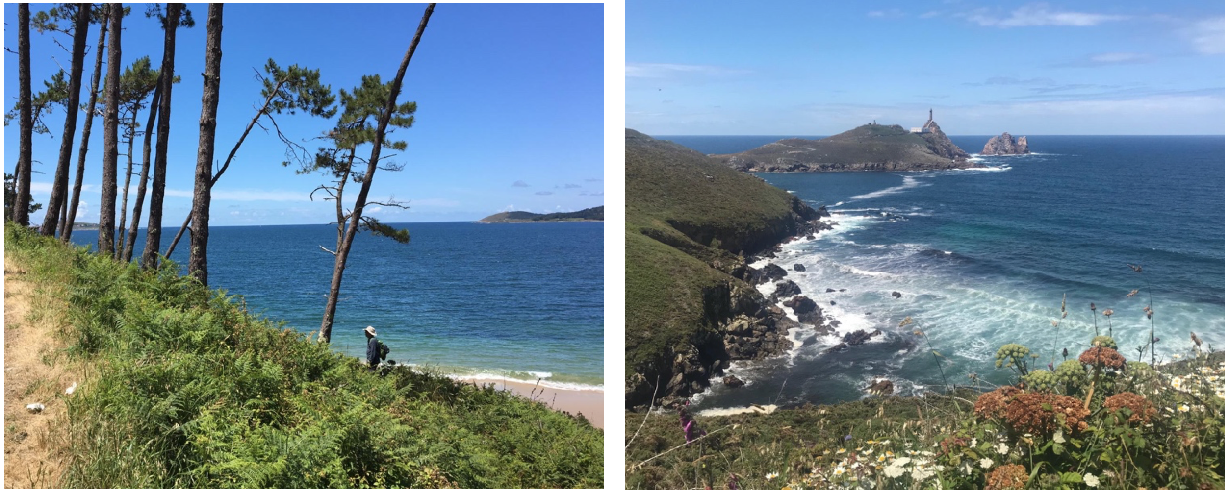
Costa da Morte. Galicia. Spain.