how was your summer? My summer was great and I am ready to present some fresh cases!
Just to warm you up, here is a case recently shown to me in Hospital Dr Peset.
This is a 38 year old male with upper respiratory symptoms.
Final solution:
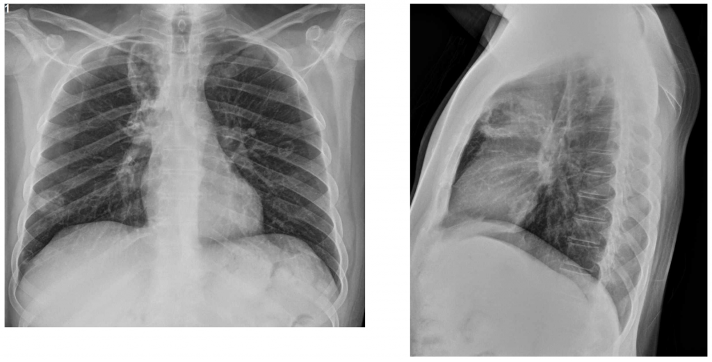
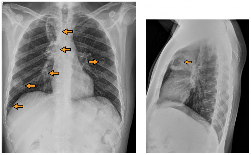
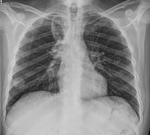
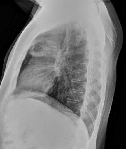
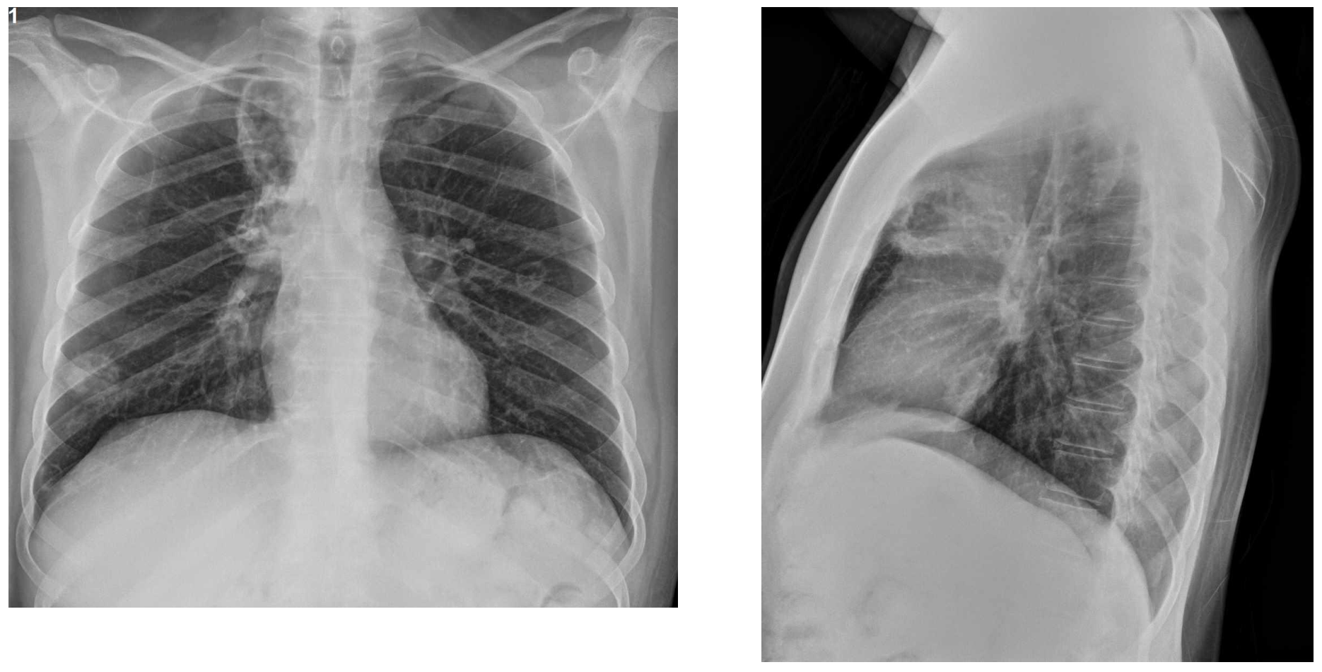
The CXR showed multiple cavitated masses/nodules in both lungs (arrows). The differential diagnosis of cavitated pulmonary lesions is mainly: Tuberculosis and other granulomatous infections, septic embolisms, rheumatoid nodules, metastasis and vasculitis.
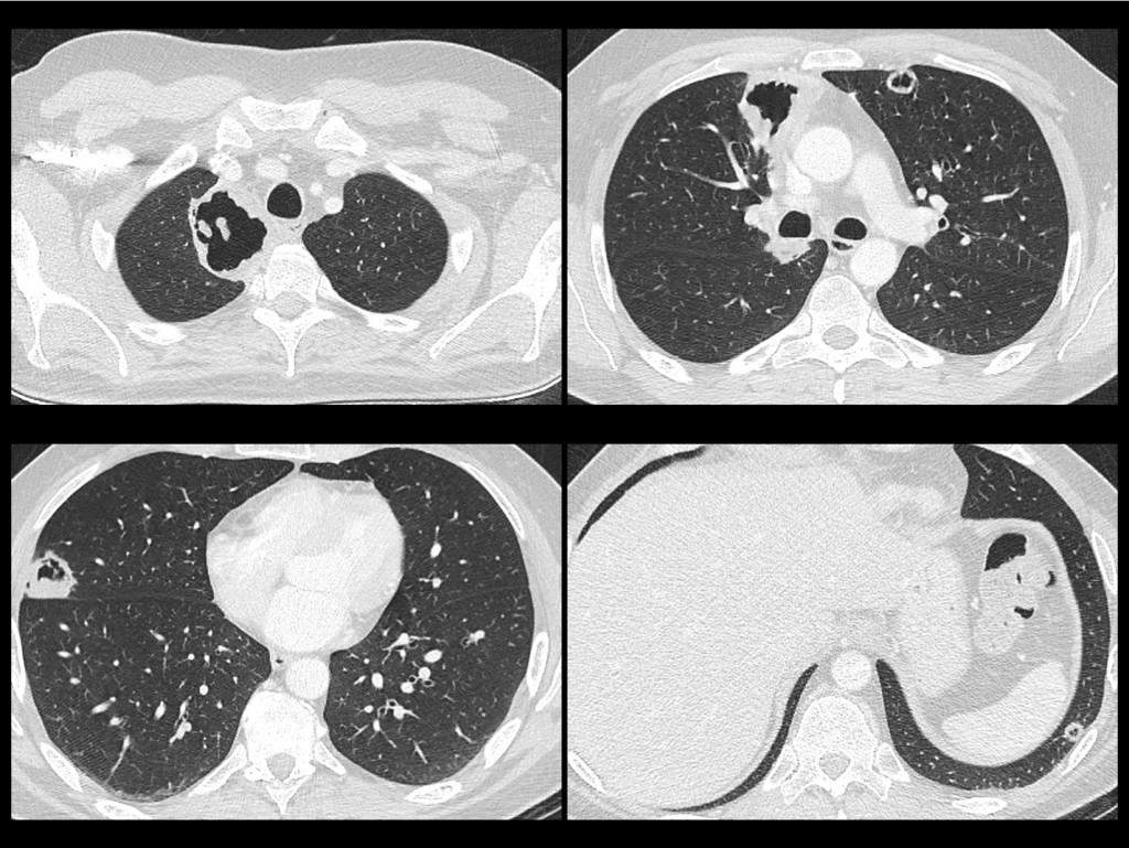
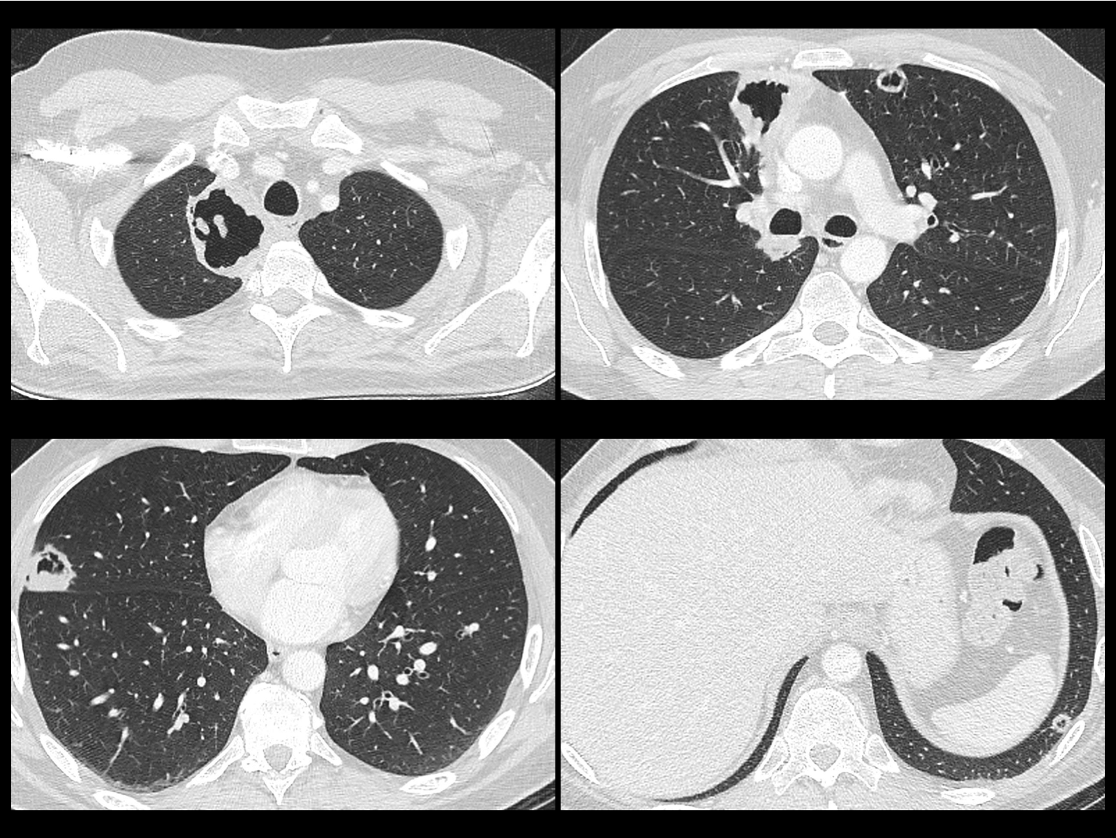
CT Demonstrated the cavitated masses/nodules in both lungs.
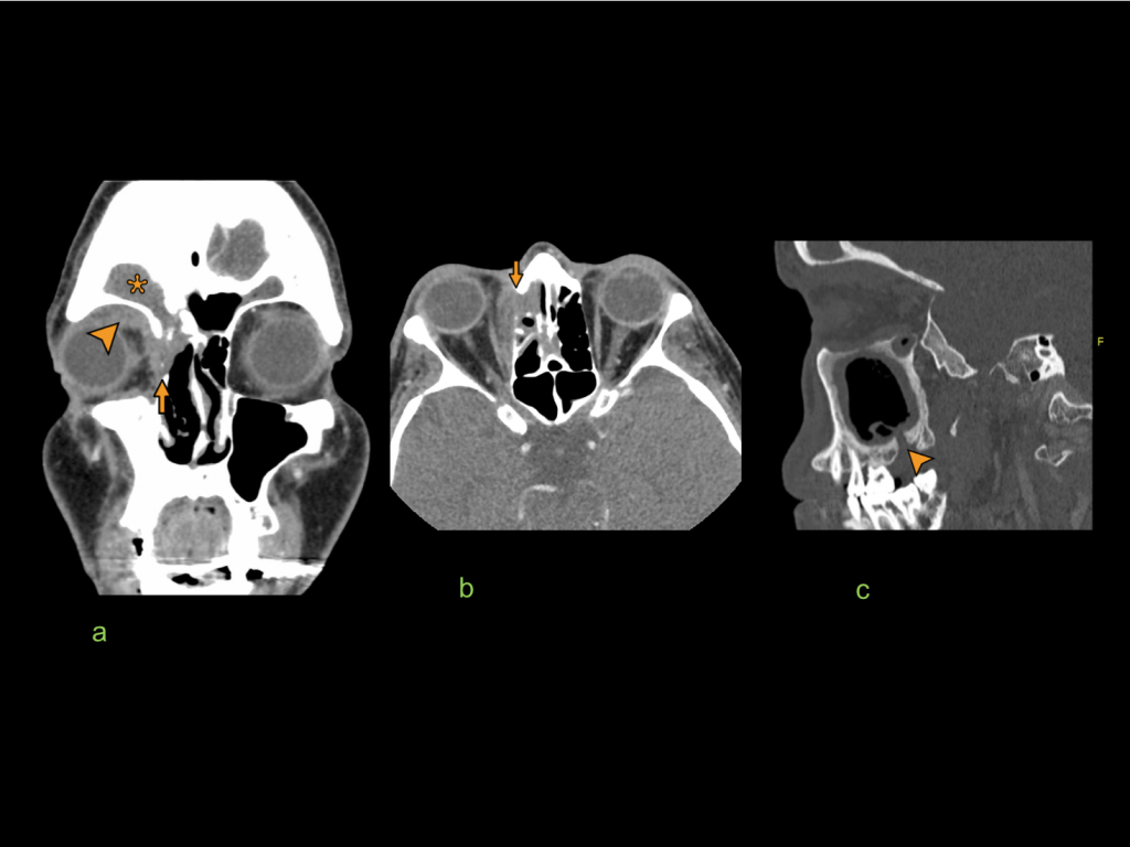
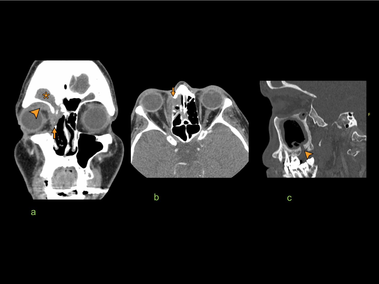
Cranial CT.
Frontoethmoidal Sinusitis with extension to the extraconal orbit (a), and rupture of the lamina propia. (b) There is also an oro-antral fistula (c).
DIAGNOSIS: The association of cavitated lesions with sinusitis and upper respiratory symptoms has to make you think of vasculitis, especially Granulomatosis with polyangiitis ( formerly Wegener).
The cANCA test was positive as it occurs in the majority of cases.
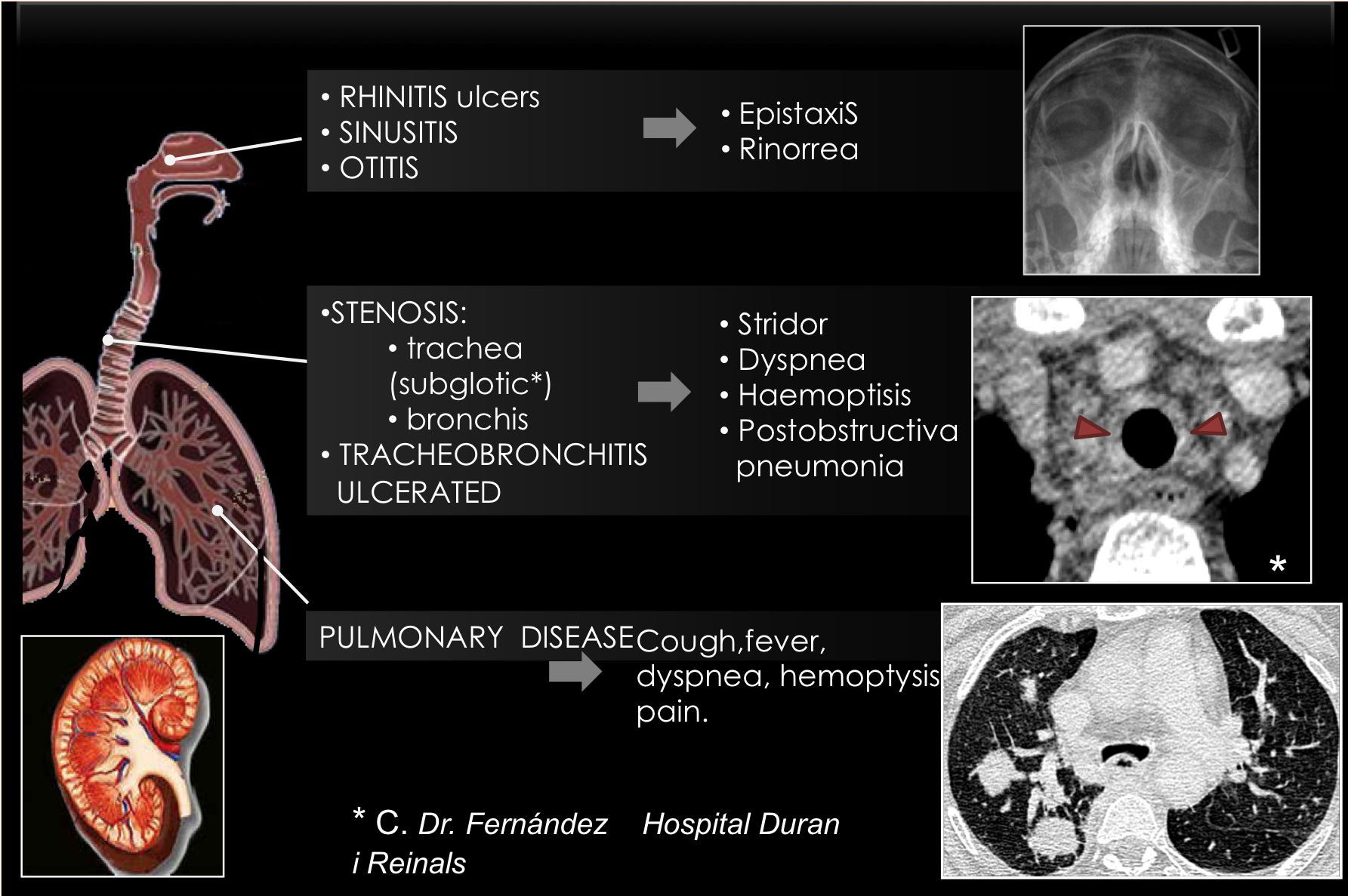
Granulomatosis with polyangiitis is a vasculitis that involves multiple structures and thus has a spectrum of clinical presentations, mainly related to the lungs and upper respiratory tract.
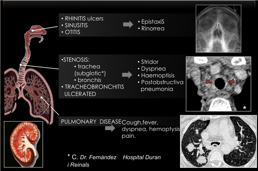
This slide from my friend and colleague Eva Castañer ( Barcelona) shows the main features of Granulomatosis with Polyangiitis (Wegener).
Point to remember:
Cavitated masses in the lungs: Pay attention to the accompanying clinical scenario.
(Remember that cavitation of masses and nodules in the lungs is different from pulmonary cysts.)