Dear friends,
The fall is here and after a hike in the Teruel mountains I collected some mushrooms (“lactarius deliciosus” and “lepista nuda”, the blue one) and now, after tasting them, and perfectly healthy, I am ready to send you a new case.

This is a 73-year-old lady with a fractured hip; she had a this preoperatory chest radiograph. No respiratory symptoms.
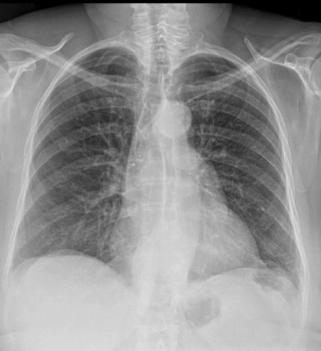
Let us see what you think and more images will follow in a couple of days.
Update:
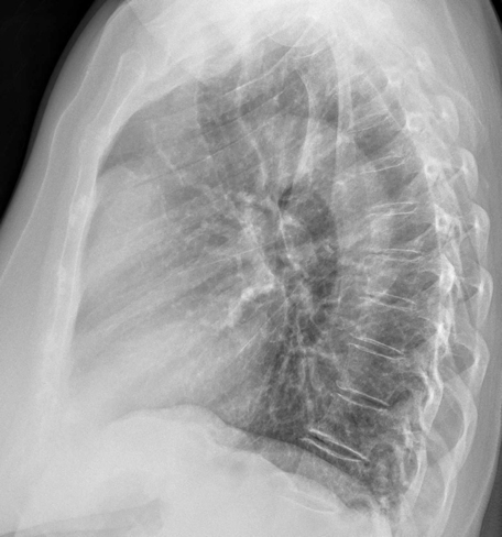
If you asked for the lateral projection here it is.
Does this clarify the case?
Click here for the answer
Final solution:
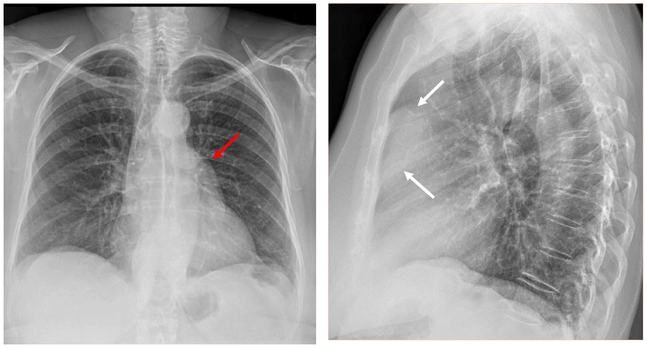
The PA chest radiograph shows a well defined convex lesion In the left border of the mediastinum (red arrow). Through the lesion we can see the hilar vessels. This is a “hilar overlay sign” that indicates that the lesion is not in the hilum and therefore must be anterior or posterior to it.
In the lateral radiograph we can see that a mass is occupying the retrosternal space ( white arrows).Thus we have an anterior mediastinal mass.
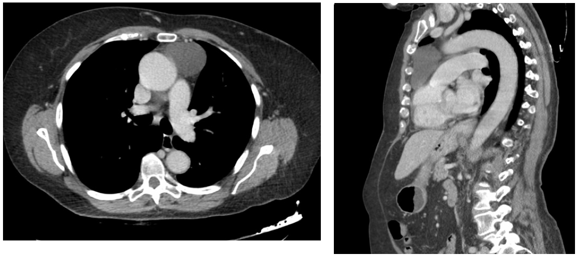
A CT demonstrated a cystic lesion in the anterior mediastinum with no solid component or contrast enhancement.
Diagnosis: Anterior mediastinal lesion; Thymic Cyst
Comment: Cysts in the mediastinum may appear in any compartment. Mediastinal cysts are benign and, unless symptomatic, do not need any treatment. We must not confuse cysts with “cystic” lesions such as Thymoma or Teratoma that should always have a solid component. (CT in cysts shows absence of contrast uptake)
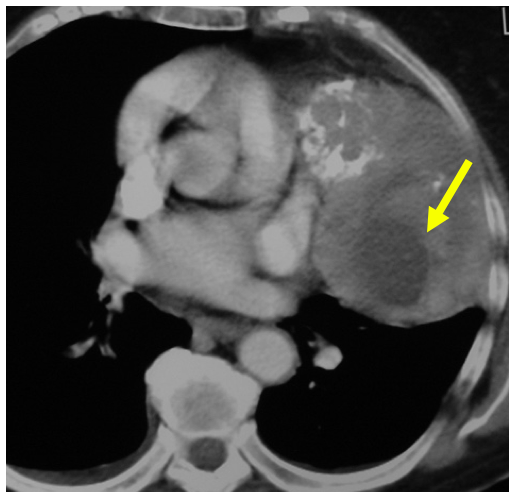
This CT shows an enhancing mass in the anterior mediastinum containing calcium and a cystic component ( yellow arrow) produced by necrosis: Thymoma.
Main points in Case 17:
- Hilar overlay sign indicates most of the time that a mediastinal lesion is situated in the anterior compartment.
- Anterior mediastinal cysts are usually thymic cysts, but pericardial cysts can also be included in the differential diagnosis.
Note: The third Mogul sign, mentioned by one of you, usually refers to the prominent left atrial appendage, but, in a wide sense, anterior mediastinal masses may also produce this sign. Well seen!


Enlarged left pulmonary artery
Lower mediastinal mass, looks calcified, could be cold abscess. Need lateral view and hystory. And a CT would be nice, also.
I see the “sign of the third Mugel” in the left side of the heart.
Hello, I am a PA student and am looking for some help with interpreting CXRs. Please email [email protected] if anyone is willing to help me, it would be much appreciated. Thank you.