 Hello everybody,
I hope you enjoyed your holidays wherever you are. I wish you a wonderful new year!
After case 20 (Tracheobronchomegaly) that was very well interpreted by those who dared to send a diagnosis, here is another challenge.
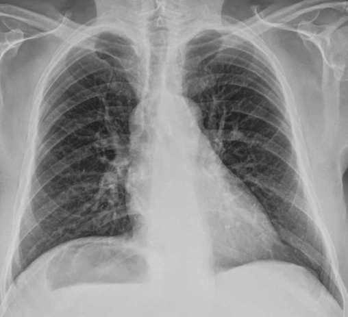
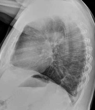
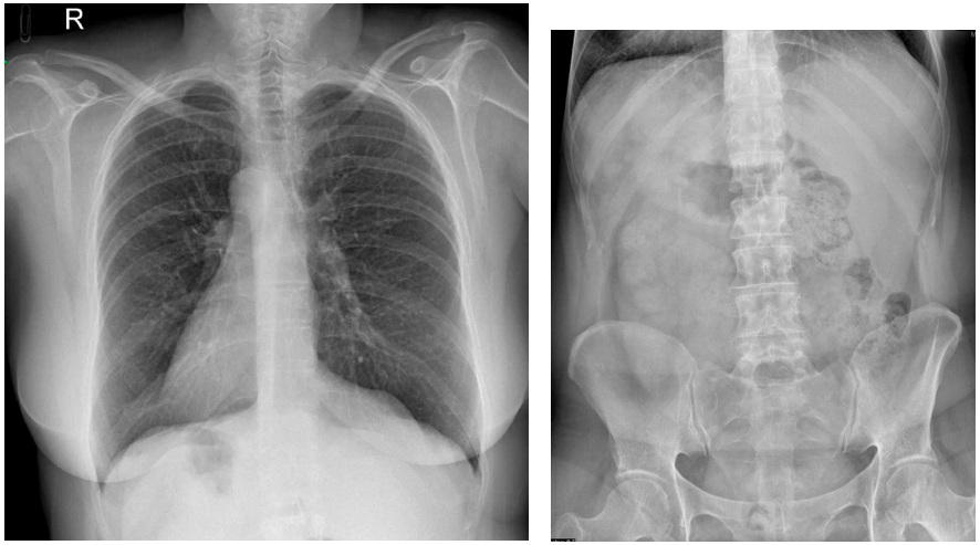
Preoperatory chest radiographic study. No significant clinical symptoms.
Additional information:
Ok friends, some of you mentioned situs inversus, so I am adding some more data to see if you can define better the findings.
Click here for the answer
Solution:
The PA and lateral chest radiographs show an air/fluid under the right hemidiaphragm. Fluid is not usual when the colon is interposed (Chilaiditi). This is the typical image of the stomach, but on the wrong side. The heart is in the normal position.
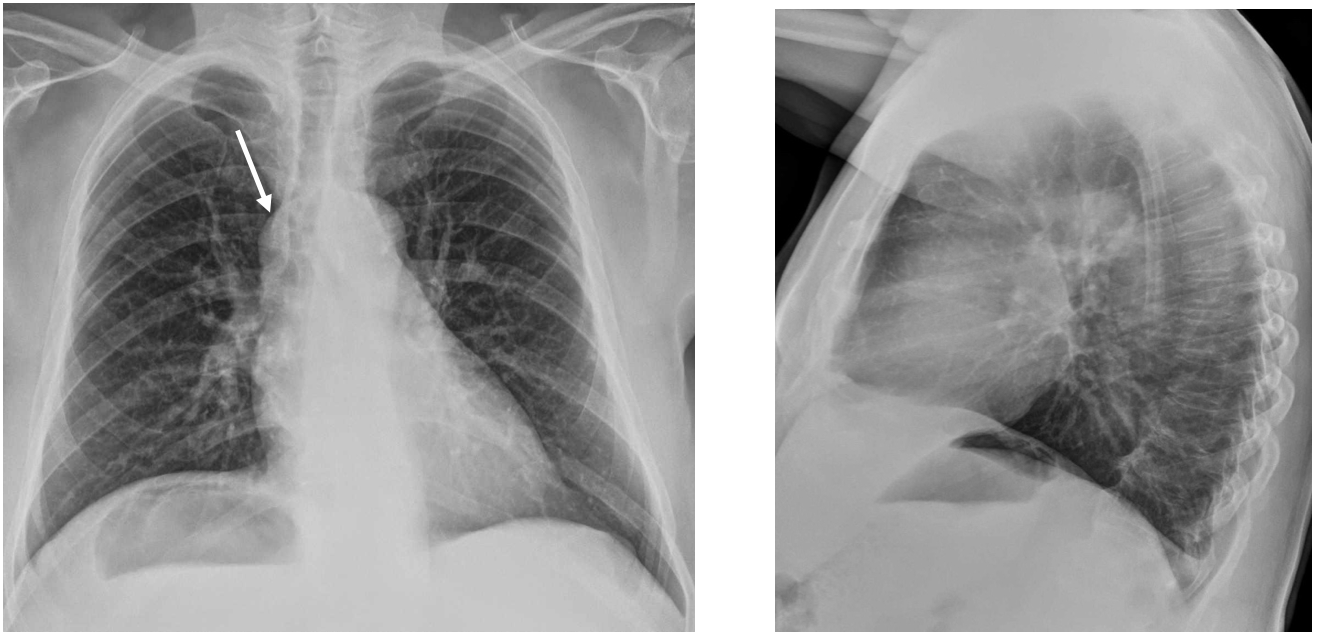
A not so obvious finding is the slight prominence of the azigos. (Arrow) and the fact that both hila seem to be at the same height.
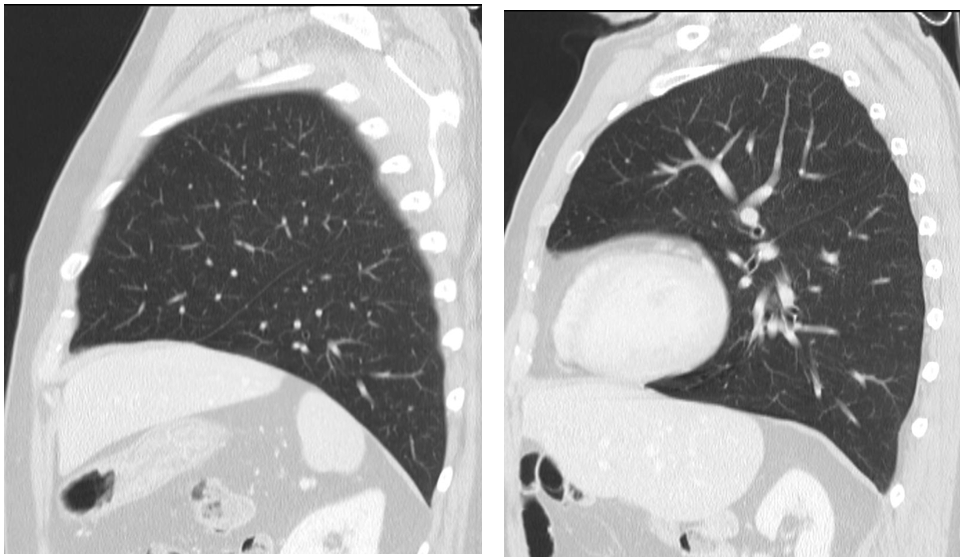
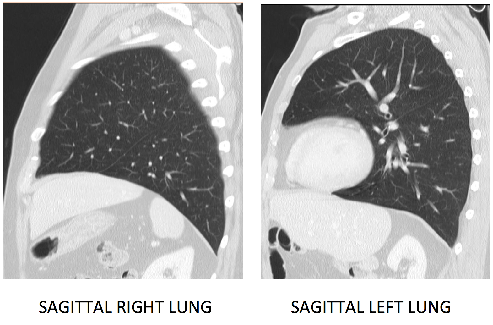
CT: The sagittal images show that both lungs are very similar with one major fissure and no minor fissure. This is, as very well indicated by Pppp in the blog, a Left Isomerism: The patient has two “left lobes”. This is why the hila are higher since in the normal left lung the pulmonary artery is above the bronchus.
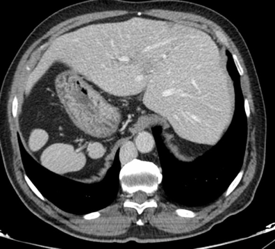
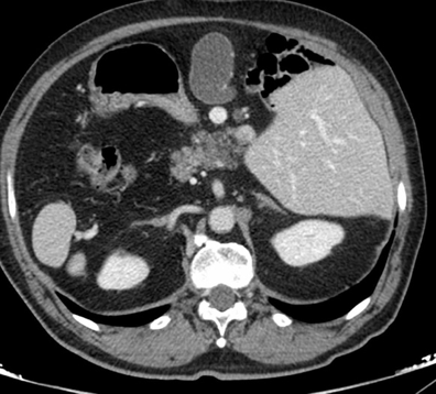
Furthermore, the abdominal CT images (that I did not show you) show several findings:
- Multiple round enhancing nodules in the left hypochondrium: These are spleens: Polisplenia. (Yellow arrows)
- The liver is in the center/right.
- The stomach is in the right side.
- The head of the pancreas, and the gallbladder( Red arrow) are in the midline.
- Interruption of inferior vena cava with azigos continuation, which explains the azigos enlarged in the PA chest radiograph. (White arrows)
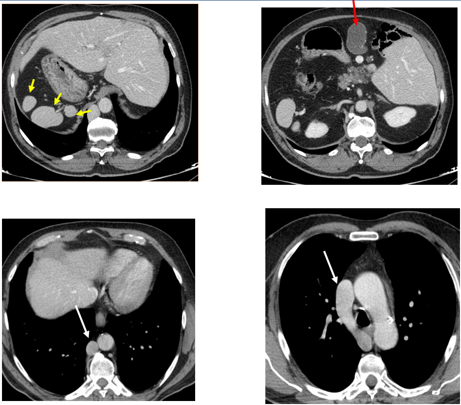
In summary; this patient has a congenital anomaly with abnormal disposition of several organs and associated malformations.
Heterotaxia or situs ambiguous. ( Heterotaxia means “different arrangement”).
In situs inversus, mentioned by some of you, the heart and the stomach are in the right side and the largest lobe of the liver in the left, (Viscero atrial situs following Van Praagh). This is seen in this other case.
If you want to know more about this topic here you have two references that could help you:
- Segmental Approach to Imaging of Congenital Heart Disease
Chantale Lapierre , Julie Déry, Ronald Guérin, Loïc Viremouneix, Josée Dubois, Laurent Garel
Radiographics: Mar 8 2010
https://doi.org/10.1148/rg.302095112
- Describing Congenital Heart Disease by Using Three-Part Segmental Notation
Erica K. Schallert, Gary H. Danton , Richard Kardon, Daniel A. Young
Radiographics: Mar 2 2013
https://doi.org/10.1148/rg.332125086


Situs inversus with levocardia
Situs inversus and levocardia
Air bubble under the right copula; possibly gastric air bubble in situs inversus,
However the right copula is higher than the left; so i might think of Chiladiti sign.
Chilaiditi syndrome
Air underneath the right hemidiaphragm raising suspicion of a gas forming hepatic abscess.
Situs ambiguous
Left isomerism
Heterotaxy syndrome
Abdominal situs inversus with bilateral bilobed lungs
levocardia and bubble in lung