
Dear Friends,
After a complicated case (Case 21: Heterotaxia) here is more straightforward one.
A young man with breathing difficulty when doing physical activity.
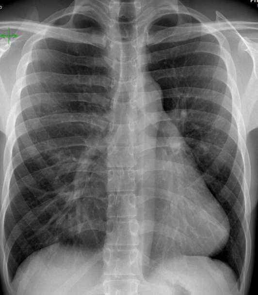
Let’s see what you think!
Click here for the answer

Congratulations to several of you who identified the case as Pectus Excavatum.
Obviously the lateral radiograph is the clue in this case where you can see a profound excavation of the anterior chest.
The most common features in pectus excavatum (as seen in this case)
- Blurring of the right heart border
- Increased density of lower right lung
- Horizontal posterior ribs
- Vertical anterior ribs
- Heart displaced to the left
Most common pitfalls in the diagnosis:
- Middle lobe consolidation or atelectasis ( Silhouette sign).
- Cardiac pathology
- Mediastinal mass
CT is used in severe cases to evaluate the possibility of surgical repair. Axial images provide excellent information regarding the AP diameter of the chest and the angle of the sternum.
In our case no need for treatment was decided.
Restrepo CS, Martinez S, Lemos DF et-al. Imaging appearances of the sternum and sternoclavicular joints. Radiographics. 29 (3): 839-59. doi:10.1148/rg.293055136 – Pubmed citation


